Copyright © 2004, American Society for Microbiology. All Rights Reserved.
Multilocus Sequence Typing Analysis of Human and Animal
Clostridium difficile
Isolates of Various Toxigenic Types
Ludovic Lemee, Anne Dhalluin, Martine Pestel-Caron, Jean-Franc¸ois Lemeland,
and Jean-Louis Pons*
Groupe de Recherche sur les Antimicrobiens et les Microorganismes (G.R.A.M. EA 2656, I.F.R. 23), Universite´ de Rouen, U.F.R. Me´decine-Pharmacie, 76183 Rouen Cedex, France
Received 10 October 2003/Returned for modification 18 December 2003/Accepted 17 February 2004
A multilocus sequence typing (MLST) scheme was developed to study the genetic relationships and
popu-lation structure of 72Clostridium difficileisolates from various hosts, geographic sources, PCR ribotypes, and
toxigenic types (determined by PCR targetingtcdAandtcdBgenes). MLST was performed by DNA sequence
analysis of seven housekeeping genes (aroE,ddl,dutA,tpi,recA,gmk, andsodA). The number of alleles ranged
from five (dutAandddl) to eleven (recA). Allelic profiles allowed the definition of 34 different sequence types
(STs). These STs lacked correlation with geographic source but were well correlated to toxigenic type. The dendrogram generated from a matrix of pairwise genetic distances showed that animal isolates did not constitute a distinct lineage from human isolates and that there was no hypervirulent lineage within the population of toxigenic human isolates (isolates recovered from pseudomembranous colitis and
antibiotic-associated diarrhea did not cluster in distinct lineages). However, AⴚBⴙvariant isolates shared the same ST
that appeared as a divergent lineage in the population studied, indicating a single evolutionary origin. The population structure was further examined by analysis of allelic polymorphism. The dendrogram generated from composite sequence-based analysis revealed a homogeneous population associated with three divergent
lineages, one of which was restricted to Aⴚ Bⴙ variant isolates. C. difficile exhibited a clonal population
structure, as revealed by the estimation of linkage disequilibrium (Ia) between loci. The analysis of alleles
within clonal complexes estimated that point mutation generated new alleles at a frequency eightfold higher than recombinational exchange, and the congruence of the dendrograms generated from separate housekeep-ing loci confirmed the mutational evolution of this species.
Clostridium difficileis a frequent cause of antibiotic-associ-ated diarrhea (AAD) and is responsible for virtually all cases of pseudomembranous colitis (PMC) (5, 48). Since it is recog-nized as a nosocomial enteropathogen (24, 34), many molec-ular typing methods such as pulsed-field gel electrophoresis (PFGE), PCR ribotyping, random amplified polymorphic DNA (RAPD) analysis, or amplified fragment length polymor-phism have been used to investigate nosocomial outbreaks of C. difficile infections (3, 10, 27, 28) or to distinguish relapse from reinfection (4, 26). Conversely, very little data specifically addresses the population genetics and long-term epidemiology of this species. Indeed, several problems remain unclear about the population genetics of this organism. (i) Are there hyper-virulent lineages, which spread by clonal diffusion? (ii) Is there human or animal host specificity? (iii) What is the evolutionary history of the toxin A-negative, toxin B-positive (A⫺B⫹) vari-ant isolates recently reported in human pathogenic situations (1)? (iv) What are the relative rates of mutations and recom-binations in evolutionary dynamics ofC. difficile?
Previous studies, although focusing on short-term molecular epidemiology, tentatively answered some of these questions. Several reports have shown that someC. difficileisolates from different countries and without any direct epidemiological link harbored the same PCR ribotype (49, 50). Furthermore, until
recently, almost all A⫺B⫹isolates also harbored the same PCR ribotype, whatever their geographical origin (2, 42). To-gether, these two results suggest a clonal population structure of this species. Likewise, Rupnik et al. (41) developed toxino-typing and found that many isolates harbored the same toxi-notype despite very different origins, suggesting a clonal diffu-sion of these strains. However, these data were obtained from methods based on DNA banding patterns, which generate re-sults difficult to compare between laboratories despite strenu-ous efforts at standardization. In addition, although PFGE or PCR ribotyping successfully identified clusters of epidemiolog-ically related isolates (7), these methods may be less suitable for global and long-term epidemiological studies and are in-adequate for population genetics analysis.
Multilocus sequence typing (MLST) has been recently de-veloped for the study of clonal relationships within bacterial populations and has been successfully used for population ge-netics and global epidemiological analysis ofNeisseria menin-gitidis(22),Streptococcus pneumoniae(18),Staphylococcus au-reus (16), Streptococcus pyogenes (19), Campylobacter jejuni (15),Salmonella enterica(29), andEnterococcus faecium(23). MLST characterizes multilocus genotypes of bacterial isolates by using 400- to 500-bp intragenic sequences of several (gen-erally seven) housekeeping genes (33). Thus, MLST is similar in principle to multilocus enzyme electrophoresis, which has been largely developed in bacterial population genetics (45, 46) but presents a higher sensitivity due to its ability to detect neutral genetic variations. MLST has also been suggested as offering several advantages over other molecular typing meth-* Corresponding author. Mailing address: U.F.R.
Me´decine-Phar-macie de Rouen, G.R.A.M. (EA 2656), 22 Boulevard Gambetta, F-76183 Rouen Cedex, France. Phone: (33) (2) 35-14-84-52. Fax: (33) (2) 32-88-80-24. E-mail: [email protected].
2609
on May 15, 2020 by guest
http://jcm.asm.org/
ods. First, the data (DNA sequences) are unambiguous and so readily comparable between different laboratories and can be stored in a shared central database to provide a broader re-source for epidemiological studies. Second, evolutionary ge-netics studies can be performed, since MLST describes varia-tions affecting housekeeping genes. In the present study, we describe an MLST analysis ofC. difficilebased on the nucleo-tide sequences of seven housekeeping genes. Using this ap-proach, we study the allelic diversity and population structure of a collection of 72 C. difficile isolates from various hosts, geographic sources, and toxigenic types.
MATERIALS AND METHODS
Bacterial isolates.A total of 72C. difficileisolates from various hosts and geographic sources and collected over a 12-year period were studied. Of these, 64 isolates were recovered from human stools: 36 from patients with AAD, 11 from patients with PMC, and 11 from patients with asymptomatic carriage (the data for 6 human isolates were unknown). Eight isolates from animal hosts suspected of clostridial intestinal infection were also included. Isolates were identified asC. difficileby Gram stain, colony morphology, and fluorescence, API 20A (Bio-Merieux, Marcy l’Etoile, France) biochemical profiles and, for some isolates, by sequencing the first 500 bp of 16S ribosomal DNA (rDNA) and of an internal fragment of thetpigene (14) to confirm their species identification. Toxigenic types were determined by PCR targeting the toxins A (25) and B genes (36). Among the total 72C. difficileisolates, 52 harbored thetcdAgene (encoding toxin A) and thetcdBgene (encoding toxin B), 8 harbored a deleted variant of
tcdAgene and thetcdBgene (A⫺B⫹variants), and 12 lacked thetcdAand the
tcdBgenes.
PCR ribotyping.PCR ribotyping was performed according to a procedure described elsewhere (7) to assess the genetic diversity of the isolates included in the present study, particularly their lack of a direct epidemiological link.
MLST.Seven housekeeping loci were selected for the characterization ofC. difficileisolates by MLST (Table 1):aroE(shikimate dehydrogenase),ddl(D -alanine:D-alanine ligase), dutA (dUTP pyrophosphatase),gmk (guanylate
ki-nase),recA(recombinase), sodA(superoxide dismutase), andtpi (triosephos-phate isomerase). The choice of these housekeeping genes was based on their use in MLST schemes of other bacterial species and/or on the availability of sequence data fromC. difficile(http://www.sanger.ac.uk/) and from other species. Only one copy of each of the seven housekeeping genes was found on theC. difficile630 genome.
To prepare a DNA sample for PCR amplification, a bacterial colony was taken
from blood agar culture and resuspended in 1 ml of distilled water in a micro-centrifuge tube. The sample was then boiled for 20 min prior to being micro-centrifuged for 2 min to settle bacterial debris, and 10l of supernatant, containing the genomic DNA, was used for subsequent PCR amplification. Internal 400- to 500-bp fragments of the selected genes were amplified with primers (Table 1) designed from sequence alignments of homologuous genes from low %G⫹C gram-positive bacteria.
PCRs were performed on a GeneAmp System 2400 thermal cycler (Applied Biosystems) in a final volume of 50l containing 0.5M concentrations of each primer, 200M concentrations of each deoxynucleoside triphosphate, and 1.25 U ofTaqDNA polymerase (Applied Biosystems) in a 1⫻amplification buffer (10 mM Tris-HCl [pH 8.3], 50 mM KCl, 2.5 mM MgCl2). The PCR mixtures were
heated for 3 min at 95°C and then a touch-down procedure followed, consisting of 30 s at 95°C, annealing for 30 s at temperatures decreasing from 60°C to 50°C during the first 11 cycles (with 1°C decremental steps in cycles 1 to 11), and ending with an extension step at 72°C for 30 s. Forty cycles were performed. PCR products were then purified with a QiaQuick gel extraction kit (Qiagen) and sequenced (200 to 500 ng of DNA) with PCR forward or reverse primers by using an ABI-PRISM BigDye terminator sequencing kit on an ABI-Prism 310 genetic analyzer (Applied Biosystems). Different sequences of a given locus were given allele numbers, and each unique combination of alleles (multilocus allelic pro-file) was assigned a sequence type (ST). Single point polymorphisms were as-sessed by sequencing both DNA strands from two separate PCR experiments.
Computer analysis of MLST data.Clustering of the 72 isolates from the matrix of pairwise similarities between the allelic profiles was performed by using the START program (http://www.mlst.net) by the unweighted pair-group method with arithmetic averages (UPGMA). Nucleotide sequences were aligned by using BioEdit sequence alignment editor (http://www.mbio.ncsu.edu/BioEdit/bioedit .html). Average numbers of nucleotide differences between alleles and ratios of nonsynonymous to synonymous substitutions (dN/dS) were calculated to test the degree of selection operating on a locus by using the START program. Gene trees were constructed by using the neighbor-joining method and bootstrapping algorithms contained in the PHYLIP package (http://www.genebee.msu.su /services/phtree㛭full.html). Nucleotide composite sequences (2,217 bp, derived from seven concatenated gene fragments) were also aligned, and a phylogenetic dendrogram was generated by using the same procedure.
[image:2.603.43.542.82.288.2]The index of association (Ia) between alleles (47) was used to test for linkage disequilibrium between alleles of the seven loci analyzed (http://www.mlst.net). The observed variance in the distribution of allelic mismatches in all pairwise comparisons of the allelic profiles was compared to that expected in a freely recombining population (linkage equilibrium). The significance of the difference in the observed and expected variances was evaluated by computing the maxi-mum variance in the distribution of allelic mismatches obtained by using 100 randomizations of the data set.
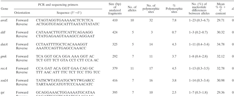
TABLE 1. Genetic polymorphism of the seven housekeeping genes analyzed by MLST
Gene
PCR and sequencing primers Size (bp) of analyzed fragments
No. of alleles
No. of polymorphic
sites
% Polymorphic
sites
No. (%) of nucleotide differences between alleles
Mean % G⫹
C content
dN/dSa Orientation Sequence (5⬘33⬘)
aroE Forward CTAGTAGGTGAAAAACTCTCTCA 410 10 32 7.8 1–23 (0.3–6.7) 29.71 0.0682 Reverse ACTGGTGTAGCATTTAATATTATATC
ddl Forward CATAAACTTGTTCATTCAGAAGG 424 5 3 0.7 1–3 (0.2–0.7) 30.32 0.1654 Reverse CTATGAGAAGTAAAGCCAGGAAT
dutA Forward CCTAATTTTGCTCACAAAGGT 325 5 14 4.3 1–11 (0.4–3.4) 34.78 0.1125 Reverse AAATCCAGTTGAGCCAAACC
gmk Forward TCA GGT GCA GGA AAA GGT AC 292 7 11 3.7 1–8 (0.4–2.8) 32.12 0.1074 Reverse TCT GTT TCT GTA CCT CTT CCA AC
recA Forward CCA GAT ACA GGT GAA CAG GC 379 11 17 4.5 1–13 (0.3–3.5) 32.78 0.0000 Reverse TTT AAC ATT TTC TCT TCC TTG TCC
sodA Forward TATSCWTATGATGCWYTWGARCC 416 7 16 3.8 1–14 (0.3–3.4) 30.98 0.0813 Reverse TARTAAGCATGYTCCCAAACATC
tpi Forward GCAGGAAACTGGAAAATGCATAA 395 7 10 2.5 1–7 (0.3–1.8) 29.36 0.2611 Reverse CAGATTGGCTCATATGCAACAAC
aRatio of nonsynonymous to synonymous substitutions.
on May 15, 2020 by guest
http://jcm.asm.org/
Another estimation of recombination rates was made by using the BURST program (http://www.mlst.net), which defines clonal complexes (groups in which every isolate shares at least five identical alleles with at least one other isolate) and characterizes ancestral genotypes and their corresponding single-locus vari-ants (SLVs; isolates that differ at only one of the seven loci) within these clonal complexes. Comparisons between the sequences of the alleles in each of the SLVs and in the corresponding putative ancestral genotype allowed to us to determine the relative rates of mutations and recombinations in the short-term evolution of the population studied (21).
Nucleotide sequence accession numbers.Nucleotide sequences of the internal fragment genes analyzed in the present study have been deposited in the GenBank database under accession numbers AY533246 to AY533255 (foraroE), AY533256 to AY533260 (fordutA), AY530799 to AY530802 (forddl), AY533261 to AY533267 (forgmk), AY533268 to AY533278 (forrecA), AY533279 to AY533285 (forsodA), and AY533286 to AY533292 (fortpi).
RESULTS
Allelic variation in C. difficile. Data reporting the allelic
variations of the seven housekeeping genes are summarized in Table 1. The number of individual alleles for each of the seven housekeeping genes ranged from 5 fordutAandddlto 11 for recA(forddl; one of these five alleles was a null allele, since several amplification attempts with various primers spanning over the whole gene remained unsuccessful for 16 strains). The
number of polymorphic sites on a given locus varied from 3 (forddl) to 32 (foraroE), and the number of nucleotide dif-ferences between alleles of a given locus varied from one to 3 for ddl and from one to 23 for aroE. The variations in the sequences extended over the whole stretch of each of the seven genes investigated (data not shown). Most polymorphisms re-sulted in synonymous substitutions, the ratios of nonsynony-mous to synonynonsynony-mous substitutions (dN/dS) varying from 0 (for recA) to 0.2611 (fortpi). These low ratios indicate a limited contribution of environmental selection to the sequence vari-ations in the seven housekeeping genes analyzed; therefore, these loci are assumed to be suitable for population genetic study.
MLST analysis ofC. difficileisolates.The lack of an
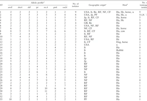
epide-miological link between the 72 isolates studied was confirmed by PCR ribotyping; a total of 62 PCR ribotypes were recorded. Among these 72 isolates, MLST generated 34 different STs (Table 2). The majority of these (22 of 34 STs) were repre-sented by single isolates. Among STs shared by several isolates, the most frequently encountered were ST1 (9 isolates), ST2 (8 isolates), ST3 (5 isolates), and ST4 (5 isolates). No correlation was found between genotype and geographic origin: for exam-TABLE 2. Characteristics of the 34 allelic profiles (STs)
ST Allelic profile
d
No. of
isolates Geographic origina Hostb
No. of toxinogenic
isolatesc
aroE dutA ddl tpi recA gmk sodA
1 2 2 2 1 2 1 2 9 USA, It, Be, RF, NF, CF Ha, Hc, horse, u 9
2 4 3 5 3 3 3 4 8 USA, Jp, PF Ha, Hc, u 8 (A⫺B⫹)
3 2 2 2 1 1 1 5 5 Jp, It, RF, CF Ha, horse 5
4 5 2 2 1 1 1 2 5 RF, NF Ha, Hc 5
5 2 2 2 2 1 1 2 4 GB, Be Ha 4
6 2 2 2 2 1 2 2 3 USA, NF, RF Ha 3
7 6 2 3 1 6 1 1 3 NF, CF Hc, horse 0
8 8 5 5 5 7 6 6 3 It, RF, CF Ha, cow 2
9 5 2 4 1 1 2 1 3 It, RF Ha 3
10 7 4 5 1 1 1 2 3 RF, NF Ha 3
11 1 1 1 1 1 1 1 2 USA, RF Hc 0
12 2 2 2 1 1 2 2 2 It, CF Dog, horse 2
13 3 2 1 1 1 2 3 1 USA u 1
14 3 2 3 1 1 4 2 1 It Ha 0
15 2 2 2 1 1 2 5 1 It Rabbit 1
16 2 2 1 2 1 1 5 1 It Ha 1
17 5 2 2 2 1 1 1 1 It Ha 1
18 6 2 3 1 4 1 1 1 Jp Ha 0
19 5 2 4 2 1 2 1 1 Jp Ha 1
20 6 2 3 1 5 1 1 1 RF Hc 0
21 2 2 1 4 1 2 2 1 RF Hc 0
22 2 2 2 1 7 5 6 1 NF Hc 1
23 2 2 2 6 1 1 2 1 It Ha 1
24 5 2 4 1 8 2 1 1 NF Ha 1
25 7 4 4 1 1 1 2 1 NF Ha 1
26 4 3 5 7 9 7 5 1 NF Hc 0
27 2 2 2 2 2 1 1 1 RF Ha 1
28 9 2 2 1 1 1 7 1 RF Ha 1
29 8 5 5 5 10 6 6 1 RF Ha 1
30 3 2 2 1 11 2 1 1 RF Ha 1
31 3 1 1 1 3 2 1 1 RF Ha 1
32 2 2 2 4 1 2 2 1 RF Hc 0
33 10 2 2 1 2 1 2 1 PF Ha 1
34 8 5 2 5 7 6 6 1 PF Ha 1
aBe, Belgium; It, Italia; Jp, Japan; NF, Nancy (France); RF, Rouen (France); CF, Caen (France); PF, Paris; USA; United States of America; GB, Great Britain. bHa, Human adult; Hc, Human child; u, unknown.
cA⫹B⫹isolates, except for ST2 (A⫺B⫹isolates). dNumbers are identification numbers of alleles.
on May 15, 2020 by guest
http://jcm.asm.org/
[image:3.603.45.545.82.426.2]ple, ST1 isolates were from Belgium, Italy, Japan, and the United States and from different French hospitals; ST2 isolates were from the United States, Japan, and France. Conversely, a good correlation was observed between STs and toxigenic types: for example, ST1, ST3, ST4, and ST5 isolates were all A⫹B⫹, whereas ST7 and ST11 isolates were all nontoxigenic, and ST2 isolates were all A⫺B⫹variant isolates. In addition, the same correlation was also observed for four clonal com-plexes (including 18, 3, 3, and 2 STs, respectively) that were characterized by BURST analysis (http://www.mlst.net; data not shown).
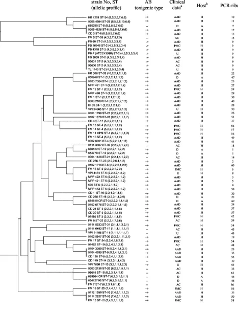
The results of clustering of the allelic profiles by UPGMA are shown in Fig. 1. The dendrogram confirms the correlation between toxigenic type and ST: only ST8 comprised both tox-igenic and nontoxtox-igenic isolates, and all A⫺ B⫹isolates are included in the ST2. This ST differed from ST26 (the nearest ST) in four of the seven alleles and from all other STs in at least six of the seven alleles. The dendrogram reveals also that animal isolates (belonging to ST1, ST3, ST7, ST8, ST12, and ST15) did not constitute a distinct subpopulation from human isolates. Toxigenic animal isolates clustered with toxigenic hu-man isolates, and nontoxigenic animal isolates clustered with nontoxigenic human isolates, with the exception of ST8 (one nontoxigenic animal with two toxigenic human isolates). Con-cerning human isolates, PMC isolates belonged to 9 STs which contain only A⫹B⫹isolates except for ST2 which contains only A⫺B⫹variant strains. They did not cluster in distinct lineages from AAD human isolates, and thus no lineage could be char-acterized as PMC specific. Clustering analysis also revealed very divergent STs (ST8, ST26, ST29, ST34, and ST2), which differed in at least six of the seven alleles from all other STs. The isolates of ST2 (all A⫺B⫹variant isolates) sharedtpi3, gmk3, andsodA4alleles, which were not found among any of the other isolates and thus could be alternative markers of this lineage.
Composite sequence-based analysis.In order to determine
the overall divergence of the sequenced gene fragments of the loci studied, the sequences of six loci (withoutddlbecause of lack of sequence data for the null allele) were spliced together to obtain a concatenated composite sequence for each of the isolates. The identity between the 72 composite sequences was found between 96.8 and 100%. A dendrogram created from the matrix of pairwise sequence divergences of composite se-quences is shown in Fig. 2. A majority of strains constituted a homogeneous population because of the very high degree of conservation observed in the housekeeping genes studied. Nevertheless, 3 divergent lineages were characterized: the first contained all A⫺B⫹variants (ST2) and a nontoxigenic isolate (ST26); the second contained ST8, ST29, and ST34; and the third was restricted to one isolate (ST22).
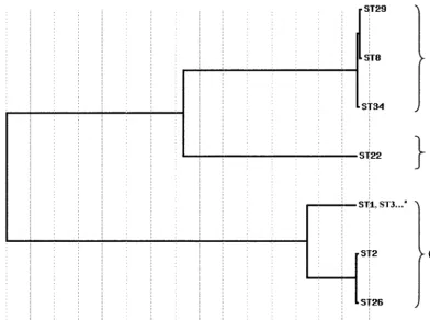
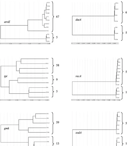
Dendrograms based on allelic variation of each
housekeep-ing gene. Dendrograms from the alleles of the six separate
housekeeping genes were found to be congruent, as shown in Fig. 3 (ddlwas not analyzed as upper justified). A clear bifur-cation into majority and minority allelic populations was ob-served for all of the six genes studied. ST8 (three isolates), ST29 (one isolate), and ST34 (one isolate) belonged only to the minority allelic population of these six housekeeping genes. ST2 (variant A⫺B⫹isolates) belonged to the majority allelic
population fordutA, recA, andsodAloci and to the minority allelic population foraroE,tpi, andgmkloci.
The five most divergent isolates (belonging to STs 8, 29, and 34), clustered in a distantly related lineage on the dendrograms constructed from both the multilocus allelic profiles (Fig. 1) and the composite sequence analysis (Fig. 2). In addition, considering the separate housekeeping genes, they differed in six of the seven alleles from all of the remaining isolates in-vestigated. To confirm that these isolates actually belonged to C. difficile, we sequenced the first 500 bp of their 16S rDNA genes and an alternative species-specific target, the tpi gene encoding triosephosphate isomerase (14). 16S rDNA analysis revealed that these isolates shared 99.4% identity with theC. difficile ATCC 9689 strain and that they shared only 93% identity with C. sordelliiand C. bifermentanstype strains. On the basis of tpi sequences, these isolates were grouped in a cluster having at least 96.2% homology with theC. difficiletype strain sequence, well separated (81% homology) fromC. sor-dellii and C. bifermentans type strain sequences (14). Thus, these isolates diverging from the whole population studied belonged unambiguously toC. difficile.
Estimation of the relative contributions of recombination
and mutation to genomic evolution ofC. difficile.A quantitative
analysis of the linkage between alleles from the different loci was performed by calculating the index of association (Ia) (47).
When all of the 72 isolates were included in the analysis, significant linkage disequilibrium was detected (Ia⫽1.78). At
the level of STs (one isolate from each ST to avoid bias due to a possible epidemic population structure), significant linkage disequilibrium was also detected (Ia⫽0.89). After exclusion of
the five genetically most divergent isolates, significant linkage disequilibrium between alleles of the remaining 67 isolates or the 31 STs was also detected (Ias⫽1.53 and 0.50 for isolates
and STs, respectively).
Another estimation of the relative contributions of recom-bination and mutation to clonal divergence was made by using the method and the criteria described by Feil et al. (21). It was estimated that point mutation generated new alleles at a fre-quency more than eightfold higher than recombinational ex-change. These analyses, together with the congruence of trees constructed from individual loci, indicate that mutation events appeared much more frequently than recombination events in the population studied.
DISCUSSION
The aim of the present study was to provide a molecular approach that should be suitable for population genetics and global epidemiology analysis ofC. difficile. Molecular methods such as PFGE (27) and PCR ribotyping (10) have been dem-onstrated accurate for short-term epidemiology ofC. difficile but are inadequate for long-term epidemiology or population genetics of this organism. Hence, we chose to develop an MLST scheme, since MLST indexes neutral variations accu-mulating relatively slowly within the nucleotide sequences of housekeeping genes, which are thus suitable for phylogenetic analysis.
We determined the degree of allelic variations in seven housekeeping genes of 72C. difficileisolates from various hosts and geographic sources. The degree of isolate differentiation
on May 15, 2020 by guest
http://jcm.asm.org/
FIG. 1. Dendrogram showing cluster analysis (UPGMA) of the 72C. difficileisolates. Clinical data: D, diarrhea; AC, asymptomatic carriage; U, unknown. Other abbreviations are as defined in the text. Host: H, Human; A, Animal.
on May 15, 2020 by guest
http://jcm.asm.org/
by MLST (34 STs among 72 isolates) appears to be adequate for use in a population genetics setting. The allelic polymor-phism (number of distinct STs among the isolates and number of polymorphic sites per locus) was comparable to that of S. aureus(16), a species whose population structure is very clonal (20), but lower than those of N. meningitidis and S. pneu-moniae, two naturally recombinant species (21, 22). Of note, PCR ribotyping generated a higher number of genotypes than MLST. However, since our study mainly aimed at population genetics, we needed to analyze epidemiologically unrelated isolates, which therefore exhibited numerous different PCR ribotypes, whereas MLST genotypes reflect more ancestral genetic relationships.
This MLST analysis allowed us to study the possible corre-lations between STs and geographic source, host, toxigenic type, and pathogenic potential. We could not detect any relation between ST and geographic origin. This lack of cor-relation was also seen forC. difficilewith other typing methods such as toxinotyping (41) and PCR ribotyping (50) and forS. aureuswith MLST (17). Thus, at least some of the STs should be regarded as stable subpopulations of C. difficile that are spread worldwide, a typical case for organisms with a clonal population structure.
C. difficile has been isolated from the feces of numerous animal species such as horses, cattle, cats, dogs, camels, don-keys, or hamsters (40). A correlation between a primary
anti-biotic treatment and the animal disease was demonstrated, as for the human disease (6, 40). However, few studies were interested in genetic relationships of human and animal C. difficile isolates (37, 50). Restriction endonuclease analysis found no correlation between C. difficile isolates recovered from pets and human isolates (37), but the isolates studied were collected from different locations. Nevertheless, based on the intestinal carriage rate ofC. difficilein cats and dogs (40), it was speculated that domestic animals could constitute an important reservoir ofC. difficileisolates and a potential source for human acquisition (37). Since the present MLST study did not characterize any host specificity, we can also hypothesize that domestic animals could constitute a source for human community infections.
[image:6.603.88.480.100.392.2]It is still unclear why certain toxigenic strains can be recov-ered from asymptomatic patients or induce moderate to severe diarrhea or even PMC. Several studies have hypothesized that C. difficileinfection is more a host-related phenomenon rather than being related to the characteristics of the organism (35, 37). Nevertheless, previous studies characterized various levels of intrinsic virulence inC. difficile, classifying toxigenic strains as highly or less virulent in the hamster model (12). These different virulence potentials of strains, however, could not be confirmed by other researchers (44), who were unable to dis-criminate in the hamster model between toxigenic strains with various levels of postulated virulence in humans. In our MLST FIG. 2. Dendrogram showing genetic relationships of the 72 C. difficile isolates based on composite sequence of six housekeeping gene fragments. Other STs of this lineage: STs 4 to 7, 9 to 21, 23 to 25, 27, 28, and 30 to 33.
on May 15, 2020 by guest
http://jcm.asm.org/
analysis, isolates recovered from PMCs did not constitute a subpopulation distinct from AAD or asymptomatic carriage associated isolates. Therefore, if hypervirulent lineages do ex-ist, the genetic determinants of virulence are probably not only vertically transmitted, but also horizontally exchanged between strains.
Toxin A variants (A⫺ B⫹) strains have been recently
[image:7.603.70.505.92.591.2]re-ported (1, 8, 9). Two types of A⫺B⫹variant strains are well characterized: the main one, similar to the reference strain of serogroup F (13), has been reported from multiple countries (25, 32), but the other one is restricted to only one isolate (8, 31). The clinical significance of these A⫺B⫹variant strains remains unclear. Although strains from serogroup F were ini-tially reported to be nonpathogenic and mainly isolated from FIG. 3. Dendrograms showing genetic relationships of the 72C. difficileisolates based on allele sequences of individual loci used for MLST. Only one ST is mentioned for each allele, except for STs 8, 29, and 34, which characterize the minority allelic population.
on May 15, 2020 by guest
http://jcm.asm.org/
asymptomatic children (12), cases of PMC or diarrhea associ-ated with A⫺B⫹variant strains have been recently reported (1, 25, 30). The majority of the A⫺B⫹variant strains share the same genetic changes in the toxin A gene (1.7-kb deletion in the repetitive 3⬘-end domain and a nonsense mutation at po-sition 47) (41), despite the further description of several other toxin A variants (43). They also share the same DNA profile using PCR ribotyping (50) and PFGE (38), although a few different genotypes were recently described (43). In the present study, A⫺B⫹variant isolates exhibited the same PCR ribotype and clustered in a very homogeneous MLST phylo-genetic lineage, although they were isolated from epidemio-logically unrelated patients. A null allele was encountered for ddlin all of these A⫺B⫹isolates, which were also found highly divergent for the six other housekeeping genes, confirming the originality of this lineage. Taken together, all of these results support the hypothesis of a low genetic diversity of A⫺B⫹ variant strains and of the international spread of this phyloge-netic lineage. However, the origin and evolutionary history of this lineage requires further investigation, since the isolates we analyzed did not allow us to identify any ST relating this A⫺ B⫹lineage to the other isolates analyzed.
Several previous observations led us to presume that the population structure ofC. difficilewas very clonal: (i) isolates from diverse geographic origins could be included in the same toxinotype (41) or PCR ribotype (50), (ii) PCR ribotyping and toxinotyping display a strong correlation (42), and (iii) effective in vitro gene transfer is difficult to perform (39). In the present work, we also observed a lack of correlation between STs and geographic sources and a strong correlation between multilo-cus genotypes (STs) and toxigenic types. In addition, the index of association (Ia) between alleles of the different loci revealed linkage disequilibrium between alleles, confirming the pre-sumed clonal population structure ofC. difficile.
In bacteria with a clonal population structure, mutation plays a more important role than recombination in the genomic evolution. We undertook two approaches to estimate the relative impact of recombination and mutation on clonal divergence in C. difficile: (i) comparison of allele sequences within clonal complexes and (ii) visual inspection of individual MLST gene trees. The analysis of clonal complexes showed that a single allele is 8- to 10-fold more likely to change by point mutation than by recombination. This result is in agree-ment with that ofS. aureus, another very clonal pathogen (20), but contrasts with those ofN. meningitidisandS. pneumoniae, in which an allele changes between 5- and 10-fold more fre-quently by recombination than by mutation (21). However, (i) it is not obvious that the microevolutionary events occurring within clonal complexes reflect the relative importance of re-combination and mutation over longer evolutionary time scales (20), and (ii) the accuracy of the method depends on the proportion of alleles that are effectively represented in the data set and then on the number of isolates analyzed. Therefore, we also examined the congruence of individual gene trees, since this approach requires a smaller number of isolates (chosen to be distantly related on the basis of source, date of isolation and MLST data) to examine the effect of recombination over the long-term. The strong congruence between single-locus phylo-genetic trees suggests a consistent phylophylo-genetic signal between the loci and gives an additional argument for a mutational
evolution of this species. However, we detected a probable recombinational event between the FM9 strain (ST22) and another uncharacterized strain: ST22 clustered with the ma-jority population onaroE, dutA,tpi, andrecAgene trees and with the divergent minority population ongmkandsodAgene trees and was located in an intermediate position between majority and minority populations by the composite sequence approach. This indicates that recombinational events occurred in some regions of gmkand sodA genes. Previous data also suggest the occurrence of recombination inC. difficile, such as strains sharing the same PCR ribotype despite very different toxinotypes (42), or the suggestion that the cytotoxin gene of serotype F C. difficile strains should be a functional hybrid between theC. difficiletoxin B gene and theC. sordelliilethal toxin gene (11). Finally, although homologous recombination may occasionally contribute to the evolution of C. difficile, there is evidence that the long-term evolution is mainly based on mutation events.
Finally, the MLST scheme proposed forC. difficileallowed us to establish the lack of host specificity and the lack of hypervirulent lineage within toxigenic strains and gives strong arguments for a clonal population structure and mutational evolution of this pathogen. Since MLST provides unambiguous and exportable sequence data, a central database could be proposed to collect allelic variation data from worldwide sources, which would be available for further population ge-netics and global epidemiology analysis ofC. difficile.
ACKNOWLEDGMENTS
We thank Anne Collignon (Chatenay-Malabry, France), Karine Maillard (Caen, France), Frederic Barbut (Paris, France), and Francine Mory (Nancy, France) for supplying strains.
REFERENCES
1. Alfa, M. J., A. Kabani, D. Lyerly, S. Moncrief, L. M. Neville, A. Al-Barrak, G. K. Harding, B. Dyck, K. Olekson, and J. M. Embil.2000. Characterization of a toxin A-negative, toxin B-positive strain ofClostridium difficile respon-sible for a nosocomial outbreak ofClostridium difficile-associated diarrhea. J. Clin. Microbiol.38:2706–2714.
2. Barbut, F., V. Lalande, B. Burghoffer, H. V. Thien, E. Grimprel, and J. C. Petit.2002. Prevalence and genetic characterization of toxin A variant strains ofClostridium difficileamong adults and children with diarrhea in France. J. Clin. Microbiol.40:2079–2083.
3. Barbut, F., N. Mario, M. Delmee, J. Gozian, and J. C. Petit.1993. Genomic fingerprinting ofClostridium difficileisolates by using a random amplified polymorphic DNA (RAPD) assay. FEMS Microbiol. Lett.114:161–166. 4. Barbut, F., A. Richard, K. Hamadi, V. Chomette, B. Burghoffer, and J. C.
Petit.2000. Epidemiology of recurrences or reinfections ofClostridium dif-ficile-associated diarrhea. J. Clin. Microbiol.38:2386–2388.
5. Bartlett, J. G., N. Moon, T. W. Chang, N. Taylor, and A. B. Onderdonk.1978. Role ofClostridium difficilein antibiotic-associated pseudomembranous co-litis. Gastroenterology75:778–782.
6. Baverud, V.2002.Clostridium difficile infections in animals with special reference to the horse: a review. Vet. Q.24:203–219.
7. Bidet, P., V. Lalande, B. Salauze, B. Burghoffer, V. Avesani, M. Delmee, A. Rossier, F. Barbut, and J. C. Petit.2000. Comparison of PCR-ribotyping, arbitrarily primed PCR, and pulsed-field gel electrophoresis for typing Clos-tridium difficile.J. Clin. Microbiol.38:2484–2487.
8. Borriello, S. P., B. W. Wren, S. Hyde, S. V. Seddon, P. Sibbons, M. M. Krishna, S. Tabaqchali, S. Manek, and A. B. Price.1992. Molecular, immu-nological, and biological characterization of a toxin A-negative, toxin B-positive strain ofClostridium difficile.Infect. Immun.60:4192–4199. 9. Brazier, J. S., S. L. Stubbs, and B. I. Duerden.1999. Prevalence of toxin A
negative/B positiveClostridium difficilestrains. J. Hosp. Infect.42:248–249. 10. Cartwright, C. P., F. Stock, S. E. Beekmann, E. C. Williams, and V. J. Gill.
1995. PCR amplification of rRNA intergenic spacer regions as a method for epidemiologic typing ofClostridium difficile.J. Clin. Microbiol.33:184–187. 11. Chaves-Olarte, E., P. Low, E. Freer, T. Norlin, M. Weidmann, C. von Eichel-Streiber, and M. Thelestam.1999. A novel cytotoxin fromClostridium diffi-cileserogroup F is a functional hybrid between two other large clostridial cytotoxins. J. Biol. Chem.274:11046–11052.
on May 15, 2020 by guest
http://jcm.asm.org/
12. Delmee, M., and V. Avesani.1990. Virulence of ten serogroupsof Clostridium difficilein hamsters. J. Med. Microbiol.33:85–90.
13. Delmee, M., M. Homel, and G. Wauters.1985. Serogrouping ofClostridium difficilestrains by slide agglutination. J. Clin. Microbiol.21:323–327. 14. Dhalluin, A., L. Lemee, M. Pestel-Caron, F. Mory, G. Leluan, J. F.
Leme-land, and J. L. Pons.2003. Genotypic differentiation of twelveClostridium
species by polymorphism analysis of the triosephosphate isomerase (tpi) gene. Syst. Appl. Microbiol.26:90–96.
15. Dingle, K. E., F. M. Colles, R. Ure, J. A. Wagenaar, B. Duim, F. J. Bolton, A. J. Fox, D. R. Wareing, and M. C. Maiden.2002. Molecular characteriza-tion ofCampylobacter jejuniclones: a basis for epidemiologic investigation. Emerg. Infect. Dis.8:949–955.
16. Enright, M. C., N. P. Day, C. E. Davies, S. J. Peacock, and B. G. Spratt.2000. Multilocus sequence typing for characterization of methicillin-resistant and methicillin-susceptible clones ofStaphylococcus aureus.J. Clin. Microbiol.
38:1008–1015.
17. Enright, M. C., D. A. Robinson, G. Randle, E. J. Feil, H. Grundmann, and B. G. Spratt.2002. The evolutionary history of methicillin-resistant Staphy-lococcus aureus(MRSA). Proc. Natl. Acad. Sci. USA99:7687–7692. 18. Enright, M. C., and B. G. Spratt.1998. A multilocus sequence typing scheme
forStreptococcus pneumoniae: identification of clones associated with serious invasive disease. Microbiology144:3049–3060.
19. Enright, M. C., B. G. Spratt, A. Kalia, J. H. Cross, and D. E. Bessen.2001. Multilocus sequence typing ofStreptococcus pyogenesand the relationships betweenemmtype and clone. Infect. Immun.69:2416–2427.
20. Feil, E. J., J. E. Cooper, H. Grundmann, D. A. Robinson, M. C. Enright, T. Berendt, S. J. Peacock, J. M. Smith, M. Murphy, B. G. Spratt, C. E. Moore, and N. P. Day.2003. How clonal isStaphylococcus aureus? J. Bacteriol.
185:3307–3316.
21. Feil, E. J., M. C. Enright, and B. G. Spratt.2000. Estimating the relative contributions of mutation and recombination to clonal diversification: a comparison betweenNeisseria meningitidisandStreptococcus pneumoniae.
Res. Microbiol.151:465–469.
22. Feil, E. J., M. C. Maiden, M. Achtman, and B. G. Spratt.1999. The relative contributions of recombination and mutation to the divergence of clones of
Neisseria meningitidis.Mol. Biol. Evol.16:1496–1502.
23. Homan, W. L., D. Tribe, S. Poznanski, M. Li, G. Hogg, E. Spalburg, J. D. Van Embden, and R. J. Willems.2002. Multilocus sequence typing scheme forEnterococcus faecium.J. Clin. Microbiol.40:1963–1971.
24. Johnson, S., C. R. Clabots, F. V. Linn, M. M. Olson, L. R. Peterson, and D. N. Gerding.1990. NosocomialClostridium difficilecolonization and dis-ease. Lancet336:97–100.
25. Kato, H., N. Kato, K. Watanabe, N. Iwai, H. Nakamura, T. Yamamoto, K. Suzuki, S. M. Kim, Y. Chong, and E. B. Wasito.1998. Identification of toxin A-negative, toxin B-positiveClostridium difficileby PCR. J. Clin. Microbiol.
36:2178–2182.
26. Kato, H., N. Kato, K. Watanabe, K. Ueno, Y. Sakata, and K. Fujita.1996. Relapses or reinfections: analysis of a case ofClostridium difficile-associated colitis by two typing systems. Curr. Microbiol.33:220–223.
27. Kato, H., N. Kato, K. Watanabe, K. Ueno, H. Ushijima, S. Hashira, and T. Abe.1994. Application of typing by pulsed-field gel electrophoresis to the study ofClostridium difficilein a neonatal intensive care unit. J. Clin. Micro-biol.32:2067–2070.
28. Klaassen, C. H., H. A. van Haren, and A. M. Horrevorts.2002. Molecular fingerprinting ofClostridium difficileisolates: pulsed-field gel electrophoresis versus amplified fragment length polymorphism. J. Clin. Microbiol.40:101– 104.
29. Kotetishvili, M., O. C. Stine, A. Kreger, J. G. Morris, Jr., and A. Su-lakvelidze.2002. Multilocus sequence typing for characterization of clinical and environmentalSalmonellastrains. J. Clin. Microbiol.40:1626–1635. 30. Limaye, A. P., D. K. Turgeon, B. T. Cookson, and T. R. Fritsche.2000.
Pseudomembranous colitis caused by a toxin A⫺B⫹strain ofClostridium
difficile.J. Clin. Microbiol.38:1696–1697.
31. Lyerly, D. M., L. A. Barroso, T. D. Wilkins, C. Depitre, and G. Corthier.
1992. Characterization of a toxin A-negative, toxin B-positive strain of Clos-tridium difficile.Infect. Immun.60:4633–4639.
32. Lyerly, D. M., L. M. Neville, D. T. Evans, J. Fill, S. Allen, W. Greene, R. Sautter, P. Hnatuck, D. J. Torpey, and R. Schwalbe.1998. Multicenter evaluation of theClostridium difficileTOX A/B TEST. J. Clin. Microbiol.
36:184–190.
33. Maiden, M. C., J. A. Bygraves, E. Feil, G. Morelli, J. E. Russell, R. Urwin, Q. Zhang, J. Zhou, K. Zurth, D. A. Caugant, I. M. Feavers, M. Achtman, and B. G. Spratt.1998. Multilocus sequence typing: a portable approach to the identification of clones within populations of pathogenic microorganisms. Proc. Natl. Acad. Sci. USA95:3140–3145.
34. McFarland, L. V., M. E. Mulligan, R. Y. Kwok, and W. E. Stamm.1989. Nosocomial acquisition ofClostridium difficileinfection. N. Engl. J. Med.
320:204–210.
35. McFarland, L. V., C. M. Surawicz, and W. E. Stamm.1990. Risk factors for
Clostridium difficilecarriage andC. difficile-associated diarrhea in a cohort of hospitalized patients. J. Infect. Dis.162:678–684.
36. Norwood, D. A., Jr., and J. A. Sands.1997. Physical map of theClostridium difficilechromosome. Gene201:159–168.
37. O’Neill, G., J. E. Adams, R. A. Bowman, and T. V. Riley.1993. A molecular characterization ofClostridium difficileisolates from humans, animals and their environments. Epidemiol. Infect.111:257–264.
38. Pituch, H., N. van den Braak, W. van Leeuwen, A. van Belkum, G. Marti-rosian, P. Obuch-Woszczatynski, M. Luczak, and F. Meisel-Mikolajczyk.
2001. Clonal dissemination of a toxin-A-negative/toxin-B-positive Clostrid-ium difficilestrain from patients with antibiotic-associated diarrhea in Po-land. Clin. Microbiol. Infect.7:442–446.
39. Purdy, D., T. A. O’Keeffe, M. Elmore, M. Herbert, A. McLeod, M. Bokori-Brown, A. Ostrowski, and N. P. Minton.2002. Conjugative transfer of clos-tridial shuttle vectors fromEscherichia colitoClostridium difficilethrough circumvention of the restriction barrier. Mol. Microbiol.46:439–452. 40. Riley, T. V., J. E. Adams, G. L. O’Neill, and R. A. Bowman.1991.
Gastro-intestinal carriage ofClostridium difficilein cats and dogs attending veteri-nary clinics. Epidemiol. Infect.107:659–665.
41. Rupnik, M., V. Avesani, M. Janc, C. von Eichel-Streiber, and M. Delmee.
1998. A novel toxinotyping scheme and correlation of toxinotypes with se-rogroups ofClostridium difficileisolates. J. Clin. Microbiol.36:2240–2247. 42. Rupnik, M., J. S. Brazier, B. I. Duerden, M. Grabnar, and S. L. Stubbs.
2001. Comparison of toxinotyping and PCR ribotyping ofClostridium difficile
strains and description of novel toxinotypes. Microbiology147:439–447. 43. Rupnik, M., N. Kato, M. Grabnar, and H. Kato.2003. New types of toxin
A-negative, toxin B-positive strains amongClostridium difficileisolates from Asia. J. Clin. Microbiol.41:1118–1125.
44. Sambol, S. P., J. K. Tang, M. M. Merrigan, S. Johnson, and D. N. Gerding.
2001. Infection of hamsters with epidemiologically important strains of Clos-tridium difficile.J. Infect. Dis.183:1760–1766.
45. Selander, R. K., D. A. Caugant, H. Ochman, J. M. Musser, M. N. Gilmour, and T. S. Whittam.1986. Methods of multilocus enzyme electrophoresis for bacterial population genetics and systematics. Appl. Environ. Microbiol.
51:873–884.
46. Selander, R. K., T. K. Korhonen, V. Vaisanen-Rhen, P. H. Williams, P. E. Pattison, and D. A. Caugant.1986. Genetic relationships and clonal struc-ture of strains ofEscherichia colicausing neonatal septicemia and meningitis. Infect. Immun.52:213–222.
47. Smith, J. M., N. H. Smith, M. O’Rourke, and B. G. Spratt.1993. How clonal are bacteria? Proc. Natl. Acad. Sci. USA90:4384–4388.
48. Spencer, R. C.1998. Clinical impact and associated costs ofClostridium difficile-associated disease. J. Antimicrob. Chemother.41(Suppl. C):5–12. 49. Spigaglia, P., R. Cardines, S. Rossi, M. G. Menozzi, and P. Mastrantonio.
2001. Molecular typing and long-term comparison ofClostridium difficile
strains by pulsed-field gel electrophoresis and PCR-ribotyping. J. Med. Mi-crobiol.50:407–414.
50. Stubbs, S. L., J. S. Brazier, G. L. O’Neill, and B. I. Duerden.1999. PCR targeted to the 16S–23S rRNA gene intergenic spacer region ofClostridium difficileand construction of a library consisting of 116 different PCR ri-botypes. J. Clin. Microbiol.37:461–463.