0095-1137/06/$08.00⫹0 doi:10.1128/JCM.44.3.881–887.2006
Copyright © 2006, American Society for Microbiology. All Rights Reserved.
Differentiating Host-Associated Variants of
Mycobacterium avium
by PCR for Detection of Large Sequence Polymorphisms
Makeda Semret,
1Christine Y. Turenne,
1Petra de Haas,
2Desmond M. Collins,
3and Marcel A. Behr
1*
McGill University Health Centre, Montreal, Quebec, Canada1; National Institute of Public Health and the Environment,3720BA Bilthoven, The Netherlands2; and AgResearch, Wallaceville Animal Research Centre, Upper Hutt, New Zealand3
Received 13 October 2005/Returned for modification 8 December 2005/Accepted 28 December 2005
TheMycobacterium aviumspecies consists of a group of organisms that are genetically related but
pheno-typically diverse, with certain variants presenting clear differences in terms of their host association and disease manifestations. The ability to distinguish between these subtypes is of relevance for accurate diagnosis and for control programs. Using a comparative genomics approach, we have uncovered large sequence polymorphisms that are, respectively, absent from bird-typeM. aviumisolates and from cattle types and sheep types ofM. avium subsp. paratuberculosis. By evaluating the distribution of these genomic polymorphisms across a panel of strains, we were able to assign unique genomic signatures to these host-associated variants. We propose a simple PCR-based strategy based on these polymorphisms that can rapidly type M. avium
isolates into these subgroups.
Mycobacterium aviumorganisms responsible for Johne’s dis-ease, avian tuberculosis, and opportunistic infections in hu-mans have been classified as belonging to one species based on numerical taxonomy (27), DNA-DNA hybridization (20), and 16S rRNA sequencing (29). This shared designation has placed emphasis on the genetic relatedness of these organisms. Yet, there is ample evidence that members of this group exhibit phenotypic differences in laboratory characteristics and have widely varying propensities to cause disease in different hosts.
In agreement with this diversity, M. avium organisms differ
according to the presence of genetic elements, such as inser-tion elements (11, 12, 14), and also vary extensively at certain genomic regions (18, 22, 23).
Of theM. aviumorganisms,M. aviumsubsp.paratuberculosis
presents a clear diagnostic concern, as it is a serious pathogen of cattle and other ruminants and a potential zoonotic agent (1, 2, 4, 5, 13, 21). Traditionally identified through phenotypic assays, and more recently by the presence of the insertion
element IS900, M. avium subsp.paratuberculosiscan now be
specifically characterized by the deletion of a large sequence
called LSPA8 that is present in otherM. aviumorganisms (22).
Whole-genome comparisons of a small number ofM. avium
subsp. paratuberculosis isolates have shown relatively few
genomic differences among strains (18, 23), suggesting genomic features specific to tested strains should lend them-selves to robust diagnostic algorithms. One source of variability
described withinM. aviumsubsp.paratuberculosisis the
exis-tence of two host-associated types: the more prevalent cattle (C) strains, also known as type II strains, and the rarer sheep (S) strains, also called type I strains. These types have been distinguished through molecular fingerprints and PCR assays exploiting the variable presence of the mobile insertion
ele-ments IS900and IS1311(7, 8, 25). The ability to both detect
and differentiate between these types of strains is of obvious importance for both accurate diagnosis and to guide control programs.
AmongM. aviummembers other thanM. aviumsubsp.
para-tuberculosis, phenotypic and genetic studies suggest the exis-tence of two principal groups of organisms. Certain strains are
characterized by the presence of the insertion element IS901
and a particular molecular fingerprint based on the closely
related insertion sequences IS1311and IS1245(6, 14) referred
to in some papers in which low-stringency hybridization was
used as based solely on IS1245 (16, 19). These strains are
associated with severe disease in domestic and wild birds and
are thought to cluster into a unique group, calledM. avium
subsp. avium (16, 19). The genetic distinction between this
group and the subspecies known asM. aviumsubsp.silvaticum
is not yet clear. In contrast, the remaining M. aviumstrains
manifest a diverse range of IS1245/IS1311 patterns and are
pathogens of pigs and opportunistic pathogens of susceptible humans. These observations suggest that nonparatuberculosis
M. aviumorganisms might be divided into two
epidemiologi-cally relevant variants, those calledM. aviumsubsp.aviumor
bird-type M. avium and the rest, recently named M. avium
subsp.hominissuis(16).
The goal of our study was to determine if these different
variants ofM. aviumcould be structured into distinct genomic
profiles. Specifically, we hypothesized that cattle and sheep
strains ofM. aviumsubsp.paratuberculosismay be associated
with unique genomic signatures, that bird-type M. avium
strains would constitute a separate cluster of organisms with their own genomic profile, and that PCR-based testing for these regions would provide a robust method of identifying
theseM. aviumvariants.
MATERIALS AND METHODS
Bacterial isolates.We assembled a panel ofM. aviumisolates, representative of each of the subspecies, selected on the basis that they were isolated from different hosts and from different geographic provenance. The isolates had been * Corresponding author. Mailing address: Division of Infectious
Diseases and Medical Microbiology, A5-156, Montreal General Hos-pital, 1650 Cedar Avenue, Montreal, QC H3G 1A4 Canada. Phone: (514) 934-1934, ext. 42815. Fax: (514) 934-8423. E-mail: marcel.behr @mcgill.ca.
881
on May 16, 2020 by guest
http://jcm.asm.org/
cultured with standard mycobacterial media with or without mycobactin J sup-plementation, and DNA extraction was performed according to standard meth-ods (30).
All isolates were initially identified asM. aviumbased on 16S rRNA sequenc-ing and were further subspeciated based on mycobactin J dependence and presence of IS900(M. aviumsubsp.paratuberculosis;n⫽21), initial mycobactin J dependence and presence of IS901(M. aviumsubsp.silvaticum;n⫽3), or mycobactin J independence and presence of IS1245/IS1311(M. aviumsubsp.
aviumorM. aviumsubsp.hominissuis;n⫽23), for a total of 47 isolates. TheM. aviumsubsp.paratuberculosisisolates were further characterized as C type (n⫽11) or as S type (n⫽10) by restriction fragment length polymorphism (RFLP) with IS900(6) and additionally by PCR testing with primers able to distinguish between these two types (7). Strains were carefully chosen to ensure that they originated from different geographic locations and, further, had non-identical S or C IS900fingerprints. TheM. aviumsubsp.aviumandM. avium
subsp.hominissuisisolates were characterized as bird type (M. aviumsubsp.
avium;n⫽13) or as multiband, hominissuis type (M. aviumsubsp.hominissuis;
n⫽10) by RFLP with IS1245/IS1311(16).
Microarray-based genomic comparisons.Comparative genomic studies were performed on a whole-genome DNA microarray composed of 70-bp oligonucle-otide probes, printed in duplicate on microarray slides (Sigmascreen; Sigma) using a microarray robot (Chipwriter model SDDC2; Virtek). The array is representative of 98% of the open reading frames (ORFs) for the genome ofM. aviumsubsp.paratuberculosisstrain K10 and 93% of the predicted ORFs for the genome ofM. aviumstrain 104. DNA fromM. avium104 (the reference se-quenced strain, a clinical isolate from a human AIDS patient in the United States) served as the comparator DNA in cohybridization experiments with the following test isolates: twoM. aviumsubsp.aviumstrains characterized as bird type by RFLP (strains R13 and D71076), twoM. aviumsubsp.silvaticumstrains (ATCC 49884 and 9800851), twoM. aviumsubsp.paratuberculosisstrains of the cattle type (sequenced strain K10, synonymous with ATCC BAA-968, and strain 17, a clinical isolate from a bison), and twoM. aviumsubsp.paratuberculosis
strains of the sheep type (strains LN20 and 4857). Fluorescent labeling of the DNA samples and hybridizations were performed according to previously described methods (23), and arrays were scanned using a confocal scanner (ScanArray Lite; Packard BioScience, Massachusetts). Fluorescence intensities were quantified using a software package (ScanArray Express version 2; Packard BioScience, Massachusetts). The fluorescence ratio for each spot was determined; this ratio was log10transformed and then normalized with respect to the mean value of the
fluorescence ratios. AZvalue [Z⫽(log10of the fluorescence ratio)⫺(mean/
standard deviation)] was calculated for each spot, and spots with aZscore of greater than 2 were flagged for study. We screened for regions of three or more contiguous ORFs which hybridized with DNA fromM. aviumstrain 104 but not with test isolates. Regions that were thus identified as divergent in the test isolates were further analyzed using a PCR approach. We first verified that these sequences were missing from test isolates using primers designed towards a sequence internal to these regions. In a second step, we performed PCR using primers designed towards the flanking regions, such that an amplicon would be obtained only if the region were missing. The resulting amplicons were se-quenced in a core sequencing facility (McGill University and Ge´nome Que´bec Innovation Centre) on a 3730XL DNA Analyzer system, using ABI dye termi-nator chemistry. Resulting sequences were aligned to the genomes ofM. avium
104 andM. aviumsubsp.paratuberculosisK10 to identify the exact site at which the sequence polymorphism occurs.
Testing for the distribution of specific LSPs across a panel of isolates.Each isolate in our panel was first tested for the presence or absence of LSPA8, a large
sequence previously shown to be specifically missing fromM. avium subsp.
paratuberculosis(22). This was done to ensure that isolates designated asM. aviumsubsp.paratuberculosisas per conventional methods (phenotypic charac-terization and presence of IS900) conformed to a uniform genomic definition of
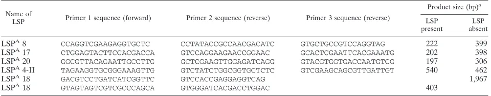
M. aviumsubsp.paratuberculosis. In a subsequent step, isolates were tested for the presence or absence of the large sequences identified by microarray as potentially associated with distinct groups of organisms. This was done with a multiplex PCR approach using a set of three primers: two primers (forward and reverse) designed towards the flanking regions (bridging primers) of the large sequence polymorphism (LSP) and a third primer designed to recognize a se-quence internal to the LSP (internal primer). The primers were designed using Primer 3 software (http://frodo.wi.mit.edu/cgi-bin/primer3/primer3_www.cgi), and primers were designed such that the resulting PCR products would be of different sizes depending on the presence or absence of the LSP under study. Primer sequences and predicted amplicon lengths are provided in Table 1.
PCRs.PCRs were performed in 50-l volumes, using 5l (equivalent to 5 ng) of DNA. For all but one set of reaction mixtures, we used 1 UTaqpolymerase (MBI Fermentas), 5l of 10⫻PCR buffer (MBI Fermentas), 2.5 mM MgCl2, 5 l acetamide 50% (wt/vol), 0.2 mM deoxynucleoside triphosphates (dNTPs), and 0.5M of each primer. In one instance where the expected product was close to 2 kb (LSPA
18), we were unable to design a reliable three-primer PCR because of the presence of an insertion element and a sequence inversion; therefore, we did separate PCRs for the presence or the absence of this genomic polymor-phism. In this case, to amplify the 2-kb product, we used 3.75 UTaqpolymerase and 5l of 10⫻buffer 3 (Expand Long template PCR system; Roche Diagnostics Corporation, Indianapolis, IN), 5l acetamide 50% (wt/vol), 0.4 mM lithium-stabilized dNTPs (dNTP set Li-salt solution; Roche Diagnostics Corporation), and 0.5M of each primer. For all reactions, PCR amplification consisted of an initial denaturation step at 94°C for 5 min followed by 35 cycles of denaturation at 94°C for 45 s, with annealing at 60°C (LSPA8) or 55°C (other LSPs) for 45 s,
elongation at 72°C for 2 min, and a final elongation step at 72°C for 10 min. PCR products were separated by electrophoresis in 1.5% (wt/vol) agarose gels con-taining ethidium bromide. When the amplicon indicated that the LSP was miss-ing, this PCR product was subjected to sequencing to ensure that the exact same polymorphism was detected in each isolate.
RESULTS
From analysis of DNA microarray-based cohybridization ex-periments, we identified three large sequences that were
poly-morphic among the four strains ofM. aviumsubsp.
paratuber-culosisstudied by microarray. Of these, two sequences, LSPA
4-II and LSPA18, were present in both of the S strains but
missing from the two C strains. Another sequence, LSPA20,
was present in the C strains but missing from the S strains. We
also identified one large sequence, LSPA17, that was missing
from theM. aviumsubsp.aviumbird-type strains andM. avium
subsp.silvaticumbut present inM. avium104 and theM. avium
[image:2.585.42.544.81.179.2]subsp. paratuberculosis strains studied by microarray. These
TABLE 1. Primers used for testing of LSPs
Name of
LSP Primer 1 sequence (forward) Primer 2 sequence (reverse) Primer 3 sequence (reverse)
Product size (bp)a
LSP present
LSP absent
LSPA8 CCAGGTCGAAGAGGTGCTC CCTATACCGCCAACGACATC GTGCTGCCGTCCAGGTAG 222 399
LSPA17 CTGGAGTACTTCCACGACCA GTCCAGGAAGAACCGGAAC GCACTCGAATTCACGAAATG 202 398
LSPA20 GGCGTTACAGAATTGCCTTG GCTCGAAGTTGGAGATCAGG GTACGTGGTGACCAATGTCG 197 306
LSPA4-II TAGAAGGTGCGGGAAAGTTG GTCTATCTGGCGGTGCTCTC GTCGAAGCAGCGTTGATTGT 540 462
LSPA18 GACGTCCTGATCATCGGTTC GTCCACCGAGGAGGTCAG 1,967
LSPA18 GTAGTAGTCGTCGCCCAGCA GTGGGATCACGACCTGGAC 403
a
For all but one LSP (LSPA
18), PCR tests were done using the three primers in a multiplex PCR; expected product sizes are listed in the last two columns. Primers 1 and 3 are the bridging primers, and primer 2 is the internal primer. For LSPA
18, PCR testing was performed using two primers only, such that a product was obtained with one set of (bridging) primers when the region was missing and one set of (internal) primers when the region was present.
on May 16, 2020 by guest
http://jcm.asm.org/
LSPs and their distribution across a panel of isolates are de-scribed in greater detail below.
Description of sequences polymorphic in strains ofM. avium
subsp.paratuberculosis. (i) LSPA
4-II.From previous genomic
comparisons of members of theM. avium complex, we had
identified LSP 4, a large sequence present inM. avium104
but missing inM. aviumsubsp.paratuberculosisK10 (23). In
the former, this 197-kb sequence is located within the
my-cobactin synthesis operon between mbtA and mbtJ. In M.
aviumsubsp. paratuberculosis K10, LSP 4 is replaced by a
different, 19-kb sequence called LSPP 12 (MAP 2179 to
MAP 2197), determined by PCR to be highly specific toM.
aviumsubsp. paratuberculosisisolates (22). Microarray data
for C and S strains ofM. aviumsubsp.paratuberculosisindicated
that LSPP12 was present in both but that a 26-kb segment of
theM. aviumLSP 4 element was present only in the S strains.
These results suggested that 171 kb of LSP 4 was present inM.
avium104 but absent from S strains and that a further 26-kb segment was deleted from C strains; we named these two
polymorphisms at the same locus LSPA 4-I and LSPA 4-II,
respectively (Fig. 1A).
(ii) LSPA
18. LSPA 18 is a 16-kb sequence present in M.
aviumsubsp.aviumand S strains ofM. aviumsubsp. paratu-berculosis that was absent in C strains of M. avium subsp.
paratuberculosis.This sequence is immediately adjacent to an
800-kb sequence that is conserved inM. avium subsp.
para-tuberculosisK10 (C type) but inverted in relation toM. avium
104 and S strains ofM. avium subsp.paratuberculosis. In M.
aviumsubsp.paratuberculosisK10, this sequence is replaced by
an IS900element, MAP4281 (Fig. 1B).
(iii) LSPA
20.LSPA20, an 8-kb sequence spanning MAP1490
to MAP1484c, was absent from S strains of M. avium subsp.
paratuberculosis. This sequence is predicted to encode proteins involved in metabolism, notably includes genes annotated as putatively encoding pyruvate dehydrogenases, and is highly
conserved in other mycobacteria, including Mycobacterium
[image:3.585.54.527.73.385.2]tuberculosis. Sequence analysis of S strains indicated that MAP1490 and MAP1484c were both truncated compared to
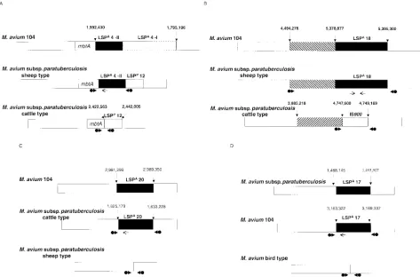
FIG. 1. Schematic representation of large sequence polymorphisms. LSPA4-II and LSPA18 are specifically absent fromM. aviumsubsp. paratuberculosiscattle type, LSPA20 is absent fromM. aviumsubsp.paratuberculosissheep type, and LSPA17 is absent fromM. aviumsubsp. aviumbird type. Coordinates on the genome are given as base pairs, starting from the first nucleotide of the start codon ofdnaAinM. avium
104 andM. aviumsubsp.paratuberculosisK10, respectively. White boxes represent homologous sequences acrossM. avium104,M. avium
subsp.paratuberculosissheep type, andM. aviumsubsp.paratuberculosiscattle type. (A) LSPA4-II is depicted by the black box, LSPA4-I is
depicted by the striped box, and LSPP12 is indicated by the gray box. (B) The striped box represents a large sequence that is conserved but
inverted inM. aviumsubsp.paratuberculosiscattle type, and the black box represents LSPA18. (C) The black box represents LSPA20. (D) The
black box represents LSPA17. Thick arrows represent primers flanking the LSP (bridging primers); a PCR product is obtained if the region
is missing. Thin arrows represent primers targeting a sequence that is within the LSP (internal primers); a PCR product is obtained if the region is present.
on May 16, 2020 by guest
http://jcm.asm.org/
the annotated ORF in C strains, with the polymorphism
oc-curring at position 1633228 of theM. aviumsubsp.
paratuber-culosisK10 genome, suggesting that the loss of LSPA20 is a
deletion event that occurred selectively in S strains ofM. avium
subsp.paratuberculosis(Fig. 1C).
(iv) Distribution of large sequence polymorphisms among strains of M. avium subsp. paratuberculosis: LSPA
8. All 21
strains ofM. aviumsubsp.paratuberculosisisolates in this study
lacked the LSPA8 sequence, while the sequence was detected
as present in all non-M. aviumsubsp.paratuberculosisisolates,
as shown by a multiplex PCR approach (Fig. 2). In a previous
report, we noted that in a small minority ofM. aviumsubsp.
paratuberculosisisolates, the absence of LSPA8 could not be
demonstrated but that intervening sequence could not be am-plified for these samples (22). In the present study, the absence
of this region was 100% sensitive forM. aviumsubsp.
para-tuberculosis, suggesting a now-resolved technical limitation in our previous report (Table 2).
LSPs absent from C strains.All 11 C strains ofM. avium
subsp.paratuberculosis lacked LSPA 4-II, and sequencing of
their PCR products revealed identical sequences with a
trun-catedmbtAgene (MAP2178). In contrast, LSPA4-II amplified
as present in S strains ofM. aviumsubsp.paratuberculosisand
all nonparatuberculosisM. aviumstrains studied. All C strains
ofM. aviumsubsp.paratuberculosisalso lacked LSPA18. With
primers flanking the LSPA18 sequence and under PCR
con-ditions that were optimized to amplify a 2-kb product across an
IS900 element, we were able to successfully obtain a PCR
product in all 11 C strains ofM. aviumsubsp.paratuberculosis
and in none of the S strains. We sequenced these amplicons
and confirmed they were identical to the sequence ofM. avium
subsp.paratuberculosisK10. In contrast, the presence of LSPA
18 was demonstrated in all other strains by PCR using primers internal to the sequence.
LSP absent from S strains. All S strains studied lacked
[image:4.585.44.540.81.490.2]LSPA20, which was present in all C strains, as demonstrated by
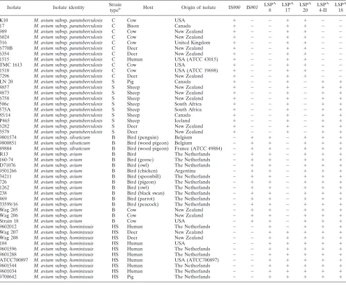
TABLE 2. PCR testing for presence or absence of LSPs across a panel ofM. aviumisolatesa
Isolate Isolate identity Strain
typeb Host Origin of isolate IS900 IS901
LSPA
8 LSPA
17 LSPA
20 LSPA
4-II LSPA
18
K10 M. aviumsubsp.paratuberculosis C Cow USA ⫹ ⫺ ⫺ ⫹ ⫹ ⫺ ⫺
17 M. aviumsubsp.paratuberculosis C Bison Canada ⫹ ⫺ ⫺ ⫹ ⫹ ⫺ ⫺
989 M. aviumsubsp.paratuberculosis C Cow New Zealand ⫹ ⫺ ⫺ ⫹ ⫹ ⫺ ⫺
6024 M. aviumsubsp.paratuberculosis C Cow New Zealand ⫹ ⫺ ⫺ ⫹ ⫹ ⫺ ⫺
316 M. aviumsubsp.paratuberculosis C Cow United Kingdom ⫹ ⫺ ⫺ ⫹ ⫹ ⫺ ⫺
6770B M. aviumsubsp.paratuberculosis C Deer New Zealand ⫹ ⫺ ⫺ ⫹ ⫹ ⫺ ⫺
6354 M. aviumsubsp.paratuberculosis C Deer New Zealand ⫹ ⫺ ⫺ ⫹ ⫹ ⫺ ⫺
1515 M. aviumsubsp.paratuberculosis C Human USA (ATCC 43015) ⫹ ⫺ ⫺ ⫹ ⫹ ⫺ ⫺
TMC 1613 M. aviumsubsp.paratuberculosis C Cow USA ⫹ ⫺ ⫺ ⫹ ⫹ ⫺ ⫺
1518 M. aviumsubsp.paratuberculosis C Cow USA (ATCC 19698) ⫹ ⫺ ⫺ ⫹ ⫹ ⫺ ⫺
7296 M. aviumsubsp.paratuberculosis C Deer New Zealand ⫹ ⫺ ⫺ ⫹ ⫹ ⫺ ⫺
LN 20 M. aviumsubsp.paratuberculosis S Pig Canada ⫹ ⫺ ⫺ ⫹ ⫺ ⫹ ⫹
4857 M. aviumsubsp.paratuberculosis S Sheep New Zealand ⫹ ⫺ ⫺ ⫹ ⫺ ⫹ ⫹
4873 M. aviumsubsp.paratuberculosis S Sheep New Zealand ⫹ ⫺ ⫺ ⫹ ⫺ ⫹ ⫹
6758 M. aviumsubsp.paratuberculosis S Sheep New Zealand ⫹ ⫺ ⫺ ⫹ ⫺ ⫹ ⫹
506c M. aviumsubsp.paratuberculosis S Sheep South Africa ⫹ ⫺ ⫺ ⫹ ⫺ ⫹ ⫹
575A M. aviumsubsp.paratuberculosis S Sheep South Africa ⫹ ⫺ ⫺ ⫹ ⫺ ⫹ ⫹
85/14 M. aviumsubsp.paratuberculosis S Sheep Canada ⫹ ⫺ ⫺ ⫹ ⫺ ⫹ ⫹
P465 M. aviumsubsp.paratuberculosis S Sheep Iceland ⫹ ⫺ ⫺ ⫹ ⫺ ⫹ ⫹
6282 M. aviumsubsp.paratuberculosis S Deer New Zealand ⫹ ⫺ ⫺ ⫹ ⫺ ⫹ ⫹
3579 M. aviumsubsp.paratuberculosis S Deer New Zealand ⫹ ⫺ ⫺ ⫹ ⫺ ⫹ ⫹
9801574 M. aviumsubsp.silvaticum B Bird (penguin) Belgium ⫺ ⫹ ⫹ ⫺ ⫹ ⫹ ⫹
9800851 M. aviumsubsp.silvaticum B Bird (wood pigeon) Belgium ⫺ ⫹ ⫹ ⫺ ⫹ ⫹ ⫹
49884 M. aviumsubsp.silvaticum B Bird (wood pigeon) France (ATCC 49884) ⫺ ⫹ ⫹ ⫺ ⫹ ⫹ ⫹
R13 M. aviumsubsp.avium B Bird The Netherlands ⫺ ⫹ ⫹ ⫺ ⫹ ⫹ ⫹
160-74 M. aviumsubsp.avium B Bird (goose) The Netherlands ⫺ ⫹ ⫹ ⫺ ⫹ ⫹ ⫹
D71076 M. aviumsubsp.avium B Bird (owl) The Netherlands ⫺ ⫹ ⫹ ⫺ ⫹ ⫹ ⫹
9501266 M. aviumsubsp.avium B Bird (chicken) Argentina ⫺ ⫹ ⫹ ⫺ ⫹ ⫹ ⫹
34211 M. aviumsubsp.avium B Bird (spoonbill) The Netherlands ⫺ ⫹ ⫹ ⫺ ⫹ ⫹ ⫹
726 M. aviumsubsp.avium B Bird (pigeon) The Netherlands ⫺ ⫹ ⫹ ⫺ ⫹ ⫹ ⫹
1262 M. aviumsubsp.avium B Bird (owl) The Netherlands ⫺ ⫹ ⫹ ⫺ ⫹ ⫹ ⫹
238 M. aviumsubsp.avium B Bird (black swan) The Netherlands ⫺ ⫹ ⫹ ⫺ ⫹ ⫹ ⫹
469 M. aviumsubsp.avium B Bird (parrot) The Netherlands ⫺ ⫹ ⫹ ⫺ ⫹ ⫹ ⫹
33599/16 M. aviumsubsp.avium B Bird (peacock) The Netherlands ⫺ ⫹ ⫹ ⫺ ⫹ ⫹ ⫹
Wag 205 M. aviumsubsp.avium B Cow New Zealand ⫺ ⫹ ⫹ ⫺ ⫹ ⫹ ⫹
Wag 206 M. aviumsubsp.avium B Cow New Zealand ⫺ ⫹ ⫹ ⫺ ⫹ ⫹ ⫹
Strain 18 M. aviumsubsp.avium B Cow USA ⫺ ⫹ ⫹ ⫺ ⫹ ⫹ ⫹
9602012 M. aviumsubsp.hominissuis HS Human The Netherlands ⫺ ⫺ ⫹ ⫺ ⫹ ⫹ ⫹
Wag 207 M. aviumsubsp.hominissuis HS Deer New Zealand ⫺ ⫺ ⫹ ⫹ ⫹ ⫹ ⫹
Wag 208 M. aviumsubsp.hominissuis HS Deer New Zealand ⫺ ⫺ ⫹ ⫹ ⫹ ⫹ ⫹
104 M. aviumsubsp.hominissuis HS Human USA ⫺ ⫺ ⫹ ⫹ ⫹ ⫹ ⫹
9601596 M. aviumsubsp.hominissuis HS Human The Netherlands ⫺ ⫺ ⫹ ⫹ ⫹ ⫹ ⫹
9601288 M. aviumsubsp.hominissuis HS Human The Netherlands ⫺ ⫺ ⫹ ⫹ ⫹ ⫹ ⫹
ATCC700897 M. aviumsubsp.hominissuis HS Human USA (ATCC700897) ⫺ ⫺ ⫹ ⫹ ⫹ ⫹ ⫹
9601544 M. aviumsubsp.hominissuis HS Human The Netherlands ⫺ ⫺ ⫹ ⫹ ⫹ ⫹ ⫹
9601034 M. aviumsubsp.hominissuis HS Human The Netherlands ⫺ ⫺ ⫹ ⫹ ⫹ ⫹ ⫹
9700642 M. aviumsubsp.hominissuis HS Pig The Netherlands ⫺ ⫺ ⫹ ⫹ ⫹ ⫹ ⫹
a⫹, sequence present;⫺, sequence absent. Results are based on the presence or absence of PCR products of different sizes. bStrain type abbreviations: C, cattle (type II); S, sheep (type I); B, bird; HS, hominissuis.
on May 16, 2020 by guest
http://jcm.asm.org/
a multiplex PCR approach. Sequences obtained from all 10 S strains were identical, with the polymorphism occurring at the exact same site and confirming the truncation of the ORFs of each end of this sequence.
Sequence polymorphic in M. avium strains isolated from birds: LSPA
17.Analysis of the cohybridization experiments of
M. avium104 with two strains ofM. aviumsubsp.avium
charac-terized as bird type and two isolates labeledM. aviumsubsp.
silvaticum revealed the consistent absence of a 6-kb sequence
called LSPA 17, spanning MAP1375c to MAP1381c. From in
silico analyses of the genomes ofM. avium 104 andM. avium
subsp. paratuberculosis K10, we determined that this region is
conserved between these genomes (sequence identity of 98%), although a 253-bp portion situated in the middle of this sequence is missing from the latter. This polymorphism occurs at the same
junction site inM. aviumsubsp.aviumas well as in strains called
M. aviumsubsp.silvaticum, corresponding to position 1493907 of the M. aviumsubsp. paratuberculosisstrain K10 genome (Fig.
1D). LSPA 17 contains several genes with homology to those
encoding short-chain dehydrogenases, in addition to a
transcrip-tional regulator of the LysR family. Of note, LSPA 17 is not
syntenous with the genome of theM. tuberculosiscomplex and
does not appear to be conserved in other mycobacteria. From testing for the presence or absence of this region using
a multiplex PCR assay, we noted that the 12M. aviumsubsp.
aviumisolates of the bird type and the 3 isolates designated as
M. aviumsubsp.silvaticumall lacked LSPA17. This region was
detected as present in all M. avium subsp. paratuberculosis
isolates and in the majority ofM. aviumsubsp.hominissuistype
isolates. Exceptionally, we determined that one M. avium
subsp.hominissuisisolate, obtained from a patient with AIDS,
also lacked this sequence. We further characterized this isolate
by amplification and sequencing of the 3⬘ end of thehsp65
gene, a genotyping method that we recently applied towards
classification ofM. aviumorganisms (28). TheM. aviumsubsp.
hominissuisisolate that lacked LSPA 17 was shown to differ
from the bird-typehsp65sequevar (code 4) by only one SNP
and belonged to the sequevar called code 3. Consistent with other strains belonging to this sequevar, the isolate did not
possess IS901, an insertion element generally found to be
present in bird strains of M. avium. To determine if the
ab-sence of LSPA 17 might be a feature of code 3 strains, we
tested five other isolates belonging to this sequevar and noted
that these isolates also lacked LSPA17. These data suggest that
the absence of LSPA17 is a shared feature of code 3 strains
and bird strains and is therefore sensitive forM. aviumsubsp.
aviumbut not perfectly specific.
DISCUSSION
M. avium organisms present many phenotypic differences between and within subspecies, leading to concerted efforts to understand their genomic diversity. In this work, we uncovered large regions of genomic differences between phenotypically
and genetically distinct subsets ofM. aviumsubsp.
paratuber-culosisand nonparatuberculosisM. aviumisolates. The recog-nition of these sequence differences facilitates their use in
determining whichM. avium subsets are associated with
hu-man and veterinary diseases. As has been done with
polymor-phisms of theM. tuberculosiscomplex (3, 15), we have adopted
a three-primer PCR strategy that is practical and immediately applicable in diagnostic and reference laboratories (17, 26).
M. avium subsp. avium strains (generally associated with severe disease in birds) consistently lack a large sequence,
LSPA17, although the absence of this region was not strictly
restricted to this cluster. Isolates previously designated asM.
aviumsubsp.silvaticumalso lacked this sequence and could not specifically be identified with a particular genomic profile. This
is in agreement with our recent typing scheme forM. avium
isolates, in which bird-type M. avium and M. avium subsp.
silvaticumcould not be distinguished (28). In thathsp65-based
study, we noted that some IS901-negative strains associated
with disseminated disease in AIDS patients had a closely
re-lated sequevar which differed from that of bird-typeM. avium
by just one SNP. Both these groups, termedhsp65code 3 and
code 4, lacked LSPA17, suggesting that this LSP event
pre-ceded the SNP that distinguishes these two lineages.
Our findings also show that S and C strains of M. avium
subsp. paratuberculosishave undergone distinct evolutionary
paths. One sequence, LSPA20, appears to represent a genomic
deletion specific to S strains ofM. aviumsubsp.
paratubercu-losis. Conversely, two other sequences, LSPA18 and LSPA4-II,
were absent from all C strains ofM. aviumsubsp.
paratuber-culosistested but present in S strains, likely indicating deletions
characteristic of the C lineage ofM. aviumsubsp.
paratuber-culosis. Because these represent complex genetic events, the
genomic evidence points to S strains as being closer to theM.
aviumsubsp.paratuberculosisancestor and C strains having a more derivative status (Fig. 1). Our data for the regions
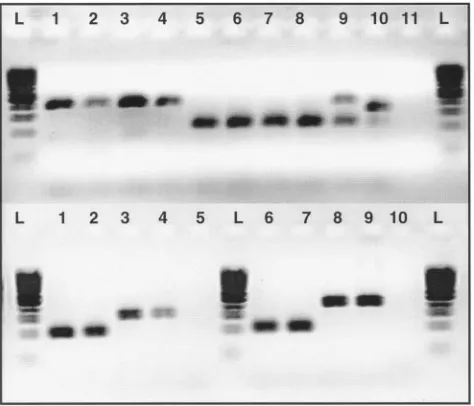
miss-FIG. 2. Detection of subspecies and subtypes ofM. aviumusing PCR for large sequences polymorphic among strains ofM. avium. On the top panel, 11 samples were tested for LSPA8 using a three-primer
PCR. Lanes: L, 100-bp ladder; 1 to 4,M. aviumsubsp.paratuberculosis
strains; 5 and 6, nonparatuberculosis strains ofM. avium; 9, mixed sample (M. avium104 andM. aviumsubsp.paratuberculosisK10); 10,
M. intracellulareATCC 13950 strain; 11, water. In the bottom panel, four samples were tested for LSPA20 and four samples are tested for
LSPA17 using three-primer PCRs. Lanes: L, 100-bp ladder; 1 and 2, M. aviumsubsp.paratuberculosisC type; 3 and 4, M. aviumsubsp.
paratuberculosisS type; 5, water; 6 and 7,M. aviumsubsp.hominissuis; 8 and 9,M. aviumsubsp.aviumbird type; 10, water.
on May 16, 2020 by guest
http://jcm.asm.org/
[image:5.585.46.283.70.273.2]ing from C strains ofM. aviumsubsp.paratuberculosisare in agreement with and expand upon findings from a representa-tional difference analysis-based study that identified three loci missing from type II (C type) strains (9). The 233-bp locus they identified as pig-RDA10 (AY266300) forms part of the 16-kb
region we called LSPA 18. The 197-bp locus pig-RDA20
(AY266301) is located within the 26-kb segment we called
LSPA 4-II. While the third locus they describe (AY266302)
also forms part of a larger segment, microarray-based com-parisons indicated that this segment was also variably
miss-ing from nonparatuberculosis M. avium isolates
(unpub-lished observations); therefore, the loss of this region does
not appear to be specifically associated withM. aviumsubsp.
paratuberculosis.
In this work we focused only on regions consistently
associ-ated with distinctM. aviumsubgroups, as these would have the
greatest applicability for diagnostic laboratories. We have not confirmed genomic differences proposed by microarrays that
distinguish among closely related M. avium organisms and,
thus, it is possible that other large sequences are missing from selected groups of strains. For instance, in previous work we identified an LSP (called LSP 11) which was missing in the S strain tested by microarray (23). Further analysis revealed that this region was missing from only a subset of S strains, indi-cating that even within subgroups or clusters of strains signif-icant genomic variability may exist and may be of value for molecular epidemiologic applications.
For any typing method, the optimal utility depends on the question being asked and the available technologies. In
com-parison to sequencing of the 3⬘ region of hps65 (28), this
LSP-based PCR method is of lower resolution but greater simplicity (Fig. 3). Reassuringly, the two methods provide
con-sistent results, in that lineages defined by hsp65 sequencing
were branded by a shared LSP profile. This PCR-based
method was not able to reliably distinguish theMycobacterium
intracellularespecies; testing for the presence or absence of
LSPA 8 on a small number of M. intracellulare strains gave
ambiguous results (Fig. 2). The little sequence information
that is currently available for this species and ourhsp65
se-quencing results provide evidence that the M. intracellulare
species is clearly separate from theM. aviumgroup and,
fur-ther, that there is genetic diversity among the former. Thefore, testing using this LSP-based PCR method should be
re-served for isolates determined to belong to the M. avium
species by AccuProbe or alternate methods.
Unlikehsp65-based sequencing, PCR for LSPs is restricted
to testing for the currently described genomic variations. How-ever, with the addition of reactions to test for newly described polymorphic regions, this modality will be readily applicable to
testing for other variants ofM. avium and can ultimately be
packaged in the form of a deligotype platform (10). An advan-tage of PCR-based testing is the capacity to detect mixed
infections (Fig. 2), a recognized concern withM. aviumdisease
(24), in which case the sequencing ofhsp65may only return the
result for the predominant clone. Finally, in settings where sequencing is not readily available or large volumes of isolates are to be screened, PCR with the primers we have described
can provide an immediate gel-based indication of which M.
[image:6.585.113.474.68.340.2]avium variant is present and stimulate additional testing by other methods as indicated.
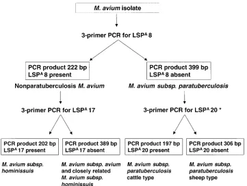
FIG. 3. Diagnostic algorithm for PCR-based identification and typing of anM. aviumisolate. *, testing for the presence or absence of LSPA
4-II or LSPA18 are other alternatives for typingM. aviumsubsp.paratuberculosisisolates into cattle or sheep types.
on May 16, 2020 by guest
http://jcm.asm.org/
ACKNOWLEDGMENTS
This work was supported by a grant from the Natural Science and Engineering Research Council (grant number GEN2282399). M.S. is funded by the Fonds de la Recherche en Sante du Quebec. M.B. is a New Investigator of the Canadian Institutes of Health Research.
None of the authors have a conflict of interest or any commercial association that may pose a conflict of interest.
We thank Fiona McIntosh for her assistance with microarray exper-iments and Murray E. Hines III for providing DNA for “strain 18,” formerly identified as “MAP 18.”
REFERENCES
1.Autschbach, F., S. Eisold, U. Hinz, S. Zinser, M. Linnebacher, T. Giese, T. Loffler, M. W. Buchler, and J. Schmidt.2005. High prevalence of Mycobac-terium avium subspecies paratuberculosis IS900 DNA in gut tissues from individuals with Crohn’s disease. Gut54:944–949.
2.Behr, M. A., M. Semret, A. Poon, and E. Schurr.2004. Crohn’s disease, mycobacteria, and NOD2. Lancet Infect. Dis.4:136–137.
3.Behr, M. A., M. A. Wilson, W. P. Gill, H. Salamon, G. K. Schoolnik, S. Rane, and P. M. Small.1999. Comparative genomics of BCG vaccines by whole-genome DNA microarray. Science284:1520–1523.
4.Bull, T. J., E. J. McMinn, K. Sidi-Boumedine, A. Skull, D. Durkin, P. Neild, G. Rhodes, R. Pickup, and J. Hermon-Taylor.2003. Detection and verifica-tion of Mycobacterium aviumsubsp.paratuberculosisin fresh ileocolonic mucosal biopsy specimens from individuals with and without Crohn’s dis-ease. J. Clin. Microbiol.41:2915–2923.
5.Chacon, O., L. E. Bermudez, and R. G. Barletta.2004. Johne’s disease, inflammatory bowel disease, and Mycobacterium paratuberculosis. Annu. Rev. Microbiol.58:329–363.
6.Collins, D. M., S. Cavaignac, and G. W. de Lisle.1997. Use of four DNA insertion sequences to characterize strains of the Mycobacterium avium complex isolated from animals. Mol. Cell Probes.11:373–380.
7.Collins, D. M., M. De Zoete, and S. M. Cavaignac.2002.Mycobacterium aviumsubsp.paratuberculosisstrains from cattle and sheep can be distin-guished by a PCR test based on a novel DNA sequence difference. J. Clin. Microbiol.40:4760–4762.
8.Collins, D. M., D. M. Gabric, and G. W. de Lisle.1990. Identification of two groups ofMycobacterium paratuberculosisstrains by restriction endonuclease analysis and DNA hybridization. J. Clin. Microbiol.28:1591–1596. 9.Dohmann, K., B. Strommenger, K. Stevenson, L. de Juan, J. Stratmann, V.
Kapur, T. J. Bull, and G. F. Gerlach. 2003. Characterization of genetic differences betweenMycobacterium aviumsubsp.paratuberculosistype I and type II isolates. J. Clin. Microbiol.41:5215–5223.
10.Goguet de la Salmoniere, Y. O., C. C. Kim, A. G. Tsolaki, A. S. Pym, M. S. Siegrist, and P. M. Small. 2004. High-throughput method for detecting genomic-deletion polymorphisms. J. Clin. Microbiol.42:2913–2918. 11.Green, E. P., M. L. Tizard, M. T. Moss, J. Thompson, D. J. Winterbourne,
J. J. McFadden, and J. Hermon-Taylor.1989. Sequence and characteristics of IS900, an insertion element identified in a human Crohn’s disease isolate of Mycobacterium paratuberculosis. Nucleic Acids Res.17:9063–9073. 12.Guerrero, C., C. Bernasconi, D. Burki, T. Bodmer, and A. Telenti.1995. A
novel insertion element fromMycobacterium avium, IS1245, is a specific target for analysis of strain relatedness. J. Clin. Microbiol.33:304–307. 13.Harris, N. B., and R. G. Barletta.2001.Mycobacterium aviumsubsp.
para-tuberculosisin veterinary medicine. Clin. Microbiol. Rev.14:489–512. 14.Johansen, T. B., B. Djonne, M. R. Jensen, and I. Olsen.2005. Distribution of
IS1311and IS1245inMycobacterium aviumsubspecies revisited. J. Clin. Microbiol.43:2500–2502.
15.Mahairas, G. G., P. J. Sabo, M. J. Hickey, D. C. Singh, and C. K. Stover. 1996. Molecular analysis of genetic differences betweenMycobacterium bovis
BCG and virulentM. bovis. J. Bacteriol.178:1274–1282.
16.Mijs, W., P. de Haas, R. Rossau, L. T. Van der, L. Rigouts, F. Portaels, and D. van Soolingen.2002. Molecular evidence to support a proposal to reserve the designation Mycobacterium avium subsp. avium for bird-type isolates and “M. avium subsp. hominissuis” for the human/porcine type of M. avium. Int. J. Syst. Evol. Microbiol.52:1505–1518.
17.Parsons, L. M., R. Brosch, S. T. Cole, A. Somoskovi, A. Loder, G. Bretzel, D. van Soolingen, Y. M. Hale, and M. Salfinger.2002. Rapid and simple ap-proach for identification ofMycobacterium tuberculosiscomplex isolates by PCR-based genomic deletion analysis. J. Clin. Microbiol.40:2339–2345. 18.Paustian, M. L., V. Kapur, and J. P. Bannantine. 2005. Comparative
genomic hybridizations reveal genetic regions within the Mycobacterium aviumcomplex that are divergent fromMycobacterium aviumsubsp. para-tuberculosisisolates. J. Bacteriol.187:2406–2415.
19.Pavlik, I., P. Svastova, J. Bartl, L. Dvorska, and I. Rychlik.2000. Relation-ship between IS901in theMycobacterium aviumcomplex strains isolated from birds, animals, humans, and the environment and virulence for poultry. Clin. Diagn. Lab. Immunol.7:212–217.
20.Saxegaard, F., and I. Baess.1988. Relationship between Mycobacterium avium, Mycobacterium paratuberculosis and “wood pigeon mycobacteria.” Determinations by DNA-DNA hybridization. APMIS96:37–42.
21.Sechi, L. A., A. M. Scanu, P. Molicotti, S. Cannas, M. Mura, G. Dettori, G. Fadda, and S. Zanetti. 2005. Detection and isolation of Mycobacterium aviumsubspeciesparatuberculosisfrom intestinal mucosal biopsies of pa-tients with and without Crohn’s disease in Sardinia. Am. J. Gastroenterol. 100:1529–1536.
22.Semret, M., D. C. Alexander, C. Y. Turenne, P. de Haas, P. Overduin, D. van Soolingen, D. Cousins, and M. A. Behr.2005. Genomic polymorphisms for
Mycobacterium aviumsubsp.paratuberculosisdiagnostics. J. Clin. Microbiol. 43:3704–3712.
23.Semret, M., G. Zhai, S. Mostowy, C. Cleto, D. Alexander, G. Cangelosi, D. Cousins, D. M. Collins, D. van Soolingen, and M. A. Behr.2004. Extensive genomic polymorphism withinMycobacterium avium. J. Bacteriol.186:6332– 6334.
24.Slutsky, A. M., R. D. Arbeit, T. W. Barber, J. Rich, C. F. von Reyn, W. Pieciak, M. A. Barlow, and J. N. Maslow.1994. Polyclonal infections due to
Mycobacterium aviumcomplex in patients with AIDS detected by pulsed-field gel electrophoresis of sequential clinical isolates. J. Clin. Microbiol. 32:1773–1778.
25.Stevenson, K., V. M. Hughes, L. de Juan, N. F. Inglis, F. Wright, and J. M. Sharp.2002. Molecular characterization of pigmented and nonpigmented isolates ofMycobacterium aviumsubsp.paratuberculosis. J. Clin. Microbiol. 40:1798–1804.
26.Talbot, E. A., D. L. Williams, and R. Frothingham.1997. PCR identification ofMycobacterium bovisBCG. J. Clin. Microbiol.35:566–569.
27.Thorel, M. F., M. Krichevsky, and V. V. Levy-Frebault.1990. Numerical taxonomy of mycobactin-dependent mycobacteria, emended description of Mycobacterium avium, and description of Mycobacterium avium subsp. avium subsp. nov., Mycobacterium avium subsp. paratuberculosis subsp. nov., and Mycobacterium avium subsp. silvaticum subsp. nov. Int. J. Syst. Bacteriol.40:254–260.
28.Turenne, C. Y., M. Semret, D. Cousins, D. M. Collins, and M. A. Behr.2006. Sequencing ofhsp65 distinguishes among subsets of the Mycobacterium aviumcomplex. J. Clin. Microbiol.44:433–440.
29.van der Giessen, J. W., A. Eger, J. Haagsma, and B. A. van der Zeijst.1993. Rapid detection and identification ofMycobacterium aviumby amplification of 16S rRNA sequences. J. Clin. Microbiol.31:2509–2512.
30.van Soolingen, D., J. Bauer, V. Ritacco, S. C. Leao, I. Pavlik, V. Vincent, N. Rastogi, A. Gori, T. Bodmer, C. Garzelli, and M. J. Garcia.1998. IS1245
restriction fragment length polymorphism typing ofMycobacterium avium
isolates: proposal for standardization. J. Clin. Microbiol.36:3051–3054.