ISSN 2250-3153
Almond leaves for the one-pot Biofabrication of silver
nanoparticles: Characterization and larvicidal
application
Ayandiran D. Ainaa*, Oluwafayoke Owoloa,d, Morenike Adeoye-Isijolaa, Folasade O. Ainab, Omeiza Favourd and Aderiike, G.Adewumif
a
Department of Microbiology, Babcock University, PMB 21244, Ikeja, Lagos, Nigeria
b
Department of Maternal and Child Health, Babcock University, PMB 21244, Ikeja, Lagos, Nigeria
c
Department of Pure and Applied Biology, Ladoke Akintola University of Technology, PMB 4000, Ogbomoso, Nigeria
d
Department of Pharmaceutical Microbiology, University of Ibadan, PMB 5116, Ibadan, Oyo State, Nigeria
f
Department of Basic and Applied Sciences, Babcock University, PMB 21244, Ikeja, Lagos, Nigeria
Corresponding author: Ayandiran, D. Aina Telephone number: +2348033565433 Email address: [email protected]
DOI: 10.29322/IJSRP.8.11.2018.p8378
http://dx.doi.org/10.29322/IJSRP.8.11.2018.p8378
Abstract
This work reports the synthesis of silver nanoparticles (AgNPs) using the leaf extract of Almond plant. Larvicidal activities of the biosynthesized AgNPs were evaluated against the fourth instar larvae of mosquito Aedes aegypti. The biosynthesised AgNPs were characterised using UV-Visible, Fourier Transmission Infrared Spectroscope (FTIR), Energy Dispersive X-ray Spectroscope (EDX) and Field Emission Scanning Electron Microscope (FESEM). The absorption spectrum of the synthesized AgNPs showed a maximum spectrum of 430 nm while FTIR analysis showed different functional groups present on the surface of the AgNPs with broad peak between 3000 and 3800 cm−1. The FESEM showed a large number of spherically shaped nanoparticles with sizes ranging from 8.34 to 78.96 nm. The 50% (LC50) lethal concentration of the nanoparticles after 12 h was 13.54 µg/ml. This study
showed that almond leaf can be used for synthesis of AgNPs thus adding to the available drugs that could be used in combating multidrug resistant pathogens.
Key words: Almond leaves, larvicidal, silver nanoparticles, green synthesis
INTRODUCTION
Nanotechnology is one of the main logical fields today since it combines learning from the fields of Physics, Chemistry, Biology, Medicine, Informatics, and Engineering. It is a rising technological field with remarkable potential to lead in great breakthrough that can be connected, all things considered. Novel nano and biomaterials, and nano gadgets are created and controlled by nanotechnology instruments and systems, which research and tune the properties, reactions, and elements of living and non-living matter, at sizes beneath 100 nm [1].
ISSN 2250-3153
nm, the properties of matter vary essentially (i.e., quantum-scale impacts assume a critical part) from that at a greater particulate scale. Nanotechnology is then the plan, the control, the building, the generation and application of structures, gadgets and frameworks of about or less than 100 nm [2-3].
Nanoparticles have been reported as having various medicinal applications. Studies such as Nasrollahi et al. [4] have shown the antifungal activities of silver nanoparticle. In another recent paper, Adelere et al. [5] reported the antimicrobial activity of a green synthesized nanoparticle. In addition, nanoparticles have been reported to have anti-inflammatory [6], antiangiogenesis [7], antiviral [8] and antiplatelet activity [9]. Plant extracts have recently been used for nanoparticles green synthesis since they are rich in bioactive compounds [5, 10-12] and hence this study was also aimed at the synthesis and application of nanoparticles gotten from plant extract.
The leaf of the almond plant is a potential wellspring of bioactives; it is intriguing to assess the viability of the aqueous extraction from almond leaves (frequently disposed of as an agrowaste) towards the biogenic synthesis of silver nanoparticle. Almond
(Prunus dulcis L.), a specie of Prunus, belongs to the subfamily Prunoideae of the family Rosaceae. Nutritiously and
therapeutically, almond has been accounted for to be a valuable food item [13-14].
As a result of lacking information from works on the effect of P. dulcis on larvicidal activity and in addition to the very little utilization of this plant in the synthesis of nanoparticles, the principal aim of this investigation is, hence, to use the aqueous extract of the plant leaves for synthesis of silver NPs and to assess the in vitro larvicidal activity of the biosynthesized silver nanoparticle.
MATERIAL AND METHODS
Sample collection
Fresh Prunus dulcis leaves were collected from the local area of the University campus. It was washed thoroughly with distilled
water several times to remove dust and dried under shade (30°C ± 2°C) for 5 days. The dried leaves were cut into small pieces, and blended into powder. The powdered samples were kept in air tight containers at room temperature for further use.
Collection and Identification of Mosquito Larva
The mosquito larvae were gotten from a stagnant drainage on the University campus and were kept in a closed container for safety precautions in case of the fast emergence of the larvae into an adult mosquito. The target mosquito larva in this study was the fourth instar larva of dengue carrying mosquito Aedes aegypti. Identified Aedes aegypti mosquito larvae (Identification was done by a medical entomologist) were separated from the other mosquito species and were placed in a water- filled plastic moulder.
Preparation of extract
ISSN 2250-3153
ions. The reaction was carried out under static condition at room temperature (30 ± 2oC) for 2hr. The formation of AgNPs was observed as a change in the solution colour [15]
Characterization of Synthesized Silver nanoparticles
The formation of the synthesized nanoparticles was confirmed by measuring its absorbance spectrum using UV–Visible spectrophotometer (Cecil, USA) operated at 190–1100 nm. The identity of the biomolecules that took part in the green synthesis was determined by FTIR spectroscopy. The measurements were performed between 4000-400 cm-1 to see the attachment of biomolecules on the surface of the AgNPs using Shimadzu FTIR spectrometer, model 8400S (Shimadzu, Japan). To achieve this, purified silver nanoparticles were dried and blended with KBr in the ratio 5: 95 to form a pellet which was used for the measurement. The size, morphology, and elemental composition of the synthesized nanoparticles were unravelled by Field Emission Scanning Electron Microscopy (FESEM) and EDX analyses. The Field Emission Scanning Electron Microscopy (FESEM) micrograph was obtained as follows. A drop of nanoparticles in suspension was placed on a 200 mesh hexagonal copper grid (3.05 mm) (Agar Scientific, Essex, UK) coated with 0.3 % formvar dissolved in chloroform. The particles were allowed to settle for 3–5 min on the grid, the excess liquid flicked off with a wick of filter paper and the grids were then air dried before FESEM viewing. Micrograph was obtained using a JEM-1400 (JEOL, USA) operating at 200 kV.
Larvicidal Activity of the synthesized Nanoparticle
The efficacy of the synthesized nanoparticle as larvicide against the dengue-vector Aedes aegypti mosquito was evaluated in a dose-response bioassay against the fourth instar larvae in accordance with the guidelines of World Health Organization [16]. The larvicidal activity was conducted in triplicate by exposing five Aedes aegypti mosquito larvae to 300µl of each of the graded concentrations (20, 40, 60, 80 and 100) of Ag-NPs at room temperature (30 ± 2oC). In the control experiment, the larvae were exposed to sterile distilled water under the same conditions. The number of death was plotted against concentration of the Ag-NPs. The effects of the synthesized nanoparticles were monitored through carefully counting the number of dead larvae after 12 hours of treatment, and the percentage mortality was computed. Probit Analysis was used to calculate LC50 value to determine lethal concentrations of the synthesized nanoparticles on Aedes aegypti mosquito larvae after 12
hours of treatment.
RESULTS AND DISCUSSION
Synthesis and Characterization of the Silver nanoparticle
ISSN 2250-3153
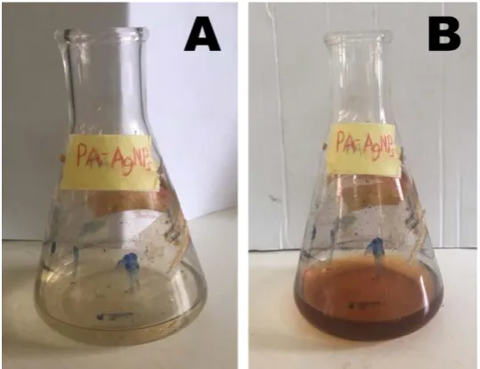
Figure 1: Synthesis of the Almond leaf extract mediated silver nanoparticles (a) immediately after the addition of the almond leaf extract to the silver nitrate; (b) Formation of deep brown colouration after 30 min.
[image:4.595.144.453.355.569.2]The biosynthesized silver nanoparticle showed a maximum absorbance wavelength at 430nm which is indicated in figure 2, a value within the range previously reported for AgNPs [17]
Figure 2: UV-Vis spectra of silver nanoparticles synthesized from almond leaf extract
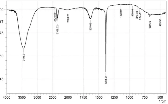
The NP shows a broad peak between 3000 and 3800 cm−1, which are identified as those of O-H vibrations and/or N-H stretching associated with N-substituted amide [18],2359 cm−1peak is that of CO2 from air, the 2000 cm−1 peak is probably from C=N
ISSN 2250-3153
Figure 3: The FTIR spectra of the silver nanoparticles synthesized from almond leaf extract
The chemical analysis of the almond leaf extract - mediated silver nanoparticle was shown (Figure 4) by the EDX spectroscopy. Elemental signals were observed around the Ag atom within the ranges of 2.5 to 3.2keV. Other elements present include copper, oxygen, and carbon). Similar observations have been reported by other researchers [23-24] with silver signals at the range of 1.5 to 5.0 keV
Figure 4: Energy dispersive spectra of the synthesized AgNPs
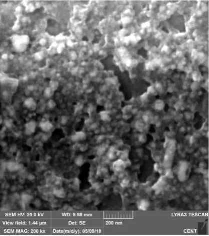
The size and morphology of the biosynthesized nanoparticle were captured by (Figure 5) FESEM. The particles which ranged from 8.34 to 78.96 nm were spherical in shape. This size range falls within the ranges reported when other plants were used in the biosynthesis of silver nanoparticle [15, 25]
AgAg Ag
O Ag
C
Cu
0 0.5 1 1.5 2 2.5 3 3.5 4 4.5 5
keV Full Scale 11232 cts Cursor: 4.799 (87 cts)
[image:5.595.145.464.403.578.2]ISSN 2250-3153
Figure 5: Field Emission Scanning Electron Micrograph of the Synthesized AgNPs
Larvicidal Activity of the synthesized Nanoparticle
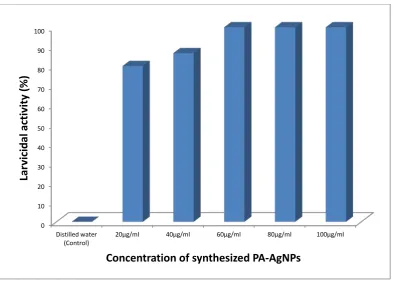
The biosynthesized silver nanoparticle was found to be highly toxic to the 4th instar larvae Aedes aegypti. After 12 hours of incubation, mortality percentage in the 20µg/ml and 40 µg/ml was found to be 80 and 86.6 respectively while the 60 µg/ml, 80 µg/ml and 100 µg/ml showed 100% mortality. The control group however had no mortality. Furthermore, the silver nanoparticles exhibited a concentration dependent activity against mosquito larvae since the percentage mortality were observed to increase with increasing concentrations of the biosynthesized nanoparticles. The 50% lethal concentration (LC50) of the nanoparticles after
12h was 13.54µg/ml. In previous study, Lateef et al. [26] also reported similar result when cell free extract of Bacillus safensis
ISSN 2250-3153
Figure 6: The larvicidal activity of the synthesized AgNPs
CONCLUSION
In this work, almond leaf extract was used to synthesize nanoparticles. The synthesised AgNPs was analysed using UV Spectrophotometer, FTIR, FESEM and EDX. The biosynthesised silver nanoparticle had larvicidal activities against Aedes aegypti
larvae. Thus almond leaf extract mediated-AgNPs could have various pharmaceutical applications.
ACKNOWLEDGEMENTS
The authors thank the department of Microbiology, Babcock University, Ilisan-Remo, Nigeria for her assistance during this study.
FUNDING
This research did not receive any specific grant from funding agencies in the public, commercial, or not-for-profit sectors
CONFLICT OF INTEREST
The authors declare that they have no conflict of interest.
REFERENCES
1.
Logothetidis, S. (2014). Nanostructured Materials and Their Applications. Berlin: Springer Berlin.2.
Sattler, K.D. (2010) Handbook of Nanophysics, Principles and Methods. CRC, New York,3.
Bhushan, B. (2004) Handbook of Nanotechnology. Springer, Berlin: Springer Berlin.4.
Nasrollahi, Y.-K., Kim, B. H., & Jung, G. (2009). Antifungal Activity of Silver Nanoparticles on some fungi. Plant Disease,0 10 20 30 40 50 60 70 80 90 100 Distilled water
(Control) 20µg/ml 40µg/ml 60µg/ml 80µg/ml 100µg/ml
La
rv
icid
al a
ct
iv
ity
(%
)
ISSN 2250-3153
5.
Adelere, A. I., Lateef, A., Oyeyemi, A. D., Ramatu, A., Usman, N., and David, B. J. (2017). Biosynthesis of silver nanoparticles using aqueous extract of Buchholzia coriacea (wonderful kola) seeds and their antimicrobial activities,Annals Food Science and Technology 18(4): 671–679.
6.
Baharara, J., Asadi-Samani, M., Ramezani, T. and Mousavi, M. (2017). Antioxidant and anti-inflammatory activity of green synthesized silver nanoparticles using Salvia officinalis extract. Annals of Tropical Medicine and Public Health, 10(5), p.1265.7.
Baharara, J., Namvar, F., Mousavi, M., Ramezani, T. and Mohamad, R. (2014). Anti-Angiogenesis Effect of Biogenic Silver Nanoparticles Synthesized Using Saliva officinalis on Chick Chorioalantoic Membrane (CAM). Molecules, 19(9), pp.13498-13508.8.
Park, I., Kwon, Y., Ryu, W. and Ahn, B. (2014). Inhibition of hepatitis B virus replication by ligand-mediated activation of RNase L. Antiviral Research, 104, pp.118-127.9.
Shrivastava S, Bera T, Singh S, Singh G, Ramachandrarao P, Dash D. (2009) Characterization of Antiplatelet Properties of Silver Nanoparticles. ACS Nano. 3, 1357-1364.10.
Ahmed, S., Saifullah, Ahmad, M., Swami, B. L., and Ikram, S. (2016). Green synthesis of silver nanoparticles usingAzadirachta indica aqueous leaf extract. Journal of Radiation Research and Applied Sciences 9(1): 1–7.
11.
Thirumurugan, A., Tomy, N. A., Ganesh, R. J., and Gobikrishnan, S. (2010). Biological reduction of silver nanoparticles using plant leaf extracts and its effect on increased antimicrobial activity against clinically isolated organisms. DerPharma Chemica 2(6): 279-284.
12.
Benakashani, F., Allafchian, A. R., and Jalali, S. A. H. (2016). Biosynthesis of silver nanoparticles using Capparis spinosa L. leaf extract and their antibacterial activity. Karbala International Journal of Modern Science2(4): 251–258.13.
Wijeratne SSK, Abou-Zaid MM, Shahidi F (2006). Antioxidantspolyphenols in almond and its coproducts. Journal of Agriculture and Food Chemistry, 54, 312–318.14.
Sarwar, S. (2012). Antioxidant characteristics of different solvent extracts from almond (Prunus dulcis L.) shell. Journal ofMedicinal Plants Research, 6(17), 3311–3316. https://doi.org/10.5897/JMPR11.1723
15.
Lateef A., Folarin B., Oladejo S., Akinola P., Beukes L. & Gueguim-Kana E. (2018). Characterization, antimicrobial, antioxidant, and anticoagulant activities of silver nanoparticles synthesized from Petiveria alliacea L. leaf extract. Preparative Biochemistry and Biotechnology. https://doi.org/10.1080/10826068.2018.147986416.
WHO. (2008). Guidelines for laboratory and field testing of mosquito larvicides. World Health Organization: 1–41.17.
Krishnaraj C., Jagan E. G., Rajasekar S., Selvakumar P., Kalaichelvan P. T., & Mohan N. (2010). Synthesis of silver nanoparticles using Acalypha indica leaf extracts and its antibacterial activity against water borne pathogens. Colloids Surf B Biointerfaces. 76: 50–56.18.
Unuabonah, E.I., Adie, G.U., Onah, L.O. and Adeyemi, O.G. (2009). Multistage optimization of the adsorption of methylene blue dye onto defatted Carica papaya seeds. Chemical Engineering Journal 155 (3): 567-57919.
Lin W., Yu D. & Yang M. (2005). pH-sensitive polyelectrolyte complex gel microspheres composed of chitosan/sodium tripolyphosphate/dextran sulfate: swelling kinetics and drug delivery properties. Colloids Surf B Biointerfaces. 44: 143-151.20.
Sun L., Du Y., Chen L., Huang R. & Chen X. (2004). The synthesis of carboxymethylchitosan hydrogel and the application in drug controlled release systems. Acta Polym Sin. 8: 191–195ISSN 2250-3153