SILC – A Newer Technique With Conventional Instruments: Does it
really need “novel ”instruments ?
Mukesh Kumar *, R.S.Jhobta *, D.K. Verma, * Sandeep Rajta*
*
Deptt of Surgery, IGMC, Shimla, Himachal, India
DOI: 10.29322/IJSRP.8.12.2018.p8445
http://dx.doi.org/10.29322/IJSRP.8.12.2018.p8445
Abstract- Gallstone surgery by laparoscopic method is most commonly performed procedure worldwide. Now a days there is more and more trend towards minimally invasive procedure. Single incision laparoscopic cholecystectomy (SILC) is becoming more popular in this field. First SILC was performed by Navarra et al1 in1997 through a single periumbilical skin incision. There were many modifications in SILC procedure also since then. Some authors have used few special devices such as designed SILSTM port Covidian), Triport TM , Single Site Laparoscopy Access System (Ethicon) and Gelpoint (Applied Medical System ) 2etc. Whereas, others have used specially desined instruments to shorten the learning curve. Aim: To evaluate the need of such costly devices or instruments over convention instruments in SILC procedure. Method:We used those instruments ,which are used for conventional laparoscopic cholecystectomy ( cLC) in 50 cases in IGMC Shimla , with 25 cases each in SILC and cLC groups .
Results: SILC was safe and cosmetically better option and was no difference in complication rates between SILC and cLC performed through conventional instruments .Duration of surgery (DOS) was prolonged in SILC group.
Conclusion: SILC procedure through conventional instruments is a safe,less costly , and feasible option in any centre where laparoscopic surgeries are regularly preformed .
Index Terms- Single incision laparoscopic cholecystectomy (SILC), Laparoscopic cholecystectomy ( cLC), Conventional, Novel
I.
INTRODUCTION
onventioally , laparoscopic cholecystectomy was performed using four or three ports. For last few years conventional laparoscopic cholecystectomy (cLC) as less invasive method, has replaced open cholecystectomy in the treatment of patients with symptomatic gallstone disease. In recent years, a search for even more minimally invasive approaches has led to innovative techniques of single incision laparoscopic surgery (SILS) and natural orifice transluminal endoscopic surgery (NOTES). While substantial drawbacks of NOTES technique including technical challenges and scarcity of instrumentation, have limited its adoption for common use.3 Whereas, SILS has met more favorable acceptance in surgical community because of its feasibility, safety and with no special instrument requirement .SILC is a good option for gall stone surgery with few drawbacks like prolonged surgical time and more learning curve.
First laparoscopic cholecystectomy by single incision was performed by Navarra et al1 in1997 with the removal of gall bladder through a single peri umbilical skin incision .He originally described a technique using transabdominal sutures to suspend the gallbladder during single incision laparoscopic cholecystectomy.
Romanelli et al4 performed the SILC with the newer devices called triport system.They were among the first surgeons to perform lap cholecystectomy through these devices .They also compared cholecystectomy through these devices .
Bresadola et al5 in1999 compared the transumbilical technique of laparoscopic cholecystectomy (2port) with standard laparoscopic cholecystectomy on 40 patients.He concluded that once the learning curve has been completed, transumbilical cholecystectomy is possible without some of difficulties associated with standard laparoscopic cholecystectomy.
A RCT was done by Lei Zhao et al in 2016,comparing SILC and conventional laparoscopic surgery by “novel” instrument ( dissector with long adjustable and rotating wrist )to that of conventional instruments .They concluded that SILC with novel instrument had fewer complications and was safer than SILC with conventional instruments .6
BV Sharath (2016) have used some special device like SILS TM and specially designed “Roticulating instrument”in his cases with no added benefit in terms of duration of surgery or complication rates.7
Aim of our study is to evaluate comparatively the real usefulness of these various instruments or devices used for SILC procedure over conventional instruments.
II.
METHOD
Present prospective study included ultrasonographically proved 50 patients of symptomatic cholelithiasis and were posted for elective cholecystectomy. These patients were admitted in Surgical Wards of Indira Gandhi Medical College, Shimla .SILC were performed on 25 (50% of patients) and cLC were conducted in rest of 25 (50%) patients. The patients were selected randomly. Relevant history, clinical examination and investigations were recorded. SILC was done by infra umblical incision and cLC was done by three /four Trocar Technique. Patients with acute cholecystitis /pancreatitis, choledocholithiasis jaundice / hypoproteinemia/malignancy, conversion of cLC to open cholecystectomy , patients on oral contraceptive pills/pregnancy , intra operative injury to adjacet organs/structures etc. are excluded from study .
III.
SILC PROCEDURE
Equipment and Instruments:
We used conventional laparoscopic instruments and equipment for performing SILC. We used 5mm laparoscope for performing the procedure. A sharp image that allows clear distinction between tissue planes and tissue texture is essential for safe dissection.
Position of the patient, team and equipment:
The patient was positioned supine on the operating table with the legs split apart and strapped firmly to the leg boards. Both arms of the patient were placed on arm boards at an angle less than 90º to the torso.
The surgeon stood on the left side of the patient, with the assistant opposite to him during the placement of the first port. For rest of the procedure, the surgeon stood between the legs and the camera person stood to his right (near the left leg of the patient). The monitor trolley was placed above the patient's right arm. The diathermy pedal was placed near the surgeon's left foot and all tubes and cables were fixed such that they do not interfere with the camera person.
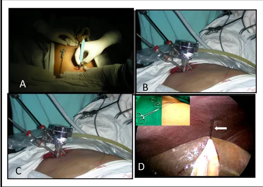
Placement of ports:
We had made an infraumbilical curved incision (Fig.1). The umbilicus was everted and held with two-toothed forceps in a cephalic and caudal position prior to making an incision of length 2-2.5 cm. This was deepened through the fat and the flaps were undermined to expose the fascia over a distance of 2-2.5 cm. The left edge of the skin incision was retracted and a fascial stab incision was made. A Veress needle was introduced through this incision and after confirmation of its intraperitoneal position, CO2 pneumoperitoneum was induced and maintained at 12 mm Hg. In the initial cases a 10 mm port was inserted at the incision line and the two five mm ports were placed 0.5 cm inferiorly and laterally on either side through the same skin incision. A grasper introduced through the right lateral port was used for fundal traction. The dissector introduced through the left lateral port was used to dissect the fine Calot’s triangle. The instrument port and the telescope port were crossed by a chopstick method to avoid “sword fighting’’ and clashing of instruments in the abdomen. At this stage, the patient's position was changed to an anti-Trendelenberg one with a left-sided tilt which helped in the better exposure of the Calot’s triangle. Later we used only two ports one five mm port for five mm camera and another was 10 mm working port through which laparoscope, needle holder, grasper and extractor were introduced at the various steps of SILC procedure( Fig.1)
Placement of traction sutures:
Dissection of the Calot's triangle:
The posterior peritoneum was divided to free the Hartmann's pouch. This was followed by further dissection of the anterior and posterior peritoneal leaves overlying the Calot's triangle with the help of a hook and/or a Maryland dissector. The cystic artery and the cystic duct were skeletonised - the endpoint of this dissection was obtaining a "critical view". (Fig.2)
Control of the cystic artery:
The two windows in the Calot's triangle were dissected more widely than during a conventional LC so as to safely observe the tips of the instruments controlling the artery and the duct. We had clipped the cystic artery with a 10-mm reusable clip applicator.
Subsequently, the cystic artery was divided.
Control of the cystic duct:
If the cystic duct appeared narrow, it was clipped thrice with 10-mm clips and divided. If the duct appeared wide, we preferred to pass a No. 1 polyglactin suture around it, exteriorize the same and fashioned an extracorporeal Meltzer’s knot. This knot was then snugged down onto the cystic duct with the help of a metal knot-pusher. The duct was divided between two extra corporeally tied ligatures. If there was a suspicion of an impacted stone in the cystic duct, it was controlled on the gallbladder side, divided partially to allow the stone to be milked out, and then the stump was ligated using extracorporeal knotting. A 10-mm port was used for introducing a spoon forceps for the retrieval of stones from cystic duct or those that may spill from the gallbladder if it was perforated during dissection. The divided ends of the cystic artery and duct were carefully inspected to confirm their secure closure.
Dissection of the gallbladder:
Alternating medial and lateral rotation of the gallbladder using the ends of the suture placed on Hartmann's pouch was done to dissect the gallbladder from the liver bed using a diathermy hook. Prior to the final detachment of the gallbladder, meticulous haemostasis in the liver bed was ensured and the subhepatic space lavaged with saline. The fundal traction suture was loosened and the gallbladder was freed from the liver ( Fig.2).
Specimen extraction:
Gall bladder was then held at neck with the grasper and extracted through the umbilical 10 mm port ( Fig.2).
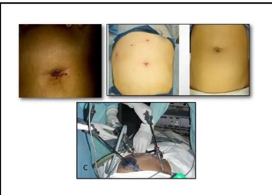
Closure of the incision:
Careful closure of the fascial incision was done to prevent formation of port-site hernia. The edges of the fascial incision were identified, grasped and elevated using fine Kocher's forceps. We closed the rectus sheath using vicryl no.1 suture. The fascia and the skin were infiltrated with a local anaesthetic and the skin was closed using sutures( Fig.3).
Postoperative course:
surgery.
IV.
RESULTS
Age of patients in the present study ranged from 15 to 58 years. In SILC group, the age ranged from 15 to 58 years and the mean age was 30.88±9.248 (standard deviation) years, whereas in cLC group, the age ranged from 15 to 54 years and the mean age was
35.20±9.923 (standard deviation) years.
Out of 50 patients, 43 patients (86%) were female and 7 patients (14%) were male. In the SILC group 24 patients (96%) were female and only patient (4%) was male, whereas in the cLC 19 patients (76%) were females and 6 patients (24%) were males.
BASAL METABOLIC INDEX (BMI):-
The BMI ranged from 18 to 30 kg/m2 with mean BMI of 21.08 whereas it was 21.04 and 21.88 in the SILC and cLC groups respectively. The p value is 0.20 which is statistically insignificant. (Table 1)
BMI(Kg/M2)
SILC cLC
No. of patients
(n=25)
%age No. of patients
(n=25)
%age
18-20 16 64 2 8
21-23 9 36 15 60
24-26 0 0 6 24
27-29 0 0 1 4
30-32 0 0 1 4
MEAN 21.04 21.88
[image:7.612.35.583.652.731.2]p value= .20 ( p >0.05- insignificant ).
Table 1:- Showing Basal Metabolic Index (BMI)
COMPARISON OF DURATION OF SURGERY (DOS):-
Only one (4%) patient of SILC group was operated during the time interval of 20-39 minutes while in cLC group 13(52%) were operated during the similar time interval. During 40-59 minutes of time interval 16(64%) patients in SILC group had undergone surgery while in cLC group 12(48%) patients were operated in this time interval. In 60-79 minutes of time duration 8 (32%) of the patients were operated in SILC group while the entire patient in cLC group had been operated before this time interval. The mean time for the duration of surgery was 53.32 min in SILC group and in cLC group mean operating time was 39.40min, with p value 0.001 showing that the operating time in the SILC group was significantly high as compared to the cLC group. (table2)
Time (in minutes)
SILC GROUP cLC GROUP
No. of patients (n =25)
%Age No. of patients
(n =25)
%Age
20-39 1 4 13 52
40-59 16 64 12 48
Table 2:- Comparison Of Duration Of Surgery (DOS)
Values of age, gender and duration of surgery (DOS) were also analysed by using t- test (2- tailed) , also showing significantly prolonged DOS in SILC with p- 0.000. (table3)
Independent Samples Test
t-test for Equality of Means
Sig. (2-tailed) p-value
Mean Difference Std. Error Difference
Age Equal variances assumed .008 -8.320 3.018
Equal variances not assumed .008 -8.320 3.018
BMI Equal variances assumed .206 -.840 .655
Equal variances not assumed .206 -.840 .655
DOS Equal variances assumed .000 13.920 2.301
[image:8.612.36.580.223.403.2]Equal variances not assumed .000 13.920 2.301
Table 3: Showing 2 tailed analysis using t –test.
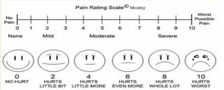
Evaluation of Post-operative Pain:
Post-operative pain among the study groups was assessed using numeric pain scale scoring system (Fig.4).Operationally the scale is usually a horizontal line, with scaling done from 0 to 10 as illustrated in figure below. The patient is asked to score his/her pain on scale from 0 to 10; a higher score signifies severer pain perceived by patient. Both set of patients were prescribed standard analgesics post-operatively. The pain scoring was done at four hours, twelve hours and twenty four hours following surgery. [image:8.612.37.473.524.699.2]
Figure 4: Showing numeric pain scale scoring system.
MEAN 53.32 35.20
Evaluation of Cosmetic Outcome:
The cosmetic outcome evaluation between conventional laparoscopic cholecystectomy and SILC group was
done using Body Image Questionnaire consisting of body image score (items 1 to 5) and cosmetic score (items
6 to 8) filled 12 weeks following surgery.( Fig.3)
Body Image Questionnaire:-
1. Are you less satisfied with your body since the operation?
□ 1 = yes, extremely
□ 2 = quite a bit □ 3 = a little bit □ 4 = no, not at all
2. Do you think the operation has damaged your body?
□ 1 = yes, extremely □ 2 = quite a bit □ 3 = a little bit □ 4 = no, not at all
3. Do you fell less attractive as a result of your operation?
□ 1 = yes, extremely □ 2 = quite a bit □ 3 = a little bit □ 4 = no, not at all
4. Do you feel less feminine/masculine as a result of your operation?
□ 1 = yes, extremely □ 2 = quite a bit □ 3 = a little bit □ 4 = no, not at all
5. Is it difficult to look at yourself naked?
□ 1 = yes, extremely □ 2 = quite a bit □ 3 = a little bit □ 4 = no, not at all
6. On a scale from 1 to 7, how satisfied are you with your scar(s)?
□ 1 = very unsatisfied □ 2 = quite unsatisfied □ 3 = a bit unsatisfied
□ 4 = not unsatisfied/not satisfied □ 5 = a bit satisfied
7. On a scale from 1 to 7, how would you describe your scar(s)?
□ 1 = revolting □ 2 = quite revolting □ 3 = a bit revolting
□ 4 = not revolting/not beautiful
□ 5 = a bit beautiful □ 6 = quite beautiful □ 7 = very beautiful
8. Could you score your own scar(s) on a scale from 1 to 10 (1 = ugliest scar imaginable, 10 = almost scarless)
Body Image Score:- Cosmetic Score:-
The pain score following four, twelve and twenty four hours after surgery was significantly lower in SILC compared to
conventional four port LC. The mean pain score after four hours for SILC was 4.46 while that for cLC was 7.72. Similarly
mean pain score after twelve hours was significantly lower for SILC (2.96) compare to cLC (5.08) and continuing the same
progression the mean pain score following 24 hours was significantly lower for SILC (1.80) compared to cLC (2.80). From it
can be concluded that SILC inflicts considerably less pain than conventional four port LC.
The cosmesis and body image score evaluated following 12 weeks after surgery had significantly better score in SILC
compared to cLC. Majority of patients 15(60%) in SILC group had score in range 41 to 48 on the other hand majority of
patients 24(96%) in cLC group had score in range 33 to 40. Mean score in for SILC patients was 40.76 while that for cLC
group was 38.28. This proves that SILC is aesthetically and cosmetically superior to conventional four ports LC.
V.
DISCUSSION
SILC being a safe, feasible and cosmetically well accepted surgery, now a days is becoming more common minimally invasive procedure.Learning curve is still an issue with SILC surgery. In recent past, many authors have been using different instruments or new operative devices or some novel instruments across the world to improvise learning curve and shorten duration of surgery. Few authors have used different types of single access ports like SILSTM port (Covidian),Triport TM , Single Site Laparoscopy Access System (Ethicon) and Gelpoint ( Applied Medical System ) etc.2
Some authors have used curved instruments for traction of gall bladder as special instruments.
Lei Zhao et al (2016), used special instrument with adjustable and rotatable wrist so called “novel” instrument in 150 cases in SILC surgery . In such instruments( novel ) , wrist movements are divided into two types in terms of relation of the directions of both the wrist at the surgeons’s hand side and instrument’s tip , ie. Syntropic and adverse wrist 6.
Technique with new instrument (novel) may be convenient to small sub group of laparoscopic surgeons but this may not be really an easy and comfortable tool to most of surgeons to work with such complexity of hand and wrist movements or co-ordinations, which may further increase operative time as well as learning curve ,which is already a big drawback of SILC procedure till date.
Whereas,complication rates were similar in patients undergoing SILC surgery or cLC with these conventional instruments or some special devise used .
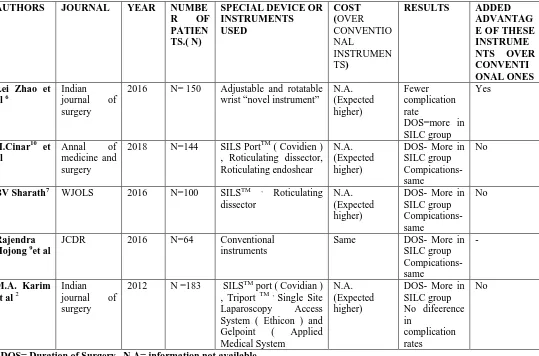
,duration of surgery( DOS) is not shortened even by novel /special instruments or devices used ( Fig.4). We have also found that with these instruments there is no added advantage.
AUTHORS JOURNAL YEAR NUMBE
R OF PATIEN TS.( N)
SPECIAL DEVICE OR INSTRUMENTS USED COST (OVER CONVENTIO NAL INSTRUMEN TS)
RESULTS ADDED
ADVANTAG E OF THESE INSTRUME NTS OVER CONVENTI ONAL ONES Lei Zhao et
al 6
Indian
journal of surgery
2016 N= 150 Adjustable and rotatable wrist “novel instrument”
N.A. (Expected higher) Fewer complication rate DOS=more in SILC group Yes
H.Cinar10 et al
Annal of medicine and surgery
2018 N=144 SILS PortTM ( Covidien ) , Roticulating dissector, Roticulating endoshear
N.A. (Expected higher)
DOS- More in SILC group Compications- same
No
BV Sharath7 WJOLS 2016 N=100 SILSTM , Roticulating
dissector
N.A. (Expected higher)
DOS- More in SILC group Compications- same
No
Rajendra Hojong 9et al
JCDR 2016 N=64 Conventional instruments
Same DOS- More in SILC group Compications- same
-
M.A. Karim et al2
Indian
journal of surgery
2012 N =183 SILSTM port ( Covidian ) , Triport TM , Single Site Laparoscopy Access System ( Ethicon ) and Gelpoint ( Applied Medical System
N.A. (Expected higher)
DOS- More in SILC group No difeerence in complication rates No
[image:11.612.41.580.137.493.2]DOS= Duration of Surgery , N.A= information not available
Table 4 : Comparative evaluation of different types of instruments used by various authors .
Safety and complications of SILC
Safety of the procedure is commonly determined by the incidence and rate of complications. SILC is considered to be a safe and effective procedure for the treatment of carefully selected patients with uncomplicated benign gallbladder disease. Although
there is a reported higher complications rate with SILC as compared to cLC, but not statistically significant. Common post-operative
fistula or bile duct injury was seen in our study. As we have experienced not much difficulty in visualizing and in order to facilitate
safe dissection of Calot’s triangle. Intraoperative blood loss was not significantly high in SILC.
Cost-effectiveness
Although, there is no such comparative data available comparing cost of these specially designed instruments.But few of devices and instruments are expensive .Whereas, some of devices used are made for one time use only, which poses extra cost to patient.This does not seem to a cost effective measure in SILC procedure, as there is no added advantage of using such instruments.
In our study , we have used instruments which are very commonly used, readily available and very convenient to almost all laparoscopic surgeons those who are doing conventional laparoscopic cholecystectomy .These conventional instruments adds no extra cost to patients as well.
User friendly
Many instruments are quite new with some specially designed features such as curved instruments, adjustable and rotatable wrist -“novel instrument”, roticulating dissector etc. Few of these instruments have unique method of its use such as wrist movements are divided into two types in terms of relation of the directions of both the wrist at the surgeons’s hand side and instrument’s tip , ie. Syntropic and adverse wrist .These type of instruments may not be convenient and user friendly to all laparoscopic surgeons across the world increasing learning curve further.
VI. CONCLUSION
SILC is relatively a newer technique in the field of minimal invasive laparoscopic surgery,which is safe and feasible option for gall stone surgery with better cosmesis and less post operative pain by using conventional instruments .However, using novel/special instruments have not shown any decrease in duration of surgery (DOS) and complication rates are also same in cLC and SILC by using those common conventional instruments. Whereas, learning curve is still another practical issue especially with newer or newly designed instruments. Cost of procedure also seems to be relatively higher with these special/novel instruments and devices used. On other hand, easy availability and good familiarity of conventional instruments among most of laparoscopic surgeons may help in reducing learning curve also in coming future.
REFERENCES
1. Navarra G, Pozza E, Occhionorelli S, et al. One-wound laparoscopic cholecystectomy. Br J Surg .1997; 84:695.
2. Muhammad Ali , Jamil Ahmed, et al .single incision vs.multiport laparoscopic cholecystectomy.ijs,(2012), 368-372.
3. Tomikawa M, Xu H, Hashizume M: Current status and prerequisites for natural orifice translumenal endoscopic surgery
(NOTES). Surg Today 2010;40:909–916
4. Romanelli JR, Mark L, Omotosho PA. Single port laparoscopic cholecystectomy with the TriPort system: A case report. Surg
Innov.2008; 15: 223–8.
5. Bresadola F, Pasqualucci A, Donini A, Chiarandini P, Anania G, Terrosu G, et al. Elective transumbilical compared with
standard laparoscopic cholecystectomy. Eur J Surg.1999; 165:29–34.
6. Lei Zhao, Zhifei Wang et al.A RCT comparing SILC using a novel instrument to that of common instrument.ijs 32( 2016)
174-178.
9. Rajendra Hajong et al.comparative study of single incision vs conventional four port laparoscopic cholecystectomy.jcdr/19982.8601
10. H.Cinar ,Koray Topgul et at.Early results of single –incision laparoscopic cholecystectomy in comparison with conventional .annals of medicine and surgery 32( 2018)1-5.
AUTHORS
First Author – Mukesh kumar ,
BSc, B.Ed, MBBS, MS( general surgery ), Deptt. Of surgery , IGMC , Shimla, drmuks1979@gmail.com
Second Author – R.S.Jhobta
MBBS, MS ,jhobtars@yahoo.co.in
Third Author – D.K.Verma , MBBS, MS, dkverma@gmail.com