(
E
)-
N
000-(2-Chlorobenzylidene)-1-methyl-4-nitro-1
H
-pyrrole-2-carbohydrazide
Jinglin Wang,* Rong He, Zhijuan Xin, Huiling Shen and Cairong Wang
Department of Chemistry, Changzhi University, Changzhi, Shanxi 046011, People’s Republic of China
Correspondence e-mail: jlwangczu@163.com
Received 8 December 2013; accepted 18 December 2013
Key indicators: single-crystal X-ray study;T= 298 K; mean(C–C) = 0.005 A˚;
Rfactor = 0.061;wRfactor = 0.164; data-to-parameter ratio = 12.8.
In the title compound, C13H11ClN4O3, the phenyl and pyrrolyl
ring are linked by an acyl–hydrazone (R2C N—N—CO—R)
group, forming a slightly bent molecule: the dihedral angle subtended by the the phenyl and pyrrolyl rings is 8.46 (12). In
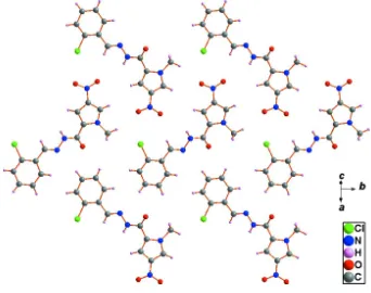
the crystal, the three-dimensional supramolecular structure is assembled by N—H O hydrogen bonding. Molecular sheets are formed parallel to (101) in a herringbone arrangement by weak van der Waals interactions; weak–[centroid–centroid phenyl–phenyl and pyrrolyl–pyrrolyl distances of 3.7816 (3) and 3.8946 (2) A˚ , respectively] interactions occur between neighbouring sheets.
Related literature
For applications and structures of aroylhydrazones, see: Raja
et al.(2012); Wanget al.(2014).
Experimental
Crystal data
C13H11ClN4O3 Mr= 306.71
a= 13.7649 (13) A˚ b= 12.4993 (11) A˚ c= 8.1263 (10) A˚
= 95.523 (1)
V= 1391.7 (2) A˚3
MoKradiation
= 0.29 mm1 T= 298 K
0.300.200.16 mm
Data collection
Bruker SMART 1000 CCD diffractometer
Absorption correction: multi-scan (SADABS; Bruker, 2005) Tmin= 0.918,Tmax= 0.955
6871 measured reflections 2452 independent reflections 1435 reflections withI> 2(I) Rint= 0.071
Refinement
R[F2> 2(F2)] = 0.061 wR(F2) = 0.164 S= 1.00 2452 reflections
191 parameters
H-atom parameters constrained
max= 0.23 e A˚
3
min=0.33 e A˚
3
Table 1
Hydrogen-bond geometry (A˚ ,).
D—H A D—H H A D A D—H A
N3—H3 O1i
0.86 2.14 2.941 (3) 154
Symmetry code: (i)x;yþ3 2;zþ
1 2.
Data collection:SMART(Bruker, 1999); cell refinement:SAINT (Bruker, 1999); data reduction:SAINT; program(s) used to solve structure:SHELXS97(Sheldrick, 2008); program(s) used to refine structure: SHELXL97 (Sheldrick, 2008); molecular graphics: DIAMOND(Brandenburg, 1999) andSHELXTL(Sheldrick, 2008); software used to prepare material for publication:PLATON(Spek, 2009) andSHELXTL.
This work was supported by the National-level College Students’ Innovative Training Plan Program of the People’s Republic of China (grant No. 201310122001) and the Scientific Research Foundation for PhDs of Changzhi University.
Supplementary data and figures for this paper are available from the IUCr electronic archives (Reference: FF2124).
References
Brandenburg, K. (1999).DIAMOND. Crystal Impact GbR, Bonn, Germany. Bruker (1999).SMARTandSAINT. Bruker AXS Inc., Madison, Wisconsin,
USA.
Bruker (2005).SADABS. Bruker AXS Inc., Madison, Wisconsin, USA. Raja, D. S., Bhuvanesh, N. S. P. & Natarajan, K. (2012).Dalton Trans.41, 4365–
4377.
Sheldrick, G. M. (2008).Acta Cryst.A64, 112–122. Spek, A. L. (2009).Acta Cryst.D65, 148–155.
Wang, J., Zhao, Y. & Yang, B. (2014).Inorg. Chim. Acta,409, 484–496.
Structure Reports
Online
supporting information
Acta Cryst. (2014). E70, o99 [https://doi.org/10.1107/S1600536813034119]
(
E
)-
N
′
-(2-Chlorobenzylidene)-1-methyl-4-nitro-1
H
-pyrrole-2-carbohydrazide
Jinglin Wang, Rong He, Zhijuan Xin, Huiling Shen and Cairong Wang
S1. Comment
A great number of aroylhydrazones (AH) have triggered wide interest because of their diverse spectra of biological and
pharmaceutical properties (Raja, et al., 2012). In our lab, the AH compound (E)-N
′-(2-hydroxybenzylidene)-1-methyl-4-nitro-1H-pyrrole-2-carbohydrazide (L) and its transition metal complexes were obtained and characterized. The
interaction of these compounds with CT-DNA and pBR322 DNA has been explored (Wang, et al., 2014). The present
report is an extension of our earlier studies in this area.
In the title compound (Fig. 1), C13H11ClN4O3, the phenyl and pyrrolyl ring are linked by acyl-hydrazone (R2C=N–N–
CO–R) to form a slightly bent molecule. The dihedral angle between the phenyl (C8—C13) and pyrrolyl rings (C2—C5,
N1) is 8.46 (12)°.
As shown in Figure 2, the herringbone molecular sheet of the title compound is formed by weak van-der-Waals
interactions along (101) plane.
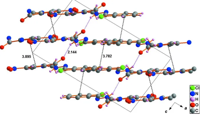
The three-dimensional supramolecular structure (Fig. 3) is assembled by N3–H3···O1i hydrogen bonding (pink dotted
lines) and weak Cg1···Cg1ii (Cg1 is the centroid of the phenyl ring) and Cg2···Cg2iii (Cg2 is the centroid of the pyrrolyl
ring) interactions (black dotted lines) between the neighbouring molecular sheets [symmetry code: (i) x, 3/2 - y, 1/2 + z;
(ii)1 - x, 2 - y, 2 – z; (iii) - x, 1 - y, 2 - z]. The data of hydrogen-bond geometry are given in Table 1.
S2. Experimental
Single crystals of the title compound were obtained accidentally in the attempted synthesis of a Ni complex. (E)-N
′-(2-hydroxybenzylidene)-1-methyl-4-nitro-1H-pyrrole-2-carbohydrazide (L) was synthesized according to literature
procedures (Wang et al., 2014). Sodium methoxide (250 µL, 3%, g/V) was added to solution of L (0.50 mmol, 0.144 g) in
15 ml MeOH and was heated to reflux. NiCl2·6H2O (0.50 mmol, 0.119 g) was then added to the refluxing mixture and
further refluxed for 2 h. The reaction mixture was cooled and was allowed to stir at room temperature overnight. The
mixture was filtered and washed with methanol. The L—Ni complex is not achieved as predicted. However, orange
single crystals of the title compound suitable for X-ray analysis were obtained after several days from the mother liquor
by slow evaporation.
S3. Refinement
H atoms attached to C atoms are placed in geometrically idealized position, with N–H=0.86 Å, C–H=0.93 and 0.96 Å, for
CH and CH3 groups, respectively, and with Uiso(H) = k × Ueq(parent C-atom), where k = 1.5 for CH3 H-atoms and =1.2 for
Figure 1
The molecular structure of the title compound with the atom numbering scheme. Displacement ellipsoids are drawn at the
30% probability level. H atoms are presented as small spheres of arbitrary radius.
Figure 2
[image:3.610.133.475.305.575.2]Figure 3
Packing of the title compound viewed along the b axis. The three-dimensional supramolecular structure is assembled by
N–H···O hydrogen bonding (pink dotted lines) and weak π···π interactions (black dotted lines) between the neighbouring
molecular sheets (all distances in Å).
(E)-N′-(2-Chlorobenzylidene)-1-methyl-4-nitro-1H-pyrrole-2-carbohydrazide
Crystal data
C13H11ClN4O3
Mr = 306.71 Monoclinic, P21/c
a = 13.7649 (13) Å b = 12.4993 (11) Å c = 8.1263 (10) Å β = 95.523 (1)° V = 1391.7 (2) Å3
Z = 4
F(000) = 632 Dx = 1.464 Mg m−3
Mo Kα radiation, λ = 0.71073 Å Cell parameters from 1539 reflections θ = 3.0–25.2°
µ = 0.29 mm−1
T = 298 K Block, yellow
0.30 × 0.20 × 0.16 mm
Data collection
Bruker SMART 1000 CCD diffractometer
Radiation source: fine-focus sealed tube Graphite monochromator
phi and ω scans
Absorption correction: multi-scan (SADABS; Bruker, 2005) Tmin = 0.918, Tmax = 0.955
6871 measured reflections 2452 independent reflections 1435 reflections with I > 2σ(I) Rint = 0.071
θmax = 25.0°, θmin = 1.5°
h = −15→16 k = −14→14 l = −6→9
Refinement
Refinement on F2
Least-squares matrix: full R[F2 > 2σ(F2)] = 0.061
wR(F2) = 0.164
S = 1.00 2452 reflections
0 restraints
Primary atom site location: structure-invariant direct methods
Secondary atom site location: difference Fourier map
w = 1/[σ2(F
o2) + (0.0787P)2]
where P = (Fo2 + 2Fc2)/3
Δρmax = 0.23 e Å−3
Δρmin = −0.33 e Å−3
Special details
Geometry. All e.s.d.'s (except the e.s.d. in the dihedral angle between two l.s. planes) are estimated using the full covariance matrix. The cell e.s.d.'s are taken into account individually in the estimation of e.s.d.'s in distances, angles and torsion angles; correlations between e.s.d.'s in cell parameters are only used when they are defined by crystal symmetry. An approximate (isotropic) treatment of cell e.s.d.'s is used for estimating e.s.d.'s involving l.s. planes.
Refinement. Refinement of F2 against ALL reflections. The weighted R-factor wR and goodness of fit S are based on F2,
conventional R-factors R are based on F, with F set to zero for negative F2. The threshold expression of F2 > σ(F2) is used
only for calculating R-factors(gt) etc. and is not relevant to the choice of reflections for refinement. R-factors based on F2
are statistically about twice as large as those based on F, and R- factors based on ALL data will be even larger.
Fractional atomic coordinates and isotropic or equivalent isotropic displacement parameters (Å2)
x y z Uiso*/Ueq
Cl1 0.29032 (9) 1.16858 (8) 1.00653 (18) 0.1044 (6)
N1 0.14340 (19) 0.51021 (19) 1.0868 (3) 0.0508 (7)
N2 −0.0430 (2) 0.5940 (3) 1.3388 (4) 0.0712 (9)
N3 0.24317 (19) 0.7725 (2) 1.0085 (3) 0.0512 (8)
H3 0.2231 0.7996 1.0964 0.061*
N4 0.30201 (18) 0.83156 (19) 0.9133 (3) 0.0470 (7)
O1 0.24317 (17) 0.62828 (16) 0.8366 (3) 0.0587 (7)
O2 −0.0656 (2) 0.6852 (3) 1.3780 (4) 0.1057 (12)
O3 −0.08399 (19) 0.5122 (3) 1.3789 (3) 0.0907 (9)
C1 0.2175 (2) 0.6716 (2) 0.9618 (4) 0.0465 (8)
C2 0.1506 (2) 0.6203 (2) 1.0708 (4) 0.0451 (8)
C3 0.0829 (2) 0.6674 (3) 1.1612 (4) 0.0514 (8)
H3A 0.0711 0.7403 1.1715 0.062*
C4 0.0352 (2) 0.5839 (3) 1.2344 (4) 0.0561 (9)
C5 0.0737 (2) 0.4884 (3) 1.1897 (4) 0.0573 (9)
H5 0.0554 0.4209 1.2235 0.069*
C6 0.2046 (3) 0.4285 (3) 1.0169 (5) 0.0659 (10)
H6A 0.2708 0.4362 1.0642 0.099*
H6B 0.2017 0.4376 0.8992 0.099*
H6C 0.1810 0.3586 1.0416 0.099*
C7 0.3117 (2) 0.9297 (2) 0.9548 (4) 0.0483 (8)
H7 0.2764 0.9569 1.0373 0.058*
C8 0.3779 (2) 1.0000 (2) 0.8743 (4) 0.0467 (8)
C9 0.3778 (2) 1.1102 (2) 0.8944 (4) 0.0571 (9)
C10 0.4438 (3) 1.1761 (3) 0.8256 (5) 0.0698 (11)
H10 0.4413 1.2498 0.8406 0.084*
C11 0.5133 (3) 1.1319 (3) 0.7348 (5) 0.0698 (11)
H11 0.5587 1.1756 0.6900 0.084*
C12 0.5154 (3) 1.0231 (3) 0.7108 (5) 0.0659 (10)
H12 0.5620 0.9933 0.6491 0.079*
C13 0.4481 (2) 0.9577 (3) 0.7784 (4) 0.0568 (9)
Atomic displacement parameters (Å2)
U11 U22 U33 U12 U13 U23
Cl1 0.1002 (9) 0.0517 (6) 0.1730 (14) 0.0028 (6) 0.0731 (9) −0.0179 (7)
N1 0.0553 (16) 0.0427 (14) 0.0560 (18) −0.0065 (13) 0.0130 (13) 0.0045 (13)
N2 0.057 (2) 0.102 (3) 0.057 (2) −0.019 (2) 0.0189 (16) 0.0023 (19)
N3 0.0646 (18) 0.0459 (15) 0.0477 (17) −0.0109 (13) 0.0286 (14) −0.0042 (12) N4 0.0549 (16) 0.0436 (15) 0.0455 (16) −0.0057 (13) 0.0199 (13) 0.0025 (11) O1 0.0821 (17) 0.0499 (12) 0.0490 (14) −0.0105 (12) 0.0310 (12) −0.0064 (11)
O2 0.088 (2) 0.121 (3) 0.117 (3) −0.010 (2) 0.056 (2) −0.027 (2)
O3 0.0637 (17) 0.134 (3) 0.077 (2) −0.0337 (18) 0.0193 (14) 0.0198 (18)
C1 0.053 (2) 0.0444 (17) 0.045 (2) −0.0027 (15) 0.0197 (15) 0.0023 (14)
C2 0.0476 (18) 0.0425 (16) 0.047 (2) −0.0063 (14) 0.0123 (15) −0.0010 (14)
C3 0.0496 (19) 0.0530 (18) 0.054 (2) −0.0032 (16) 0.0153 (16) −0.0007 (16)
C4 0.0516 (19) 0.069 (2) 0.050 (2) −0.0095 (18) 0.0160 (16) 0.0016 (17)
C5 0.059 (2) 0.059 (2) 0.055 (2) −0.0168 (18) 0.0113 (17) 0.0151 (17)
C6 0.075 (3) 0.0447 (18) 0.079 (3) 0.0036 (18) 0.014 (2) 0.0010 (18)
C7 0.0515 (19) 0.0442 (18) 0.053 (2) −0.0001 (15) 0.0227 (16) −0.0005 (15)
C8 0.0473 (18) 0.0431 (16) 0.052 (2) −0.0039 (15) 0.0150 (15) 0.0006 (14)
C9 0.056 (2) 0.0442 (18) 0.074 (3) −0.0032 (16) 0.0199 (18) 0.0008 (17)
C10 0.068 (2) 0.0459 (19) 0.098 (3) −0.0110 (18) 0.021 (2) 0.0092 (19)
C11 0.061 (2) 0.076 (3) 0.074 (3) −0.013 (2) 0.016 (2) 0.018 (2)
C12 0.063 (2) 0.075 (3) 0.064 (3) −0.002 (2) 0.0284 (19) 0.003 (2)
C13 0.064 (2) 0.0502 (19) 0.059 (2) −0.0029 (17) 0.0220 (18) 0.0008 (16)
Geometric parameters (Å, º)
Cl1—C9 1.739 (3) C5—H5 0.9300
N1—C5 1.359 (4) C6—H6A 0.9600
N1—C2 1.387 (4) C6—H6B 0.9600
N1—C6 1.472 (4) C6—H6C 0.9600
N2—O3 1.227 (4) C7—C8 1.465 (4)
N2—O2 1.232 (4) C7—H7 0.9300
N2—C4 1.439 (4) C8—C9 1.387 (4)
N3—C1 1.354 (4) C8—C13 1.402 (4)
N3—N4 1.386 (3) C9—C10 1.383 (5)
N3—H3 0.8600 C10—C11 1.379 (5)
N4—C7 1.276 (4) C10—H10 0.9300
O1—C1 1.234 (3) C11—C12 1.374 (5)
C1—C2 1.483 (4) C11—H11 0.9300
C2—C3 1.374 (4) C12—C13 1.388 (4)
C3—C4 1.397 (4) C12—H12 0.9300
C3—H3A 0.9300 C13—H13 0.9300
C4—C5 1.369 (5)
C5—N1—C2 108.5 (3) N1—C6—H6B 109.5
C5—N1—C6 124.2 (3) H6A—C6—H6B 109.5
O3—N2—C4 118.2 (4) H6B—C6—H6C 109.5
O2—N2—C4 117.1 (3) N4—C7—C8 120.8 (3)
C1—N3—N4 119.4 (2) N4—C7—H7 119.6
C1—N3—H3 120.3 C8—C7—H7 119.6
N4—N3—H3 120.3 C9—C8—C13 116.7 (3)
C7—N4—N3 114.6 (2) C9—C8—C7 122.4 (3)
O1—C1—N3 123.5 (3) C13—C8—C7 120.9 (3)
O1—C1—C2 123.1 (3) C10—C9—C8 122.4 (3)
N3—C1—C2 113.3 (3) C10—C9—Cl1 118.5 (3)
C3—C2—N1 108.5 (3) C8—C9—Cl1 119.1 (2)
C3—C2—C1 128.8 (3) C11—C10—C9 119.6 (3)
N1—C2—C1 122.6 (3) C11—C10—H10 120.2
C2—C3—C4 106.1 (3) C9—C10—H10 120.2
C2—C3—H3A 126.9 C12—C11—C10 119.8 (3)
C4—C3—H3A 126.9 C12—C11—H11 120.1
C5—C4—C3 109.2 (3) C10—C11—H11 120.1
C5—C4—N2 124.3 (3) C11—C12—C13 120.2 (3)
C3—C4—N2 126.4 (3) C11—C12—H12 119.9
N1—C5—C4 107.6 (3) C13—C12—H12 119.9
N1—C5—H5 126.2 C12—C13—C8 121.3 (3)
C4—C5—H5 126.2 C12—C13—H13 119.4
N1—C6—H6A 109.5 C8—C13—H13 119.4
C1—N3—N4—C7 171.3 (3) C2—N1—C5—C4 1.7 (4)
N4—N3—C1—O1 −0.5 (5) C6—N1—C5—C4 177.5 (3)
N4—N3—C1—C2 −177.2 (3) C3—C4—C5—N1 −1.2 (4)
C5—N1—C2—C3 −1.6 (4) N2—C4—C5—N1 178.0 (3)
C6—N1—C2—C3 −177.2 (3) N3—N4—C7—C8 175.1 (3)
C5—N1—C2—C1 −177.7 (3) N4—C7—C8—C9 168.3 (3)
C6—N1—C2—C1 6.7 (5) N4—C7—C8—C13 −14.7 (5)
O1—C1—C2—C3 −147.4 (4) C13—C8—C9—C10 −0.6 (6)
N3—C1—C2—C3 29.3 (5) C7—C8—C9—C10 176.4 (4)
O1—C1—C2—N1 27.9 (5) C13—C8—C9—Cl1 178.0 (3)
N3—C1—C2—N1 −155.5 (3) C7—C8—C9—Cl1 −4.9 (5)
N1—C2—C3—C4 0.8 (4) C8—C9—C10—C11 −0.7 (6)
C1—C2—C3—C4 176.6 (3) Cl1—C9—C10—C11 −179.4 (3)
C2—C3—C4—C5 0.3 (4) C9—C10—C11—C12 1.2 (6)
C2—C3—C4—N2 −178.9 (3) C10—C11—C12—C13 −0.4 (6)
O3—N2—C4—C5 −6.7 (6) C11—C12—C13—C8 −1.0 (6)
O2—N2—C4—C5 175.1 (4) C9—C8—C13—C12 1.5 (5)
O3—N2—C4—C3 172.3 (3) C7—C8—C13—C12 −175.6 (3)
O2—N2—C4—C3 −5.9 (6)
Hydrogen-bond geometry (Å, º)
N3—H3···O1i 0.86 2.14 2.941 (3) 154