In experiments with obligate plant parasites, detached leaf segments on an agar medium are often used under non-sterile conditions: for example, if living spores of powdery mildew or leaf rust are sampled outdoors onto leaf segments in a spore trap, or in inoculation experiments when no laminar air-flow boxes are available. The agar may become accidentally contaminated with airborne bacteria or moulds that sometimes overgrow the sampled parasite on the leaf segments or kill the leaf tissue. Therefore an-tibiotics are added to the medium in some laboratories. This is costly and only partly solves the problem, since the spectrum of sensitive micro-organisms is limited. An alternative solution would be to utilise the oligodynamic effect of silver ions which kill bacteria within hours at 0.05
to 0.5 ppm (GRIER1983; THURMAN & GERBA1989). Since silver ions are precipitated by Cl-ions, present in the tis-sue, to non-soluble AgCl and finally reduced by light to nanoclusters of metallic silver, they are probably not tak-en up by the plant tissue and therefore are not expected to interfere with the studied host–parasite interactions or harm the plant tissue, unlike chemical disinfectants or antibiotics. The aim of the presented experiments was therefore to study the efficacy of different concentra-tions of ionic silver in suppressing the contamination of a mineral–agar medium and whether or not the growth of powdery mildew on leaf segments or the interactions be-tween the mildew culture and the varieties with or with-out the mlo gene was affected.
S
HORT
C
OMMUNICATION
The Effects of Silver on Microbial Contamination of Agar Medium
and on Interactions between Mildew and Barley Leaf Segments
with and without the
mlo
Gene
E
RIKSCHWARZBACH
Miroslav, Czech Republic
Abstract: Segments of primary leaves of several barley varieties with and without the mlo gene were placed in Petri dishes on an
agar medium containing benzimidazole, mineral nutrients and 0, 0.1, 0.3, 1 and 3 ppm of AgNO3. Three Petri dishes were prepared of each concentration. The segments were uniformly inoculated with 103 conidia/cm2 of the partially mlo-virulent powdery mildew culture PV-32. Subsequently, one open Petri dish of each Ag-concentration was exposed for 1 hour to a different potential con-tamination environment: one in the laboratory (low load), one in a humid cellar close to stored vegetables (medium load) and one on the top of a compost heap of decaying garbage (heavy load). Germination of mildew spores on the medium surface declined slightly with increasing concentration of AgNO3. Mildew infection was evaluated 7 days after inoculation. The number of mildew colonies per leaf segment and the differential interaction of the Mlo- and mlo-varieties with the mildew culture was apparently not affected by the AgNO3 concentration. Contamination of the medium by airborne micro-organisms was evaluated 12 days after exposure both microscopically and by eye. The contamination of the medium increased with environmental load and with decreasing AgNO3 concentration. 0.1 ppm AgNO3 markedly retarded the growth of contaminant colonies from all three environ-ments, but did not prevent contamination. At 1 ppm AgNO3, no contamination was observed on the media exposed to low and medium load, but several dozen small contaminant colonies developed on the medium exposed to heavy load. At 3 ppm AgNO3, only three small contaminant colonies developed on the medium exposed to heavy load, while the media exposed to medium and low load remained clean. It can be concluded that adding 1 ppm AgNO3 to a mineral-agar medium efficiently suppresses its contamination under low and medium load,without apparently affecting the growth of mildew or the interaction between mildew and mlo-barley on leaf segments placed on the medium.
MATERIALS AND METHODS
Medium. Petri dishes of 12 cm inner diameter with 60 ml of a medium containing 0.4% agar, 20 ppm benzimidazole (commonly used to retard leaf senescence in vitro), 50 ppm of Scherings WUXAL-SUPER® mineral nutrient
solution and a variable amount of AgNO3 were used in the experiments. The medium was prepared by suspend-ing agar powder in a small amount of demineralised wa-ter, pouring it into hot demineralised water and boiling for 10 minutes. When the solution cooled down to ap-prox 50°C, benzimidazole and mineral nutrients were add-ed from 2000 ppm stock solutions. A stock solution of 1000 ppm AgNO3 was prepared separately, of which 0.0, 0.1, 0.3, 1.0, and 3.0 ml/l were added to the still liquid agar medium. Thus the final concentration of AgNO3 in the dishes was 0.0, 0.1, 0.3, 1 and 3.0 ppm. Three Petri dishes of each Ag-concentration were prepared.
Plant material. In each Petri dish, 25 mm long seg-ments of primary leaves of ten days old seedlings of Dia-mant, Apex and the Pallas near isogenic lines P8 and P11 were placed on the medium and uniformly inoculated with the partially mlo-virulent mildew culture PV-32, described earlier (SCHWARZBACHet al. 2002). The varieties, except Apex, do not have the mlo gene and are susceptible to the mildew culture PV-32. Cv. Apex has the mlo11 allele for resistance against almost all existing mildew patho-types and is partially resistant to PV-32. The segments were arranged side-by-side in the order P8– APEX–Dia-mant–Apex–P11–Apex–P8.
Inoculation. The Petri dishes were uniformly inoculat-ed with conidia of PV-32 at a density of approximately 103 spores per cm2 in a settling tower as described earlier
(SCHWARZBACH 2001). Three Petri dishes with the same Ag-concentration were inoculated at a time. The unifor-mity of inoculation was tested by counting the spores within two random microscope view-fields of 2 mm2 on
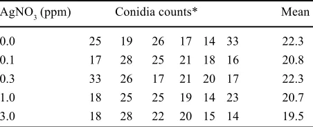
[image:2.595.57.283.592.684.2]the agar surface of each Petri dish. The spore counts are summarised in Table 1.
Table 1. Test of inoculation uniformity
AgNO3 (ppm) Conidia counts* Mean
0.0 25 19 26 17 14 33 22.3
0.1 17 28 25 21 18 16 20.8
0.3 33 26 17 21 20 17 22.3
1.0 18 25 25 19 14 23 20.7
3.0 18 28 22 20 15 14 19.5
*Conidia counts on the medium in 2 mm2 view-fields after
inoc-ulation in a settling tower
The spore counts were compared with the expected Poisson distribution, in which the variance equals the mean.
The fit was tested by the relation of the observed vari-ance to the mean, using the F-test. The calculated F -val-ue of 1.33 is far below a significant deviation from the expectation, even at the weak probability level of α = 0.05. Therefore, the inoculation can be regarded as fairly uni-form, although some small differences between the se-ries cannot be excluded.
Incubation. All series of experiments were incubated in a thermostat at 18°C under continuous light of a 10 W daylight fluorescent tube, fitted 56 cm above the leaf segments.
Conidial germination was examined 24 h after inocu-lation in two random microscope view-fields of 2 mm2 in
each Petri dish. The error was calculated from the differ-ences between the six view-fields within each Ag-con-centration.
Mildew infection was evaluated 6 days after inocula-tion by counting the number of mildew colonies on each leaf segment, using a 6× magnifying lens. Since there were no significant differences in colony counts between the non-mlo barleys, these were pooled together as “Mlo -barley”, in contrast to cv. Apex as the mlo-barley.
Contamination. The open Petri dishes containing the freshly inoculated leaf segments were exposed for 1 h to different contaminating environments. One Petri dish of each Ag-concentration was exposed to laboratory air (lowest load), the second in a humid mouldy basement close to stored potatoes and vegetables (medium load), and the third outdoors on a compost heap of decaying garden and kitchen garbage (heavy load). The dishes were protected against insects by a fine sieve. Since the contaminating load was heavy and very mixed, no deter-mination of the involved micro-organisms was carried out. After 12-days of incubation, the contamination of the medium was examined within each Petri dish under the microscope at a 10 × 10 magnification at 12 spots dis-tributed around the laid out segments. At 6 spots the length of hyphae, relative to the view-field diameter was estimated. At the other 6 spots, the total area of non-hy-phal colonies, relative to the view-field, was estimated.
RESULTS
The germination of PV-32 conidia at different concen-trations of AgNO3 in the medium is summarised in Table 2. Since there were no significant differences in germina-tion between the different contaminagermina-tion loads, the data from the different loads were pooled together (Fig. 1).
expected to be smaller than that of the contaminating micro-organisms.
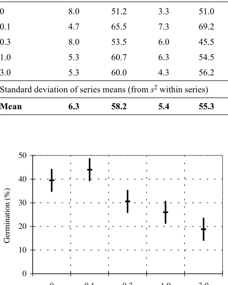
The effect of silver on infection efficiency and on the interaction between the PV-32 culture and the mlo gene is summarised in Table 2.
No significant effects of silver or contamination load on the infection efficiency of PV-32 or mildew colony number on mlo-segments, relative to Mlo-segments, could be observed. The slight decrease in infection of mlo-segments relative to Mlo-segments with increasing Ag-concentration was not significant and therefore most likely to be accidental. There is no evidence, therefore, that the addition of AgNO3 to the medium affected the infection efficiency of the mildew culture PV-32 on seg-ments with or without the mlo gene.
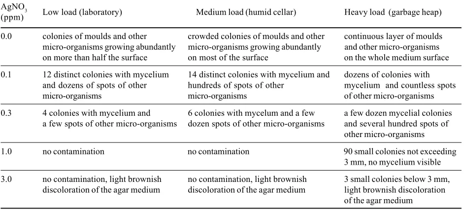
[image:3.595.57.287.151.438.2]The effects of silver on contamination of the agar-me-dium, observed microscopically, are summarised in Ta-bles 3 and 4.
Table 2. The effect of AgNO3 concentration in the medium on the colony number on leaf segments with and without the mlo gene, inoculated with the mildew culture PV-32
AgNO3 Low load Medium load Heavy load Mean mlo/Mlo
(ppm) mlo Mlo mlo Mlo mlo Mlo mlo Mlo (%)
0 8.0 51.2 3.3 51.0 6.7 53.7 6.0 52.0 11.5
0.1 4.7 65.5 7.3 69.2 9.7 67.5 7.2 67.4 10.7
0.3 8.0 53.5 6.0 45.5 4.0 67.5 6.0 55.5 10.8
1.0 5.3 60.7 6.3 54.5 3.3 48.7 5.0 54.6 9.2
3.0 5.3 60.0 4.3 56.2 4.3 57.0 4.6 57.5 8.1
Standard deviation of series means (from s2 within series) 1.12 3.36 1.93
[image:3.595.59.283.294.443.2]Mean 6.3 58.2 5.4 55.3 5.6 58.9 5.8 57.4 10.1
Table 4. The average per cent coverage of non-hyphal micro-organisms observed in a microscope view-field 12 days after exposure to different contaminating environments
AgNO3 Low load Medium load Heavy load
(ppm) (laboratory) (humid cellar) (garbage heap)
0.0 2 65 83
0.1 17 31 20
0.3 0 0 26
1.0 0 0 0
3.0 0 0 0
The figures are means of 6 estimates at different spots within the same Petri dish
Table 3. The average per cent extension of mould hyphae across microscope view-fields 12 days after exposure to different contaminating environments
AgNO3 Low load Medium load Heavy load
(ppm) (laboratory) (humid cellar) (garbage heap)
0.0 51 65 100
0.1 40 83 100
0.3 17 17 87
1.0 0 0 26
3.0 0 0 0
The figures are means of 6 estimates at different spots within the same Petri dish
0 10 20 30 40 50
0 0.1 0.3 1.0 3.0 AgNO3(ppm) confidence intervals given forα = 0.05
Germination
(%)
Fig. 1. Conidia germination on the medium with different AgNO3 concentration
[image:3.595.297.525.350.454.2] [image:3.595.297.524.544.647.2]DISCUSSION
The results show clearly that AgNO3 is a powerful means of protecting agar media containing detached leaves against contamination from non-sterile environ-ments. Since NO3– is a basic plant nutrient, the effects are
due to silver ions or nano-particles of metallic silver. The very light brownish discoloration of the agar medium at 3 ppm is obviously an effect of the photo-reduction of silver ions to nano-clusters of metallic silver and is of no consequence for the experiments. Apart from being very cheap, silver is, according to the US Environmental Pro-tection Agency (EPA), nontoxic and non-carcinogenic. The only undesirable clinical effect of silver is an irrevers-ible skin discoloration (argyrosis) after very high i.v. doses in the order of grams or oral doses in dozens of grams (GAUL & STAUD 1935; FURCHNER et al. 1968). If the contamination load is not extremely high, 1 ppm AgNO3 added to the medium protects it safely against contamination. In routine work with this concentration, ocassionally yeasts ocurred on exudates from the cutting edges of the leaf segments, spreading onto the agar me-dium, or moulds developed from particles of organic im-purities or dirt. This is not surprising, since the exudates or particles of impurities are not expected to contain pro-tective amounts of silver.
Acknowledgement: The help of the Research Institute for Plant Production at Prague-Ruzyně, which lent me a microsco-pe, was greatly appreciated. Thanks to JOHN CLARKSON, NIAB Cambridge, for correcting the English.
References
GAUL L.E., STAUD A.H. (1935): Clinical spectroscopy. Sev-enty cases of generalized argyrosis following organic and colloidal silver medication including a biospectrometric anal-ysis of ten cases. J. Am. Med. Assoc., 104: 1387–1390. FURCHNER J.E., RICHMOND C.R., DRAKE G.A. (1968):
Comparative metabolism of radionuclides in mammals – IV. Retention of silver – 110m in the mouse, rat, monkey, and dog. Health Phys., 15: 505–514.
GRIER N. (1983): Silver and its compounds. In: BLOCK S. (ed.): Disinfection, Sterilization and Preservation. Lea & Febiger, Philadelphia: 380–428.
SCHWARZBACH E. (2001): Heat induced susceptibility of mlo -barley to powdery mildew (Blumeria graminis D.C. f.sp. hordei Marchal). Czech J. Genet. Plant Breed., 37: 82–87. SCHWARZBACH E., SLATER S.E., CLARKSON J.D.S. (2002):
Occurrence of partially mlo-virulent isolates of barley powdery mildew in agricultural environments in Europe. Cereal Rusts and Powdery Mildews Bull., 30
[www.crpmb.org/]2001/0208schwarzbach/.
THURMAN R., GERBA C. (1989): The molecular mechanisms of copper and silver ion disinfection of bacteria and virus-es. Crit. Rev. Envir. Control., 18: 295–315.
US Environ. Protection Agency (EPA), document: Silver, CASRN 7440-22-4, updated 1996.
WUXAL® is a registered trademark of Hoechst Schering
AgrEvo G.m.b.H. Berlin, Germany.
Received for publication November 16, 2001
[image:4.595.56.527.109.322.2]Accepted after corrections February 28, 2002
Table 5. Visual evaluation of contamination 12 days after exposure of the agar medium to different contaminating environments
AgNO3 Low load (laboratory) Medium load (humid cellar) Heavy load (garbage heap)
(ppm)
0.0 colonies of moulds and other crowded colonies of moulds and other continuous layer of moulds
micro-organisms growing abundantly micro-organisms growing abundantly and other micro-organisms
on more than half the surface on most of the surface on the whole medium surface
0.1 12 distinct colonies with mycelium 14 distinct colonies with mycelium and dozens of colonies with
and dozens of spots of other hundreds of spots of other mycelium and countless spots
micro-organisms micro-organisms of other micro-organisms
0.3 4 colonies with mycelium and 6 colonies with mycelum and a few a few dozen mycelial colonies
a few spots of other micro-organisms dozen spots of other micro-organisms and several hundred spots of other micro-organisms
1.0 no contamination no contamination 90 small colonies not exceeding
3 mm, no mycelium visible
3.0 no contamination, light brownish no contamination, light brownish 3 small colonies below 3 mm,
discoloration of the agar medium discoloration of the agar medium light brownish discoloration
Abstrakt
SCHWARZBACH E. (2002): Vliv stříbra na mikrobiální kontaminaci agarového média a na interakce mezi padlím a listo-vými segmenty ječmene s genem mlo a bez něho. Czech J. Genet. Plant Breed., 38: 82–86.
Segmenty primárních listů linií ječmene lišících se genem mlo byly vyloženy v Petriho miskách na agarovém médiu s benzimida-zolem, minerálními živinami a s AgNO3 v koncentracích 0, 0,1, 0,3, 1 a 3 ppm. Od každé koncentrace byly připraveny tři Petriho misky. Segmenty byly rovnoměrně infikovány parciálněmlo-virulentní kulturou padlí PV-32 v množství 103 konidií/cm2. Poté byla vždy jedna otevřená Petriho miska z každé koncentrace vystavena po 1 hodinu jinému prostředí s odlišným kontaminačním potenciálem: jedna v laboratoři (mírná zátěž), jedna ve vlhkém sklepě blízko skladované zeleniny (střední zátěž) a jedna na hroma-dě kompostu s rozkládajícími se odpadky (silná zátěž). Klíčení spor padlí na povrchu média mírně klesalo se zvyšující se koncen-trací AgNO3. Napadení padlím bylo hodnoceno 7 dní po infekci. Počet kolonií padlí na listových segmentech a interakce kultury padlí s geny mlo a Mlo nebyly znatelně ovlivněny koncentrací AgNO3. Kontaminace média mikroorganismy ze vzduchu byla hodnocena mikroskopicky i vizuálně 12 dní po expozici a narůstala se zátěží prostředí a se snižující se koncentrací AgNO3. Kon-centrace 0,1 ppm AgNO3 zřetelně omezila růst kontaminujících kolonií, ale nezabránila kontaminaci v žádném prostředí. Při kon-centraci 1 ppm AgNO3 nebyla pozorována kontaminace média vystaveného mírné nebo střední zátěži, avšak desítky malých kontaminujících kolonií se vyvinuly na médiu vystaveném silné zátěži. Při koncentraci 3 ppm AgNO3 vznikly pouze tři malé kontaminující kolonie na médiu vystaveném silné zátěži, zatímco média vystavená střední a mírné zátěži zůstala čistá. Lze učinit závěr, že přidání 1 ppm AgNO3 k médiu s minerálním agarem účinně zabraňuje jeho kontaminaci za nesterilních podmínek při mírné a střední zátěži, avšak neovlivňuje na listových segmentech, vyložených na médiu, růst padlí ani interakci padlí s geny Mlo
a mlo.
Klíčová slova: kontaminace; stříbro; AgNO3; ječmen; listové segmenty; padlí travní; mlo
Corresponding author: