Tetrakis(dimethylammonium)
trans
-di-chloridobis[5,5
000-(pyrazine-2,3-diyl)-bis(1
H
-tetrazol-1-ido-
j
N
1)]copper(II)
Ju-Hsiou Liao* and Pei-Shan Shi
Department of Chemistry and Biochemistry, National Chung Cheng University, 168 University Road, Min-Hsiung, Chia-Yi, Taiwan
Correspondence e-mail: chejhl@ccu.edu.tw
Received 7 August 2012; accepted 27 August 2012
Key indicators: single-crystal X-ray study;T= 293 K; mean(C–C) = 0.002 A˚;
Rfactor = 0.030;wRfactor = 0.085; data-to-parameter ratio = 17.3.
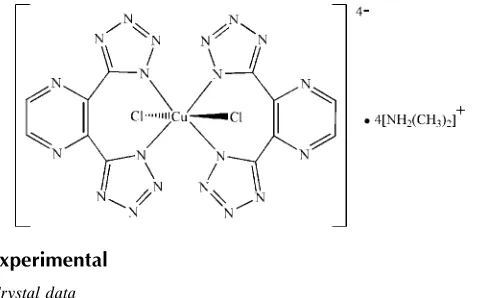
The title compound, (C2H8N)4[Cu(C6H2N10)2Cl2], consists of
an anionic complex which is composed of a CuII ion surrounded by four N atoms from two pyrazine-2,3-diylbis(1H-tetrazol-1-ide) ligands, and two Cl atoms in a
trans-Cl2N4coordination geometry; the CuIIatom lies on a site
of symmetry 2/m. The Cu—Cl distance of 2.8719 (5) A˚ is long due to the Jahn–Teller distortion of thed9electron
configura-tion of CuIIion. The tetrazole and pyrazine rings make an N— C—C—N torsion angle of 38.25 (17). The charge of the
anionic complex is balanced by four dimethylammonium cations, which interact with the anionic complexes via N— H N and N—H Cl hydrogen bonds.
Related literature
For the coordination compound of 2,3-di-1H -tetrazol-5-yl-pyrazine, see: Liet al.(2008). For related structure, see Taoet al.(2010).
Experimental
Crystal data
(C2H8N)4[Cu(C6H2N10)2Cl2] Mr= 747.17
a= 20.613 (2) A˚
b= 10.5671 (9) A˚
c= 15.0687 (12) A˚
V= 3282.3 (5) A˚3
MoKradiation
= 0.89 mm1
T= 293 K
0.060.060.05 mm
Data collection
Bruker SMART APEX diffractometer
Absorption correction: multi-scan (SADABS; Bruker, 1999)
Tmin= 0.947,Tmax= 0.959
18389 measured reflections 2079 independent reflections 1888 reflections withI> 2(I)
Rint= 0.024
Refinement
R[F2> 2(F2)] = 0.030
wR(F2) = 0.085
S= 1.08 2079 reflections 120 parameters
H atoms treated by a mixture of independent and constrained refinement
max= 0.74 e A˚
3
min=0.26 e A˚
3
Table 1
Selected bond lengths (A˚ ).
[image:1.610.51.293.546.695.2]Cu1—N1 2.0029 (10) Cu1—Cl1 2.8719 (5)
Table 2
Hydrogen-bond geometry (A˚ ,).
D—H A D—H H A D A D—H A
N6—H6B Cl1i 0.88 (2) 2.32 (2) 3.1731 (13) 162.7 (18) N6—H6A N4ii
0.93 (2) 1.91 (2) 2.8381 (17) 175 (2)
Symmetry codes: (i)xþ1;y;zþ1; (ii)x;yþ1 2;z
1 2.
Data collection:SMART(Bruker, 1998); cell refinement:SAINT (Bruker, 1999); data reduction:SAINT; program(s) used to solve structure:SHELXS97(Sheldrick, 2008); program(s) used to refine structure: SHELXL97 (Sheldrick, 2008); molecular graphics: ORTEP-3(Farrugia, 1997); software used to prepare material for publication: SHELXTL (Sheldrick, 2008) and PLATON (Spek, 2009).
The authors thank the National Science Council of Taiwan for financial support.
Supplementary data and figures for this paper are available from the IUCr electronic archives (Reference: PK2440).
References
Bruker (1998).SMART. Bruker AXS Inc., Madison, Wisconsin, USA. Bruker (1999).SAINTandSADABS. Bruker AXS Inc., Madison, Wisconsin,
USA.
Farrugia, L. J. (1997).J. Appl. Cryst.30, 565.
Li, J.-R., Tao, Y., Yu, Q., Bu, X.-H., Sakamoto, H. & Kitagawa, S. (2008).Chem. Eur. J.14, 2771–2776.
Sheldrick, G. M. (2008).Acta Cryst.A64, 112–122. Spek, A. L. (2009).Acta Cryst.D65, 148–155.
Tao, Y., Li, J.-R., Chang, Z. & Bu, X.-H. (2010).Cryst. Growth Des.10, 564– 574.
Structure Reports Online
supporting information
Acta Cryst. (2012). E68, m1234 [https://doi.org/10.1107/S1600536812036896]
Tetrakis(dimethylammonium)
trans
-dichloridobis[5,5
′
-(pyrazine-2,3-diyl)bis(1
H
-tetrazol-1-ido-
κ
N
1)]copper(II)
Ju-Hsiou Liao and Pei-Shan Shi
S1. Comment
Multifunctional tetrazolate ligands have recently been of great interest for the formation of metal-organic frameworks
(MOFs). Thus far, the di-topic tetrazolate-based ligand, 2,3-di-1H-tetrazol-5-ylpyrazine (H2dtp) has only been found in a
chiral, porous and thermally robust MOF, Zn(dtp) (Li, 2008). In our laboratory, the reaction of H2dtp and CuCl2 in
di-methylformamide (DMF) under acidic conditions afforded the title compound (I).
In the title complex anion, the CuII ion is six-coordinated in a distorted octahedral environment, surrounded by two Cl
-anions and four N-atoms from two chelating (dtp)2- anionic ligands, forming a trans-Cl
2N4 coordination geometry (Fig.
1). The bonding mode is quite different from that observed in Zn(dtp). The asymmetric unit of the [CuCl2(dtp)2]4- anion
contains one quarter of the complex, with the CuII ion located at a site of 2/m symmetry, and the two Cl- anions lie in a
mirror plane. The Cu—Cl bond length, 2.8719 (5) Å, is unusually long due to Jahn-Teller distortion of the d9 electron
configuration of CuII ion, while the Cu—N distance is normal at 2.0029 (10) Å. The tetrazolyl and pyrazinyl rings are not
coplanar, with a torsion angle of 38.25 (17)°, in accord with the single-bond character of C1—C2 bond, 1.4678 (17) Å. In
the aromatic CN4- tetrazolate ring, the N2—N3 bond, 1.3071 (16) Å, has slightly more double bond character than those
of N1—N2 and N3—N4 bonds, 1.3455 (15) Å and 1.3450 (17) Å.
Four equivalents of [(CH3)2NH2]+ cations are present to balance the charge, as shown in the packing diagram (Fig. 2).
Slabs parallel to the bc-plane are formed by hydrogen bonding networks, which are constructed by the N—H bonds of
[NH2(CH3)2]+ cations interacting with the Cl- atoms and tetrazolate-N atoms of anionic complexes. Such slabs are stacked
along the a-axis through van der Waals interactions among the methyl groups of the dimethylammonium cations.
S2. Experimental
4.3-mg (0.025 mmol) CuCl2.2H2O and 10.5-mg (0.05 mmol) H2dtp were dissolved in 1-ml dimethylformamide (DMF)
respectively. The solutions were mixed in a reaction vial, adding 50-ml 3M HCl to adjust the pH value to ~1.5. The
mixture was ultrasonicated to form a homogeneous yellowish green solution, and was kept at 120°C for three days. The
product was washed with a small amount of DMF and acetone, and then dried in air. 18.2 mg of blue plate-like crystals
were collected in 97.7% yield, based on Cu.
S3. Refinement
H atoms, except for H6A and H6B, were positioned geometrically and allowed to ride on their respective parent atoms
with C—H = 0.96 Å [methyl, Uiso(H) = 1.5Ueq(C)] and C—H = 0.93 Å [aromatic, Uiso(H) = 1.2Ueq(C)]. H6A and
Figure 1
The structure of the title compound, with displacement ellipsoids drawn at the 50% probability level for non-H atoms.
Unlabelled atoms are related to the reference atoms by the symmetry operations (1 - x, -y, 1 - z), (x, -y, 1 - z) and (1 - x, y,
Figure 2
A packing diagram of the title compound. All H-atoms except for those involved in hydrogen bonds are omitted for
clarity. Hydrogen-bonding interactions are drawn with dashed lines.
Tetrakis(dimethylammonium) trans-dichloridobis[5,5′-(pyrazine-2,3-diyl)bis(1H-tetrazol- 1-ido-κN1)]copper(II)
Crystal data
(C2H8N)4[Cu(C6H2N10)2Cl2] Mr = 747.17
Orthorhombic, Cmca
Hall symbol: -C 2bc 2
a = 20.613 (2) Å
b = 10.5671 (9) Å
c = 15.0687 (12) Å
V = 3282.3 (5) Å3
Z = 4
F(000) = 1548
Dx = 1.512 Mg m−3
Mo Kα radiation, λ = 0.71073 Å Cell parameters from 999 reflections
θ = 5–23.5°
µ = 0.89 mm−1 T = 293 K Hexagonal, blue 0.06 × 0.06 × 0.05 mm
Data collection
Bruker SMART APEX diffractometer
Radiation source: fine-focus sealed tube Graphite monochromator
φ–ω scan
18389 measured reflections 2079 independent reflections 1888 reflections with I > 2σ(I)
Rint = 0.024
Refinement on F2
Least-squares matrix: full
R[F2 > 2σ(F2)] = 0.030 wR(F2) = 0.085 S = 1.08 2079 reflections 120 parameters 0 restraints
Primary atom site location: structure-invariant direct methods
Secondary atom site location: difference Fourier map
Hydrogen site location: inferred from neighbouring sites
H atoms treated by a mixture of independent and constrained refinement
w = 1/[σ2(F
o2) + (0.0517P)2 + 1.6198P]
where P = (Fo2 + 2Fc2)/3
(Δ/σ)max = 0.001
Δρmax = 0.74 e Å−3
Δρmin = −0.26 e Å−3
Special details
Geometry. All e.s.d.'s (except the e.s.d. in the dihedral angle between two l.s. planes) are estimated using the full covariance matrix. The cell e.s.d.'s are taken into account individually in the estimation of e.s.d.'s in distances, angles and torsion angles; correlations between e.s.d.'s in cell parameters are only used when they are defined by crystal symmetry. An approximate (isotropic) treatment of cell e.s.d.'s is used for estimating e.s.d.'s involving l.s. planes.
Refinement. Refinement of F2 against ALL reflections. The weighted R-factor wR and goodness of fit S are based on F2,
conventional R-factors R are based on F, with F set to zero for negative F2. The threshold expression of F2 > σ(F2) is used
only for calculating R-factors(gt) etc. and is not relevant to the choice of reflections for refinement. R-factors based on F2
are statistically about twice as large as those based on F, and R- factors based on ALL data will be even larger.
Fractional atomic coordinates and isotropic or equivalent isotropic displacement parameters (Å2)
x y z Uiso*/Ueq
Atomic displacement parameters (Å2)
U11 U22 U33 U12 U13 U23
Cu1 0.01762 (17) 0.0475 (2) 0.02182 (18) 0.000 0.000 −0.01293 (12) Cl1 0.0268 (2) 0.0411 (3) 0.0402 (3) 0.000 0.000 0.0032 (2) C1 0.0213 (6) 0.0300 (6) 0.0231 (6) −0.0018 (4) 0.0021 (4) −0.0041 (5) C2 0.0256 (7) 0.0273 (6) 0.0231 (6) −0.0004 (5) 0.0013 (5) −0.0038 (4) C3 0.0442 (8) 0.0416 (8) 0.0316 (7) −0.0034 (7) 0.0052 (6) 0.0105 (6) C4 0.0503 (10) 0.0485 (9) 0.0433 (9) 0.0003 (7) 0.0054 (8) 0.0000 (7) C5 0.0405 (8) 0.0494 (9) 0.0428 (8) 0.0033 (7) −0.0050 (7) −0.0014 (7) N1 0.0206 (5) 0.0374 (6) 0.0219 (5) 0.0022 (4) −0.0014 (4) −0.0042 (4) N2 0.0238 (5) 0.0458 (7) 0.0274 (6) 0.0061 (5) −0.0025 (4) −0.0016 (5) N3 0.0257 (6) 0.0451 (7) 0.0319 (6) 0.0078 (5) 0.0006 (4) −0.0013 (5) N4 0.0270 (6) 0.0386 (6) 0.0279 (5) 0.0055 (4) 0.0045 (4) −0.0020 (5) N5 0.0324 (6) 0.0413 (6) 0.0309 (6) −0.0036 (5) 0.0041 (5) 0.0050 (5) N6 0.0244 (6) 0.0565 (8) 0.0282 (6) 0.0034 (5) 0.0017 (5) 0.0067 (5)
Geometric parameters (Å, º)
Cu1—N1i 2.0029 (10) C4—H4A 0.9600
Cu1—N1ii 2.0029 (10) C4—H4B 0.9600
Cu1—N1 2.0029 (10) C4—H4C 0.9600 Cu1—N1iii 2.0029 (10) C5—N6 1.469 (2)
Cu1—Cl1 2.8719 (5) C5—H5A 0.9600 C1—N4 1.3309 (16) C5—H5B 0.9600 C1—N1 1.3374 (15) C5—H5C 0.9600 C1—C2 1.4678 (17) N1—N2 1.3455 (15) C2—N5 1.3405 (17) N1—N2 1.3455 (15) C2—C2ii 1.402 (3) N2—N3 1.3071 (16)
C3—N5 1.3319 (19) N3—N2 1.3071 (16) C3—C3ii 1.378 (3) N3—N4 1.3450 (17)
C3—H3 0.9300 N6—H6A 0.93 (2) C4—N6 1.473 (2) N6—H6B 0.88 (2)
N1i—Cu1—N1ii 180.0 H4B—C4—H4C 109.5
N1i—Cu1—N1 91.77 (6) N6—C5—H5A 109.5
N1ii—Cu1—N1 88.23 (6) N6—C5—H5B 109.5
N1i—Cu1—N1iii 88.23 (6) H5A—C5—H5B 109.5
N1ii—Cu1—N1iii 91.77 (6) N6—C5—H5C 109.5
N1—Cu1—N1iii 180.0 H5A—C5—H5C 109.5
N1i—Cu1—Cl1 88.82 (3) H5B—C5—H5C 109.5
N1ii—Cu1—Cl1 91.18 (3) C1—N1—N2 105.74 (10)
N1—Cu1—Cl1 91.18 (3) C1—N1—N2 105.74 (10) N1iii—Cu1—Cl1 88.82 (3) C1—N1—Cu1 133.78 (9)
C2ii—C2—C1 124.68 (7) C1—N4—N3 105.50 (10)
N5—C3—C3ii 121.98 (8) C3—N5—C2 116.86 (13)
N5—C3—H3 119.0 C5—N6—C4 113.59 (13) C3ii—C3—H3 119.0 C5—N6—H6A 108.9 (13)
N6—C4—H4A 109.5 C4—N6—H6A 110.8 (13) N6—C4—H4B 109.5 C5—N6—H6B 111.8 (13) H4A—C4—H4B 109.5 C4—N6—H6B 110.5 (13) N6—C4—H4C 109.5 H6A—N6—H6B 100.5 (18) H4A—C4—H4C 109.5
N4—C1—C2—N5 38.25 (17)
Symmetry codes: (i) x, −y, −z+1; (ii) −x+1, y, z; (iii) −x+1, −y, −z+1.
Hydrogen-bond geometry (Å, º)
D—H···A D—H H···A D···A D—H···A
N6—H6B···Cl1iii 0.88 (2) 2.32 (2) 3.1731 (13) 162.7 (18)
N6—H6A···N4iv 0.93 (2) 1.91 (2) 2.8381 (17) 175 (2)