www.wjpr.net Vol 6, Issue 15, 2017. 756
FORMULATION AND EVALUATION OF LORNOXICAM TABLET BY
PRESS COATING METHOD FOR PULSATILE DRUG DELIVERY
SYSTEM
Deepa Kantekar*, K. Divya Lakhmi and H. Padmalatha
Gyana Jyothi College of Pharmacy, Uppal Bus Depot, Hyderabad-500089, Telangana, India.
ABSTARCT
In the present research work pulsatile drug delivery system of Lornoxicam tablets were formulated by employing compression coating technology. Initially the core tablets were prepared by using various concentrations of super disintegrates, the formulated core tablets were coated with the polymers by using compression coating technology. All the core and press coated tablet formulations were subjected to various physical and chemical evaluation tests for core and press coated tablets. The thickness, hardness and weight variation shown by all the tablet formulations were found within the official pharmacopoeial limits. In vitro release of Lornoxicam of core tablet formulations F1-F3, F1showed faster drug release after 20 mins. Faster drug release can be correlated with the high disintegration and friability observed in this study. The enteric coated formulations C1, C2 showed maximum drug release after 4 hour. C3 and C7showed maximum drug release after 7thhr. Time dependent pulsatile drug delivery system has been achieved from tablet of formulation C7 with 99.9%.
KEYWORDS: Lornoxicam, Super disintegrates, Ethyl cellulose, Pulsatile tablets.
INTRODUCTION
The pulsatile release tablets that can suppress release of the drug in the stomach and can release the drug rapidly after a predetermined time of about 3 hr in the intestine. The system consists of a core, a swelling agent of cross-linked PVP and a coating film of ethyl cellulose / Eudragit L. But Eudragit L dissolves in an environment of pH above 6 and creates pores in the coating film. So Penetration of water molecules from the surroundings through the
Volume 6, Issue 15, 756-774. Research Article ISSN 2277–7105
Article Received on 26 Sept. 2017,
Revised on 16 October 2017, Accepted on 06 Nov. 2017
DOI: 10.20959/wjpr201715-10100
*Corresponding Author Deepa Kantekar Gyana Jyothi College of
Pharmacy, Uppal Bus Depot,
Hyderabad-500089,
www.wjpr.net Vol 6, Issue 15, 2017. 757
pores into the core causes expansion of the swelling agent, bursting the film and releasing the drug with a single pulse. Manipulation of the thickness of the coating film can control the lag time.
Site specific pulsatile drug delivery system
Generally, the aim of site specific and receptor release system refers to targeting of the drug directly to a certain biological location that means the drug must release at targeted site with sufficient amount to maintain peak plasma concentration for the desired time period. Environmental factors like pH or enzymes present in the intestinal tract and also transit time control the release of a site-controlled system where as the drug release from time-controlled systems is controlled primarily by the delivery system and not by the environment. Over the past two decades the major challenge for scientist is to target the drugs especially to the colonic region of GIT. Previously colon was considered as a innocuous organ that responsible for the water absorption, electrolytes and stool storage but it‟s accepted as important site for the delivery of drugs.
Inflammation induced pulsatile drug delivery system
Any physical or chemical stress like-injury, fracture etc. which acts as a stimulus in the case of inflammation due to hydroxyl radicals produced from inflammation responsive cells. Yui et al., (1992), designed and prepared stimuli based inflammation responsive chronotropic system which responded to hydroxyl radicals and degraded in a limited manner. Basically with the system utilized hyaluronic acid (HA) which is specifically hydrolyzed by hyaluronidase or free radicals that present at inflammatory site. Degradation of HA via the hyaluronidase is very low in a normal state of health. Hence it became possible to treat patient with inflammatory diseases like rheumatoid arthritis, NSAIDS incorporated into hyaluronic acid gels as a new implantable drug delivery system.
MATERIALS AND METHODS
www.wjpr.net Vol 6, Issue 15, 2017. 758
chemical ltd.
METHOD
Formulation of core tablets by direct compression: The inner core tablets were prepared by using direct compression method as shown in the table no6.3. Powder mixtures of Lornoxicam, microcrystallinecellulose, polyplasdone XL, talc, ingredients were dry blended for 20 min. followed by addition of Magnesium Stearate. The mixtures were then further blended for 10 min., 50mg of resultant powder blend was manually compressed using, Lab press with a 5mm punch and die to obtainthe core tablet.
Formulation of mixed blend for barrier layer: The various formulation compositions containing Ethyl cellulose N 50, methocel k100, magnesium stearte, talc, pvpk-30 and microcrystalline cellulose. Different compositions were weighed dry blended at about 10 min. and used as press coating material to prepare press-coated pulsatile tablets respectively by directcompression method.
Preparation of press-coated tablets: The core tablets were press-coated with 200 mg of mixed blend as given in Table.No6.4. 100mg of barrier layer material was weighed and transferred into a 10mm die then the core tablet was placed manually at the center. The remaining 100mg of the barrier layer materiel was added into the die and compressed by using Lab press Limited, India
EVALUATION OF PRESS COATED TABLET
Disintegration time of core tablets
Disintegration test was carried out using the tablet disintegration test apparatus (Servewell Instruments pvt. Ltd., Electrolab ED-2L, India) specified in indian pharmacopoeia. Distilled water at 37 ± 0.5 ºC was used as the disintegration media and the time in second taken for complete disintegration of the tablet with no palpable mass remaining on the screen was measured in seconds.
In vitro drug release of pulsatile Lornoxicam core tablets
In vitro dissolution studies were carried out using USP XXIII Type II (paddle method)
www.wjpr.net Vol 6, Issue 15, 2017. 759
spectrophotometer.
Application of Release Rate Kinetics to Dissolution Data:
Various models were tested for explaining the kinetics of drug release. To analyze the mechanism of the drug release rate kinetics of the dosage form, the obtained data were fitted into zero-order, first order, Higuchi, and Korsmeyer-Peppas release model.
Zero order release rate kinetics
To study the zero–order release kinetics the release rate data ar e fitted to the following equation.
F = Ko t
Where, „F‟ is the drug release at time„t‟, and „Ko‟ is the zero order release rate constant. The
plot of % drug release versus time is linear.
First order release rate kinetics: The release rate data are fitted to the following equation Log (100-F) = kt
A plot of log cumulative percent of drug remaining to be released vs. time is plotted then it gives first order release.
Higuchi release model: To study the Higuchi release kinetics, the release rate data were fitted to the following equation.
F = k t1/2
Where, „k‟ is the Higuchi constant.
In higuchi model, a plot of % drug release versus square root of time is linear.
Korsmeyer and Peppas release model
The mechanism of drug release was evaluated by plotting the log percentage of drug released versus log time according to Korsmeyer- Peppas equation. The exponent „n‟ indicates the mechanism of drug release calculated through the slope of the straight Line.
Mt/ M∞ = K tn
Where, Mt/ M∞ is fraction of drug released at time „t‟, k represents a constant, and „n‟ is the
www.wjpr.net Vol 6, Issue 15, 2017. 760 Hixson-Crowell release model
(100-Qt)1/3= 1001/3– KHC.t
Where, k is the Hixson-Crowell rate constant.
Hixson-Crowell model describes the release of drugs from an insoluble matrix through mainly erosion. (Where there is a change in surface area and diameter of particles or tablets).
RESULTS AND DISCUSSION
Present study was done on pulsatile tablets with different formulations C1 to C8. Formulations had weight ratio of polymers like Ethyl cellulose, Tulson, polyplasdone, Explotab along with various excipients. Ethyl cellulose is an insoluble. Aim of the formulation was to get effect of drug in inflammatory and anti pyratic action. This section gives detailed description of the results and discussion on pulsatile Lornoxicam tablets.
PREFORMULATION STUDIES
Determination of λ max of Lornoxicam
The λ max of Lornoxicam was estimated by carrying out UV scan between the wavelength 200 to 400 nm which gave a highest peak at 271 nm and the same was selected for Lornoxicam.
Standardization method for estimation of Lornoxicam
Standard curves of Lornoxicam were prepared in 0.1N HCl, and phosphate buffer (pH 6.8).
Standard graph of Lornoxicamin 0.1N HCl
Lornoxicam showed maximum absorbance in 0.1N HCl at 271 nm. The solution obeyed Beer-Lambert‟s law for concentration range of 1 μg / mL to 10 μg / mL with regression coefficient of 0.996. Standard curve of Lornoxicam prepared in 0.1N HCl is shown below in Table 1 and Figure 1.
Table 1: Calibration data of Lornoxicamin 0.1N HCl.
Sl.No. Conc [mg/l] Abs
0 0 0
www.wjpr.net Vol 6, Issue 15, 2017. 761 Fig 1: Standard Graph of Lornoxicamin 0.1N Hcl.
Standard graph of Lornoxicam in phosphate buffer (pH 6.8)
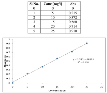
Lornoxicam showed maximum absorbance in phosphate buffer (pH 6.8) at 270 nm. The solution obeyed Beer-Lambert‟s law for concentration range of 1 to 10 μg / mL with regression coefficient of 0.998. Standard curve of prepared Lornoxicamin phosphate buffer pH 6.8 is shown below.
Table 2: Calibration data of Lornoxicam in pH 6.8 phosphate buffer.
Sl.No. Conc [mg/l] Abs
0 0 0
1 5 0.215 2 10 0.372 3 15 0.560 4 20 0.714 5 25 0.910
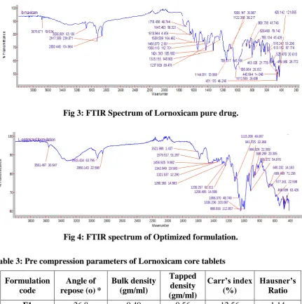
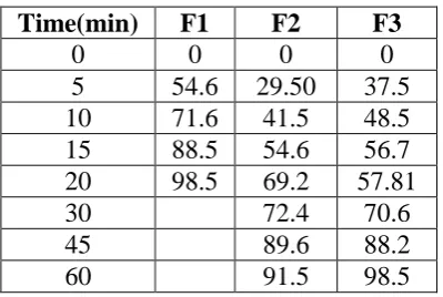
[image:6.595.120.471.434.740.2]www.wjpr.net Vol 6, Issue 15, 2017. 762 Drugs Polymer Interaction Study by FTIR spectrophotometer
In order to investigate the possible interaction between drug and selected polymers, FTIR studies were carried out. IR spectrum for pure drug and physical mixture of drug- polymers were obtained and analyzed for principle peaks The studies suggest that there is no incompatibility between drug and polymer.
Fig 3: FTIR Spectrum of Lornoxicam pure drug.
3561.467 30.647
2915.434 63.795 2850.143 22.596
1621.988 1.437 1579.517 51.287 1456.925 9.982
1342.949 19.583 1321.537 12.290
1280.360 14.863
1239.257 61.011 1208.495 14.598
1115.208 49.067
1066.070 48.749 1036.236 103.097
988.916 112.857
941.725 32.366
908.372 54.976 866.928 22.009
849.299 20.305
669.489 71.158 640.232 14.163
577.161 22.598 468.699 63.426 L-optimized formulation
[image:7.595.82.514.183.617.2]3800 3600 3400 3200 3000 2800 2600 2400 2200 2000 1800 1600 1400 1200 1000 800 600 400 200 100 90 80 70 60 Wavenumber % T ra n s m it ta n c e
Fig 4: FTIR spectrum of Optimized formulation.
Table 3: Pre compression parameters of Lornoxicam core tablets
Formulation code
Angle of repose (o) *
Bulk density (gm/ml) Tapped density (gm/ml) Carr’s index (%) Hausner’s Ratio
F1 26.8 0.49 0.56 12.56 1.14
F2 25.6 0.52 0.59 11.86 1.13
F3 29.8 0.48 0.55 12.7 1.15
www.wjpr.net Vol 6, Issue 15, 2017. 763
index of all the formulations was found to be ranging between 11 to 13 which shows that the powder has good flow properties. All the formulations has shown the hausner ratio ranging between 0 to 1.2 indicating the powder has good flow properties.
Table 4: Post compression parameters of Core tablet.
Formulation codes Weight variation (mg) Hardness(kg/c m2) Friability (%loss) Thicknes s (mm) Drug content (%) F1 47.2 2.5 0.45 4.2 98.5
F2 52.6 2.4 0.51 4.3 99.5
F3 48.96 2.4 0.43 4.5 102.2
7.5. Invitro quality control parameters for tablets
All the parameters such as weight variation, friability, hardness, thickness and drug content were found to be within limits.
Table 5: Pre compression Parameters of Lornoxicam coated Tablets.
Formulation code
Angle of repose (o) *
Bulk density (gm/ml) Tapped density (gm/ml) Carr’s index (%) Hausner’s Ratio
C1 27.01 0.51 0.56 16.21 1.09
C2 25.8 0.55 0.63 16.87 1.14
C3 26.74 0.56 0.68 17.1 1.21
C4 25.33 0.55 0.65 17.67 1.18
C5 24.24 0.56 0.68 16.92 1.21
C6 23.12 0.54 0.61 17.65 1.12
C7 22.08 0.49 0.58 16.43 1.18
C8 24.12 0.51 0.56 17.97 1.09
www.wjpr.net Vol 6, Issue 15, 2017. 764 Table 6: Post compression parameters of Coated tablet.
Formulation codes
Weight variation (mg)
Hardness
(kg/cm2) Friability Thickness
Drug content (%)
C1 255.5 4.1 0.53 4.7 99.76 C2 246.4 4.2 0.55 4.8 99.45 C3 251.6 4.0 0.59 4.9 99.34 C4 248.6 4.3 0.56 4.6 99.87 C5 247.4 4.5 0.57 4.8 99.14 C6 246.7 4.4 0.49 4.4 98.56 C7 253.3 4.3 0.57 4.7 98.42 C8 246.2 4.4 0.51 4.8 99.65
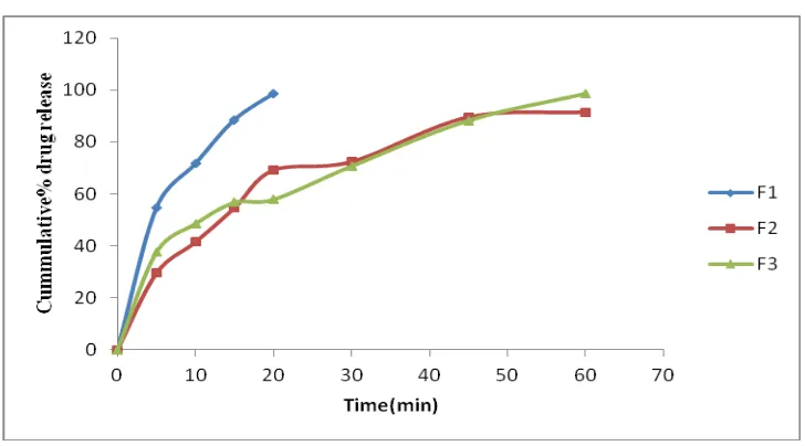
In-Vitro Drug Release Studies of Lornoxicam core tablet
In vitro dissolution studies of Lornoxicam core tablets were performed using USP XXIII
Type II rotating paddle dissolution apparatus by using phosphate buffer (pH 6.8) as a dissolution medium. From formulation F1-F3 Lornoxicam core tablets, F1 showed faster drug release after 20 mins than the other formulations. Faster drug release can be correlated with the high disintegration and friability observed in this study. So, F1 Lornoxicam core tablet formulation was selected as best formulation for further press coating and enteric coating formulations. In vitro drug release profiles of all Lornoxicam core tablets were shown in Table 7 and Figure.5.
Table no 7: Dissolution data of care tablets.
Time(min) F1 F2 F3
0 0 0 0
[image:9.595.200.400.468.602.2]www.wjpr.net Vol 6, Issue 15, 2017. 765 Fig 5: Cumulative % drug release of Lornoxicam core tablets.
In vitro drug release study of Lornoxicam pulsatile tablets
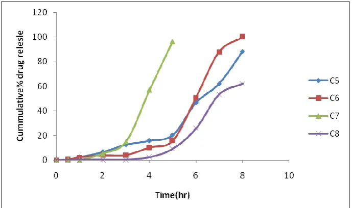
Based on the above characters formulation F1 was selected as best formulation and press coated and enteric coated to find out the changes in the release rate of the Lornoxicam from enteric coated tablets. This enteric coat has enabled us to achieve definite non release lag phase for 8 hours. The formulations C1, C2, and C3 showed maximum drug release after 3th hour. Time dependent pulsatile drug delivery system has been achieved from tablet of formulation C5 and C8 with 90.1% and 99.9% drug release which meets demand of chronotherapeautic drug delivery. The formulations containing M and Ethylecellulose N 50, Methocel k 100 in 40 mg and 60 mg respectively were found to be optimum as enteric coating polymers. The data were shown below.
Table 8: Cummulative Percentage Release Of Coated Lornoxicam Tablets.
Sl.No Time(min) C1 C2 C3 C4 C5 C6 C7 C8
0 0 0 0 0 0 0 0 0 0
[image:10.595.81.516.543.709.2]www.wjpr.net Vol 6, Issue 15, 2017. 766 Fig 6: Cumulative % release study of Lornoxicam pulsatile tabletsusing ethyl cellulose.
Fig 7: Cumulative % release study of Lornoxicam pulsatile tabletsmethocel k 100.
CONCLUSION
[image:11.595.126.471.311.515.2]www.wjpr.net Vol 6, Issue 15, 2017. 767
peak symptoms are observed in the early morning.
Standard plots of Lornoxicam in 0.1N HCl, phosphate buffer (pH 6.8) were prepared by UV Spectrophotometry which showed good correlation coefficient (R2) values. The drug-polymer interaction studies were performed by FTIR Spectrophotometry and it was found that there was no interaction between the drug and various polymers used in the formulation. The coated tablets were prepared by direct compression method.
The pulsatile tablet of Lornoxicam tablets are composed of three components, a drug containing core tablet (rapid release function), the press coated with swellable hydrophobic polymer (SSG) and hydrophobic polymer (ethyl cellulose) alone and / or in different weight ratio. An enteric coating layer.
The formulation mixtures of powders for core tablets (F1-F3) prior to the compression step have been evaluated for pre-compression parameters namely angle of repose, bulk density, tapped density and Carr‟s Index for the flow ability nature was determined. All the formulation mixtures showed good to excellent compressibility.
All the core and press coated tablet formulations were subjected to various physical and chemical evaluation tests for core and press coated tablets. The thickness, hardness and weight variation shown by all the tablet formulations were found within the official pharmacopoeial limits. All the formulations showed favourable drug loading with uniformity of drug content in the tablets and disintegration time. Based on the friability and disintegration time, formulation A1 was selected as best formulation and press coated and enteric coated for further evaluations studies. All the formulations of press coated tablets showed almost uniform size, shape and appearance. The physico-chemical properties of all the formulations (C1-C8) like thickness, friability, hardness and drug content ranged lies within pharmacopoeia limits.
www.wjpr.net Vol 6, Issue 15, 2017. 768 REFERENCES
1. Davis SS, Illum L. Drug delivery systems for challenging molecules. International Journal of Pharmaceutics, 1998; 176: 1-8.
2. Gennaro AR, ed. Remington. The Science and Practice of Pharmacy 20th ed. USA: Lippincott, Williams & Wilkins, 2000; P. 903-905.
3. Burnside BA, GO X, Fiske K, Couch RA, Treacy DJ, Chang RK, Mc Guinness CM, Rudnic EM : US20036605300 2003.
4. Bussemer T, Otto I, Bodmeier R. Pulsatile drug delivery systems Crit Rev. Therapeutic Drug Carrier Systems, 2001; 18(5): 433-458.
5. Yoshida R, Sakai K, Okano T, Sakurai Y. Pulsatile drug delivery systems using hydrogels. Advanced Drug Delivery Reviews, 1993; 11: 85-108.
6. Kikuchi A, Okano T. Pulsatile drug release control using hydrogels. Advanced Drug Delivery Reviews, 2002; 54: 53-77.
7. Gazzaniga A, Maroni A, Sangalli ME, Zema L. Time-controlled oral delivery systems for colon targeting. Expert Opinion on Drug Delivery, 2006; 3: 583-597.
8. Peppas NA, Leobandung W. Stimuli-sensitive hydrogels, ideal carriers for chronobiology and chronotherapy. Journal of Biomaterials Science, Polymer Edition, 2004; 15: 125-144.
9. Stubbe BG, DeSmedt SC, Demeester J. Programmed polymeric devices for pulsed drug delivery. Pharmaceutical Research, 2004; 21: 1732-1740.
10.Gazzaniga A, Palugan L, Foppoli A, Sangalli ME. Oral pulsatile delivery systems based on swellable hydrophilic polymers. European Journal of Pharmaceutics and Biopharmaceutics, 2008; 68: 11-18.
11.Shiwani S, Anshul DS, Roopa S. Pulsatile Drug Delivery System: A Review An Advanced Approach. International Journal of Pharmacy and Technology, 2011; 3: 1179-1188.
12.Lachman L, Lieberman HA, Kanig JL. The Theory and Practice of Industrial Pharmacy. Verghese Publishing House, 1991; 3.
13.Rubinstein A, Tirosh B, Baluom M, Nassar T, David A. The rationale for peptide drug delivery to the colon and the potential of polymeric carriers as effective tools. Journal of Controlled Release, 1995; 46: 59-73.
14.McNeil ME, Rashid A, Stevens HNE, Dispensing Device. WO Patent No. 90/09168, 1990.
www.wjpr.net Vol 6, Issue 15, 2017. 769
rupturable coated hard gelatin capsules. Journal of Control Release, 2003; 3: 331-339. 16.Krogel I, Bodmeier R. Floating or pulsatile drug delivery systems based on coated
effervescent cores. International Journal of Pharmaceutics, 1999; 187: 175-184.
17.Qureshi J, Amir M, Ahuja A, Baboota S, Ali J. Chronomodulated drug delivery system of Salbutamol Sulphate for the treatment of nocturnal Asthma. Indian Journal of Pharmaceutical Sciences, 2008; 70(3): 351–356.
18.Gazzaniga A, Paluga L, Foppoli A, Maria ES. Oral pulsatile delivery systems based on swellable hydrophilic polymers. European Journal of Pharmaceutics and Biopharmaceutics, 2008; 68: 11-18.
19.Kanakal MM, Sakeena MHF, Azmin MN, Yusrida D. Effect of coating solvent ratio on the drug release lag time of coated Theophylline osmotic tablets. Tropical Journal of Pharmaceutical Research, 2009; 8(3): 239-245.
20.Fan TY, Wei SL, Yan WW, Chen DB, Li J. An investigation of pulsatile release tablets with ethylcellulose and eudragit L as film coating materials and cross-linked polyvinylpyrrolidine in the core tablets. Journal of Controlled Release, 2001; 77: 245. 21.Crison JR, Siersma PR, Amidon GL. A novel programmable oral release technology for
delivering drugs: human feasibility testing using gamma scintigraphy. Proceed International Symposium on Controlled Release of Bioact Mater, 1996; 23: 51-52.
22.Linkwitz A, Magruder JA, Merrill S. Osmotically driven delivery device with expandable orifice for pulsatile delivery effect. US Patent No: 5,318,558, 1994.
23.http://www.rivm.nl/bibliotheek/rapporten/623860010.pdf.
24.Kadam, Vinayak D, Gattani, Surendra G. Development of colon targeted multiparticulate pulsatile drug delivery system for treating nocturnal asthma. Drug Delivery, 2010; 17(5): 343-351.
25.Takaya T, Ikada C, Imagawa N, Niwa K, Takada K. Development of a colon delivery capsule and pharmacological activity of recombinant human granulocyte colony timulating factor in beagle dogs. Journal of Pharmacy and Pharmacology, 1995; 47: 474-478.
26.Medlicott, NJ, Tucker IG. Pulsatile release from subcutaneous implants. Advance Drug Delivery Review, 1999; 139-149.
27.Bae YH, Okano T, Hsu R, Kim SW. Thermo-sensitive polymers as on-off switches for drug release. Macromolecular Rapid Communications, 1987; 8: 481-485.
www.wjpr.net Vol 6, Issue 15, 2017. 770
design, characterization and biological significance. Advance Drug Delivery, 2001; 47: 113-131.
29.Obaidat AA, Park K. Characterization of protein release through glucose-sensitive hydrogel membranes. Biomaterials, 1997; 18: 801-806.
30.30. Obaidat AA and Park K. Characterization of protein release through glucose sensitive hydrogel membranes. Pharmaceutical Research, 1996; 13: 989- 995.
31.Yui N, Okano T and Sakurai Y. In flammation responsive degradation of cross linked hyaluronic acid gels. Journal of Controlled Release, 1992; 22: 105-116.
32.Saslawski O, Weigarten C, Beniot JP, Couvereur P. Magnetically responsive microspheres for the pulsed delivery of insulin. Life Science, 1988; 42: 1521-1528. 33.Miyazaki S, Yokouchi C, Takada M. External control of drug release: control release of
insulin from a hydrophobic polymer implant by ultrasound irradiation in diabetic rats. Journal of Pharmacy and Pharmacology, 1988; 40: 716-717.
34.Averitt RD, Sarkar D and Halas NJ. Plasmon Resonance Shifts of Au-Coated Au2S Nanoshells: Insight into Multicomponent Nanoparticle Growth. Physical Review Letters, 1997; 78: 4217-4220.
35.Korenma, Charles DP. New strategies in the medical management of asthma. American Family Physician, 1998; 58(1).
36.Gwen SS. Nocturnal asthma: mechanisms and management. The Mount Sinai Journal of Medicine, 2002; 69(3): 140-1477.
37.Sarasija S, Stutie P. Chronotherapeutics: Emerging role of biorhythms in optimizing drug therapy. Indian Journal of Phrmaceutical Sciences, 2005; 67(2): 135-40.
38.Gwen SS. Nocturnal asthma: mechanisms and management. The Mount Sinai Journal of Medicine, 2002; 69(3): 140-7.
39.Peep V. Chronopharmaceutical Drug Delivery. Institute of pharmacy. University of Tartu, Estonia.
40.Rhind, GB, Connaughton JJ, Mcfie J, Douglas NJ, Flenley DC. Sustained release choline Theophylline in Nocturnal Asthma. British Medical Journal, 1985; 291: 1605- 1607.
41.Government of India Ministry of Health & Family Welfare. Indian Pharmacopoeia. Delhi: Controller of Publications, 1996; P. 750, 151.
42.Tripathi KD. Essential of Medical Pharmacology. 5th ed. New Delhi: Jaypee Brothers Medical Publishers (P) Ltd, 2003; P. 202-203.
www.wjpr.net Vol 6, Issue 15, 2017. 771
Pharmaceutical Press, P. 765-773.
44.Bronchodilators. In: Drug facts and comparisons 2001. Pocket version. 5th ed. Missouri: Facts and comparisons, 2001; P. 331-350.
45.Raymond C, Rowe H, Kibbe. Handbook of pharmaceutical excipients, 4th ed publisher-science and practice, royal pharmaceutical society of Great Britain, London, 2003.
46.Bowe R. “Hand book of pharmaceutical excipients”, 5th ed. Published by Pharmaceutical press, P. 211-214 and 278-282.
47.Lin S, Lin K, Li M. Hydrophilic excipients modulate the lag time of time controlled disintegrating press coated tablets. American Association of Pharmaceutical Science Journal, 2004; 5(4): 54.
48.Lin S, Lin K, Li M. Influence of excipients, drug and osmotic agents in the inner core on the time controlled disintegration of compression coated ethyl cellulose tablets. Journal of Pharmaceutical Science, 2002; 91(9): 2040-2046.
49.Fan T, Wei S, Yan W, Chen D, Li J. An investigation of pulsatile release tablets with ethylcellulose and Eudragit L as film coating materials and cross-linked polyvinyl pyrrolidine in the core tablets. Journal of Controlled Release, 2001; 77: 245- 251.
50.Gayatri PC and Madhabhai MP. A comparative in vitro evaluation of enteropolymers for pulsatile drug delivery system. Acta Pharmaceutica Sciencia, 2009; 51: 243-250.
51.Li B, Zhu J, Zheng C, Gong W. A novel system for three-pulse drug release based on “tablets in capsule” device. International Journal of Pharmaceutics, 2008; 352: 159-164. 52.Krogel I, Bodmeier R. Floating or pulsatile drug delivery systems based on coated
effervescent cores. International Journal of Pharmaceutics, 1999; 187: 175-184. 53.Midha KK, Teicher MH. US Patent No., US6217904, 2001.
54.Chen CM. US Patent No., US5260068, 1993.
55.Efentakis M, Koligliati S, Vlachou M. Design and evaluation of a dry coated drug delivery system with an impermeable cup, swellable top layer and pulsatile release. International Journal of Pharmaceutics, 2006; 311: 147-156.
56.Bussemer T, Bodmeier R. Formulation parameters affecting the performance of coated gelatin capsules with pulsatile release profiles. International Journal of Pharmaceutics, 2003; 267: 59-68.
www.wjpr.net Vol 6, Issue 15, 2017. 772
217: 33-43.
58.Sawada T, Kondo H, Nakashima H. Time-release compression-coated core tablet containing nifedipine for chronopharmacotherapy. International Journal of Pharmaceutics, 2004; 280: 103–111.
59.Dashevsky A, Mohamad A. Development of pulsatile multiparticulate drug delivery system coated with aqueous dispersion Aquacoat ECD. International Journal of Pharmaceutics, 2006; 318: 124–131.
60.Mastiholimath V, Dandagi P, Jain S. Time and pH dependent colon specific pulsatile delivery of Theophylline for nocturnal asthma. International Journal of Pharmaceutics, 2007; 328: 49-56.
61.Kao C, Chen S, Sheu M. Lag time method to delay dug release to various sites in the gastrointestinal tract. Journal of Controlled Release, 1997; 44(90): 263-270.
62.Bussemer T, Dashevsky A, Bodmeier R. A pulsatile drug delivery system based on upturable coated hard gelatin capsules. Journal of Controlled Release, 2003; 93: 331- 339.
63.Sungthongjeen S, Puttipipatkhachorna S, Paeratakulc O. Development of pulsatile release tablets with swelling and rupturable layers. Journal of Controlled Release, 2004; 95: 47-159.
64.Nayak U, Shavi G, Nayak Y. Chronotherapeutic drug delivery for early morning surge in blood pressure: A programmable delivery system. Journal of Controlled Release, 2009; 136: 125-131.
65.Zhang Y, Zhang Z, Wu F. A novel pulsed-release system based on swelling and osmotic pumping mechanism. Journal of Controlled Release, 2003; 89: 47-55.
66.Benberga R, Kimb J, Amidon G. Pharmacokinetics of an immediate release, a controlled release and a two pulse dosage form in dogs. European Journal of Pharmaceutics and Biopharmaceutics, 2005; 60: 17-23.
67.Ghimire M, McInnes F, Watson D. In-vitro / in-vivo correlation of pulsatile drug release from press-coated tablet formulations: A pharmacoscintigraphic study in the beagle dog. European Journal of Pharmaceutics and Biopharmaceutics, 2007; 67: 515-523.
www.wjpr.net Vol 6, Issue 15, 2017. 773
69.Freichel O, Lippold B. New oral erosion controlled drug delivery system with a late burst in the release profile. European Journal of Pharmaceutics and Biopharmaceutics, 2000; 50: 345-351.
70.Bussemer T, Peppas N, Bodmeier R. Evaluation of the swelling, hydration and rupturing properties of the swelling layer of a rupturable pulsatile drug delivery system. European Journal of Pharmaceutics and Biopharmaceutics, 2003; 56: 261-270.
71.Dashevsky A, Bussemer T, Mohamad A. Process and formulation variables affecting the performance of a rupturable capsule-based drug delivery system with pulsatile drug release. Drug development and industrial pharmacy, 2004; 30(2): 171-179.
72.Jessy S, Patole V. Novel floating pulsatile approach for chronotherapeutic release of indomethacin. Dhaka University. Journal of Pharmaceutical Science, 2007; 6(1): 37-41. 73.Sangalli M, Maroni A, Foppoli A. Different HPMC viscosity grades as coating agents for
an oral time and / or site-controlled delivery system: a study on process parameters and in vitro performances. European Journal of Pharmaceutical Sciences, 2004; 22: 469-476. 74.Maradny H. Modulation of a pulsatile release drug delivery system using different
swellable / rupturable materials. Drug Delivery, 2007; 14: 539-546.
75.Roy P, Shahiwala A. Statistical optimization of ranitidine HCl floating pulsatile delivery system for chronotherapy of nocturnal acid breakthrough. European Journal of Pharmaceutical Sciences, 2009; 37: 363–369.
76.Janugade B, Patil S, Patil S. Formulation and evaluation of press-coated montelukast sodium tablets for pulsatile drug delivery system. International Journal of Chem Tech Research, 2009; 1(3): 690-691.
77.Shobhit, K., Satish, KG., Pramod, KS. Dissolution rate enhancement of aceclofenac by solid dispersion technique Asian Journal of Pharmacy and Life Science, 2011; 1(4): 396-400.
78.Fan TY, Wei SL, Yan WW, Chen DB, Li J. An investigation of pulsatile release tablets with ethylcellulose and Eudragit L as film coating materials and cross-linked polyvinylpyrrolidone in the core tablets. Journal of Controlled Release, 2001; 77: 245– 251.
79.Lachmann L, Libberman HA, Kaing JL. The theory and practice of industrial pharmacy. 3rd ed. Mumbai: Varghese publishing house, 1990; P. 296-302.
80.Indian pharmacopoeia. Ghaziabad: The Indian pharmacopoeial commission, 2007; 1: 177-183. (Delhi: Ministry of health and family welfare, Government of, India).
www.wjpr.net Vol 6, Issue 15, 2017. 774
pulsatile drug delivery system for chronobiological disorder: Asthma. International Journal of Drug Delivery, 2011; 3: 348-356.