Anjum et al. World Journal of Pharmaceutical Research
IN VITRO EVALUATION OF PROTEIN SYNTHESIS OF LIPOSOMES
A COMPLETE PROCESS OF EVOLUTION
Saba Anjum* and Md. Sahil
University Dept. of Pharmaceutical Sciences, Utkal University, Vanivihar, Bhubaneswar,
Odisha, India.
ABSTRACT
Projected evolution of proteins is a process used to modify protein
functions. In vitro compartmentalization (IVC) is an in vitro gene
screening system for directed evolution of proteins. IVC sets up the
connection between hereditary data (genotype) and the protein
interpreted from the data (phenotype), which is basic for all
coordinated development strategies, by typifying both in a nonliving
microcompartment. In this, we present another liposome-based IVC
framework comprising of a liposome, the protein union utilizing
recombinant components (PURE) framework and a
fluorescence-actuated cell sorter (FACS) utilized as a microcompartment, in vitro
protein synthesis, and high-throughput screen, separately. Liposome
based IVC is portrayed by in vitro protein union from a solitary duplicate of a quality in a
cell-sized unilamellar liposome and quantitative useful assessment of the orchestrated
proteins. Cases of liposome-based IVC for screening proteins, for example, GFP and
β-glucuronidase are portrayed. We talk about the future headings for this strategy and its
applications.
1. INTRODUCTION
Protein engineering is a knowledge that tailors a protein to task in the desired way. Rational
design and directed evolution are two major approaches for introducing a change into the
amino acid sequence of proteins. As a small change in the protein sequence can induce
critical functional changes in proteins, altering the amino acid sequence is a crucial step in
these approaches; the amino acid sequences are primarily altered by introducing mutations in
the gene that encodes the protein of interest. In site-directed mutagenesis, specific mutations
World Journal of Pharmaceutical Research
SJIF Impact Factor 8.074
Volume 7, Issue 9, 342-359. Conference Article ISSN 2277– 7105
Article Received on 19 March 2018,
Revised on 09 April 2018, Accepted on 29 April 2018,
DOI: 10.20959/wjpr20189-12114
8533
*Corresponding Author
Saba Anjum
University Dept. of
Pharmaceutical Sciences,
Utkal University, Vanivihar,
Bhubaneswar, Odisha,
Anjum et al. World Journal of Pharmaceutical Research
in the DNA sequence are introduced, which yields the desired function if the relationship
between protein structure and function is clearly understood. However, directed evolution of
proteins is based on Darwinian selection and thus does not necessarily require knowledge of
the relationship between protein sequence and function.[1,2] Using this method, mutations are
generated through Methods such as random mutagenesis, recombination, or site-directed
diversification.[3] Subsequently, the protein variants are synthesized from the mutated genes
using living hosts (cells) or an in vitro transcription-translation system (IVTT), and they are
screened for the desired function. Therefore, the methods used for directed evolution can be
[image:2.595.94.510.280.416.2]categorized as “in vivo” and “in vitro” approaches.
Figure 1: Genotype (genetic information)-phenotype (protein synthesized from the gene
and its function) linkage and screening techniques for directed evolution of proteins.
Screening techniques are categorized as in vivo and in vitro approaches.
Figure 2: The underlying concept for in vitro compartmentalization (IVC) using double
[image:2.595.98.502.507.707.2]Anjum et al. World Journal of Pharmaceutical Research
In both cases, a fluorescence-activated cell sorter (FACS) (centre) is used for high throughput
screening. exposure to a colourimetric detection reagent, and selectively sorted using a
fluorescence-activated cell sorter (FACS).[4,5] Phage display is another in vivo display
technology that uses a phage for gene storage and protein display. In this technique, target
proteins are fused with phage coat proteins (g8p or g3p) and displayed on a phage surface.
These in vivo screening techniques have been applied to the directed evolution of proteins.
However, these techniques are applicable to a limited number of proteins that are not toxic to
growth of the host cell. Low transformation efficiency also limits genetic diversity (library
size) by up to 10.[8]
To overcome these technical drawbacks in vivo techniques, in vitro display was proposed as a
new display technique.[6,7] In this technique, protein variants are synthesized from the gene
using an IVTT, and the gene (genotype) is physically or covalently tethered to the translated
protein (phenotype) via an adaptor or linker, such as ribosomes (ribosome display),[8] Rep A
(CIS display),[9] and puromycin (mRNA display).[10] The proteins linked to the mutant gene
are screened for the desired function. These in vitro display methods are suitable for
improving protein equilibrium affinity, off rate, stability, and folding.[8]
However, these
display techniques are not suitable for improving the catalytic activity of enzymes because they rely on binding affinity between the displayed protein and an immobilized ligand for the
screen.[11] Invitro compartmentalization (IVC) is a solution to directscreening for enzymatic
reaction turnover entirely in vitro. The primary idea underlying IVC is that a DNA, an IVTT,
and a fluorogenic detection reagent are encapsulated in a cell-like compartment to form a
genotype-phenotype linkage (Figure 2, left). Proteins are translated from a single gene using
an IVTT in each compartment, and they yield a fluorescent product that is screened directly
for the catalytic activity of interest using a FACS.[11] We introduce herein the earlier studies
on IVC-based directed evolution of proteins, where water-in-oil (W/O) emulsions were used
as microcompartments. We then introduce the IVC using cell-sized lipid vesicles, liposomes.
Firstly, the technology underlying protein synthesis using an IVTT inside liposome is
described. Then, construction of the liposome-based gene screening system using FACS and
examples of the application of the liposome-based IVC to directed evolution of proteins are
described. Finally, we remark on the future directions for liposome-based IVC in directed
Anjum et al. World Journal of Pharmaceutical Research
2. In Vitro Compartmentalization (IVC)
2.1. Emulsion-Based IVC. In vitro compartmentalization (IVC) is a technique for linking
genotype to phenotype. Unlike other techniques used in conventional in vitro display, IVC
does not connect directly the gene and encoded protein. IVC utilizes microcompartments for
genotype-phenotype linkage. A single DNA and an IVTT are encapsulated in a
microcompartment (Figure 2, left). Proteins encoded by the gene accumulate inside the
microcompartment through in vitro protein synthesis. Colocalization of the gene and protein
links the genotype and phenotype W/O emulsion was first utilized for microcompartments in
IVC-based genetic screening. With this technique, genes encoding the DNA
methyltransferase M. HaeIII were enriched from a mixture containing 107-fold excess genes
encoding dihydrofolate reductase.[12] Furthermore, toward high-throughput gene screening
using a FACS, microbead display using IVC (Figure 2, right) was performed to screen
catalytic activity of enzymes with a soluble non-DNA substrate.[13] This technique enables us
to evaluate the catalytic activity of enzyme encapsulated in cell-size microcompartments
under a variety of conditions that can inhibit the in vitro protein synthesis because the
evaluation of catalytic activity is separated from the protein synthesis. As the next
advancement of IVC, water-in-oil-in-water emulsion (double emulsion) was adapted and
enabled direct sorting of intact emulsion droplets. This double emulsion technique was first
demonstrated through the model selection of emulsion droplets encapsulating FolA genes
from a droplet mixture with two separate W/O emulsions: a positive emulsion containing
FolA genes and a fluorescent marker as well as a negative emulsion containing M. HaeIII
genes and no fluorescent marker.[14] Reemulsification of W/O emulsion droplets in the
aqueous phase creates double emulsion droplets, which can be directly analyzed and sorted
using a FACS. Using the emulsion-based IVC and in vitro protein synthesis, Ebg, which is an
E. coli protein of unknown function, was evolved into mutant proteins with β-galactosidase
catalytic activity.[15] Single genes from the mutation library for Ebg as well as an IVTT and a
fluorogenic substrate were compartmentalized in a W/O emulsion droplet. In an emulsion
droplet, Ebg variants are translated from the mutant gene and yield fluorescent product if the
variants express β-galactosidase catalytic activity. After emulsification of the W/O emulsion
in the aqueous phase, double emulsion droplets were screened directly for β-galactosidase
activity (through the fluorescent signal from turnover reaction products).
2.2. Advantages and Limitations of Emulsion-Based IVC. Emulsion-based IVC is suitable for
Anjum et al. World Journal of Pharmaceutical Research
yields a fluorescent signal, which reflects the enzymatic activity of each variant. Other in
vitro display techniques involve screening based on a binding event between a displayed
protein and immobilized ligand and are not adapted for observing a catalytic turnover event.
Although emulsion-based IVC has been useful and successful for directed evolution of
enzymes, this method has two technical limitations. The first limitation concerns the
stringency of the genotype-phenotype link (Figure 3, right). Double emulsion droplets
containing multiple compartments are formed when W/O emulsion is emulsified in an
aqueous phase. During the emulsification process, two types of microcompartments can be
entrapped in a double emulsion droplet; one microcompartment can encapsulate the gene of
interest and the other can encapsulate an unrelated gene. The genotype-phenotype link would be severed if two different mutant genes were in a double emulsion droplet for sorting using a
FACS.[16] One approach to overcome the issues from multiple compartments is through a
high-throughput screening platform using droplet-based microfluidics [16]. This screening
platform comprises a droplet generation device (droplets for gene amplification), droplet
fusion device (electrocoalescence between droplet pairs of a gene-containing droplet and an
IVTT-containing droplet for the genotype-phenotype link), and sorting device (for recovery
of the genes of interest). The second limitation of the emulsion-based IVC is a technical
hurdle for its application using a variety of protein classes, such as membrane proteins
(Figure 3, left), which cannot be overcome by the use of the aforementioned droplet-based
microfluidics.
For the technical issue of the single droplet containing substructures, and that precludes
membrane protein use in directed evolution of proteins (Figure 3), cell-sized
micro-compartments with a phospholipid bilayer membrane are an ideal solution for both
issues.[17,19] We have been studying in vitro protein synthesis in liposomes.[20–23] and
constructed a high-throughput gene screening system using liposomes (liposome-based IVC)
and a FACS.[24,25] Our experimental system for protein synthesis in liposomes comprises a
liposome as the bioreactor, chemical components for protein synthesis, and analytical tools
for quantitation of the proteins produced. The following sections survey the liposomes used
in preparation methods for cell-sized compartments (Section 3), in vitro protein synthesis
using a PURE system (Section 4), high-throughput analysis using a FACS (Section 5), and
Anjum et al. World Journal of Pharmaceutical Research
3. Liposomes as Cell-Sized Microcompartments
3.1. Liposomes. A phospholipid vesicle is a spherical hollow capsule that has an inner
aqueous phase surrounded by a phospholipid bilayer membrane. The vesicular structure is
formed spontaneously by dispersing phospholipids in an aqueous medium (Figure 4). Vesicle
[image:6.595.130.474.190.421.2]formation from egg lecithin was first reported by Bangham and Horne in
Figure 3: The technical limitations of emulsion-based IVC due to the “W/O emulsion”
and “double emulsion” structures.
Figure 4: Liposomes as a platform for in vitro protein synthesis. The reaction mixture
for protein synthesis constitutes an inner aqueous phase of the liposome, in which
protein is synthesized from a DNA. Membrane protein synthesized inside liposome can
[image:6.595.158.435.487.662.2]Anjum et al. World Journal of Pharmaceutical Research
1964.[26] They observed dried samples from an aqueous dispersion of lecithin by electron
microscopy and discovered a spherical structure with a 4.4 nm thick lipid layer of lamellae.
"Liposome" is a term for a phospholipid vesicle and was proposed by Sessa and Weissmann
in 1968.[27] This term is generally accepted. Since the first report by Bangham, liposomes
have been utilized in various biophysical and biochemical studies, including model
membranes, microreactors, supramolecular assemblies for biomimetic systems, and drug
carriers for drug delivery systems.[28] Currently, liposome-related studies are motivated by a
growing interest in "synthetic cells",[29] and the "origin of life",[29] both of which are intended
to address how living things might emerge from nonliving matter.[30] The recent trend in
liposome-related studies regards liposomes as a protocell model in which biochemical
reactions inside a living cell are executed by filling liposomes with the required components.
In vitro protein synthesis in liposomes and its application to genetic screening
(liposome-based IVC) are examples of bioengineering as well as liposome-related studies.
3.2. Preparation Methods for Cell-Sized Liposomes. Liposomes are diverse in size (from
several tens of nm to hundreds of μm in diameter), lamellarity (singly lamellar or
multilamellar), and internal structure (single compartment or multiple compartments). This
diverse structure depends on the liposome preparation methods. However, not all sizes of
liposomes are applicable as microcompartments for IVC-based gene screening due to
detection limits (approximately 1 μm in diameter) in FACS measurements (see Section 5 for
details). Therefore, the liposome size suitable for this experiment is similar to a cell size
(larger than 1 μm in diameter), and such a cell-sized liposome is referred to as a "giant
liposome".[31] Giant liposomes are primarily generated using the "hydration of thin film
method," "rehydration of freeze-dried empty liposome (FDEL) method," or
"inverted-emulsion method".[32]
In liposome-based IVC, a single DNA and an IVTT are encapsulated in the same giant
liposome to link genotype and phenotype (Figure 4). Feasibility of liposome preparation,
encapsulation of the reactants for in vitro protein synthesis, and the internal structure of the
liposome are significantly influenced by the preparation method for the giant liposome. For
the hydration of thin film or rehydration of freeze-dried empty liposome methods of giant
liposome preparation, the liposomes are formed by reconstituting dried lipid film with a
reaction buffer for protein synthesis. The advantage of the hydration of thin film method is
Anjum et al. World Journal of Pharmaceutical Research
However, the disadvantage of the method is that a relatively large quantity of reaction
mixture is necessary for swelling the dried thin film of the lipids during liposome preparation,
and macromolecular compounds in the reaction mixture are difficult to trap in the
liposomes.[32] When giant liposomes are prepared by the rehydration of freeze-dried empty liposomes, the liposome structure is sufficiently strong to withstand the osmotic pressure
change in the outer solution and the sorting operation during the FACS screen.[33] However,
the giant liposomes generated by this method are unsuitable for quantitative evaluation of
in-liposome protein synthesis using a FACS because certain giant in-liposomes have multiple
compartments and lamella; thus, the liposome size measured using a FACS does not
represent the compartment size for protein synthesis.[34] Consequently, liposomes with a
single compartment and lamella (e.g., giant unilamellar liposomes) are required for
quantitative evaluation of in-liposome protein synthesis (liposome size and product quantity)
by high-throughput analysis using a FACS.
The inverted-emulsion method is a preparation method for giant unilamellar liposomes.[35]
This method comprises the following steps. The aqueous phase is emulsified in an oil phase
containing phospholipids to prepare a water-in-oil emulsion. The emulsion is layered on an
outer aqueous solution and centrifuged to sediment the emulsion droplets towards the
oil-water interface where a lipid monolayer forms. The emulsion droplets generate a second lipid
layer upon crossing the interface and transform into unilamellar liposomes in the outer
aqueous solution. When this type of giant unilamellar liposome is applied to in vitro protein
synthesis, the compartmentalized reaction mixture is separated from the outer aqueous phase
until the liposomes are formed. Thus, researchers can control the composition of both inner
and outer aqueous phases. The inverted-emulsion method is promising for construction of a
suitable microcompartment to quantitatively evaluate in-liposome protein synthesis and
reconstitution of membrane proteins.
4. Protein Synthesis in Liposomes
4.1. The PURE System for In Vitro Protein Synthesis. The primary components of in vitro
protein synthesis are DNA encoding the protein of interest, an IVTT, and a detection reagent.
These components should be encapsulated firmly in a liposome when in vitro protein
synthesis is performed in liposomes (Figure 4). An IVTT is a multimolecular machine that
facilitates protein synthesis from DNA in a test tube. Cell extracts from E. coli, wheat germ,
Anjum et al. World Journal of Pharmaceutical Research
However, cell extracts comprise certain unknown constituents. Furthermore, proteases,
DNase, RNase, and intrinsic enzymes (e.g., β-galactosidase) remain in the cell extracts, and
these remnants considerably decrease the production of protein and interfere with the
detection of protein function. These problems are inevitable as long as cell extracts are used
for in vitro protein synthesis. To overcome these problems, we have been using the IVTT
developed by reassembling the individual components for protein synthesis, which were
extracted from E. coli cells overexpressing the protein factors with a histidine tag and
thoroughly purified. This new IVTT is referred to as a “protein synthesis using recombinant
elements (PURE) system”.[37]
In vitro protein synthesis is a coupled reaction system
comprising transcription, aminoacylation of tRNA, translation, and energy source
regeneration. The PURE system includes the entire reaction system and is prepared by
reconstituting protein factors, ribosomes, tRNA mixture, and substrates (20 amino acids and four nucleoside triphosphates) in a buffer solution. The protein factors are T7 RNA
polymerase, pyrophosphatase, 20 aminoacyl-tRNA synthetases, creatine kinase, myokinase,
and nucleoside diphosphate kinase in addition to 10 translation factors (three initiation factors
(IF), three elongation factors (EF), three release factors (RF), and a ribosome recycling factor
(RRF)).
4.2. Protein Synthesis from a Single Gene in a Liposome. Usingthe experimental system with
liposomes, DNA, and an IVTT, in vitro protein synthesis in liposomes has been studied by a
number of groups.[31] The review article by Stano et al.[31] is a comprehensive survey of
biomacromolecule synthesis in liposomes for the creation of semisynthetic minimal cells and
provides the most recent and comprehensive list of publications on protein synthesis inside
liposomes. Thus, our primary focus is on protein synthesis that begins with a single gene in a
liposome, which is a crucial part of liposome-based IVC, because genotype and phenotype
must be linked for the gene screening process. Our strategy, which links genotype and
phenotype inside a liposome, includes DNA that encodes the protein of interest and is
encapsulated at a single molecule level with the PURE system in liposomes. Liposomes in
which green fluorescent protein (GFP) was translated from a single gene were successfully
detected, analyzed, and sorted for a fluorescence signal from GFP using a FACS.[24] β
-glucuronidase catalytic activity expressed from a single gene inside the liposomes was also
Anjum et al. World Journal of Pharmaceutical Research
5. High-Throughput Analysis of Liposomes Using a FACS
5.1. Application of a FACS to Liposome Measurement. In the liposome-based IVC, an
extremely large number (more than 108) of liposomes are created for in vitro protein
synthesis beginning with a single DNA. Liposomes that encapsulate a gene of interest are
screened from the large population of liposomes for the desired protein function. The protein
function expressed inside an individual liposome should be detected and quantitatively
analyzed to identify the liposomes for sorting. Protein production and function inside the
liposome are often measured and quantified using analytical tools such as a fluorescence
spectrometer and fluorescence microscope.[31] A fluorescence spectrometer detects an
averaged fluorescence signal from an ensemble of liposomes. A fluorescence microscope
measures a fluorescence signal from an individual liposome where proteins are synthesized
from genes. Microscopy measurements for liposomes provide data on the morphology (shape
and size) and fluorescence intensity of a liposome (internal reaction). However, this technique is only effective for a much smaller liposome population than the population
required for statistical analysis and gene screening in the IVC-based directed evolution of
proteins. FACS is a promising technique for observing a large population of liposomes
because of its capacity for high-throughput analysis and simultaneous measurement of
multiple characteristics.
A FACS is a powerful experimental apparatus for analyzing and sorting live cells
simultaneously. The apparatus comprises a fluidics system for transporting one cell at a time,
an interrogation system for detecting the cell by laser illumination, and a sorting system for
collecting the cells of interest from one to millions of cells. Using this technique, cells
exhibiting a specific biological characteristic are separated from a heterogeneous population
of cells using fluorescence and light scattering from individual cells in the population. The
FACS was invented in the late 1960s, commercialized in the early 1970s, and has been
utilized since then for basic studies in cell biology as well as clinical applications such as
diagnosis, disease classification, and in vivo therapies.[38] Recently, FACS measurements
have not only been used for cell-oriented applications but also for molecular screening in
directed evolution of proteins.[15,39] In addition, the FACS has been utilized for measuring
nonbiological particles such as submicron-size liposomes,[40] and double emulsion
droplets.[41] for particle size and fluorescent marker entrapment. We have used FACS to
Anjum et al. World Journal of Pharmaceutical Research
We first successfully detected a GFP synthesis in liposomes using FACS based on
fluorescence signals from the synthesized GFP.[22] For liposome structure, the internal
aqueous volume and membrane volume of individual liposomes were quantitatively
evaluated using light scattering intensity data from a FACS measurement.[42] The liposome
population selected using these structural parameters was sorted using a FACS and observed
by optical microscopy. The structural parameters generated using the FACS correlated with
liposome structural heterogeneity, as demonstrated by microscopy observations. Population
analysis of giant liposomes with a FACS was used to identify the subpopulation of
unilamellar liposomes in a 2D contour map of the surface area and internal aqueous volume
generated for giant liposomes.[43] (Figure 5). Furthermore, the substructure of the
multilamellar giant liposomes has been identified by encapsulating β-glucuronidase synthesis
in the liposome and analyzed by a FACS.[34]
5.2. Evaluation of an In-Liposome Reaction Using FACS. Analysis of biochemical reactions
in liposomes using a FACS is based on a quantitative evaluation of liposome size and
reaction products in liposomes. Liposome size is evaluated by measuring the fluorescence
intensity of a fluorescent protein as a volume marker molecule, which is encapsulated in a
liposome at a high concentration.[24,34] The fluorescence intensity of the marker protein is
converted to the number of marker molecules in a liposome and then to the volume of the
internal aqueous phase in the liposome. The reaction product is quantified by measuring the
fluorescence intensity of newly synthesized proteins or the fluorescent product of expression
of a protein function.[20,25] Through this analytical method using a FACS, a large population
of liposomes can be measured for size and reaction as well as analyzed throughout a
population or subpopulation that is defined by reactivity and a specific size.[33] For
liposome-based IVC enzyme screening, a fluorescent volume marker and fluorogenic substrate are
encapsulated in liposomes for a screening assay using a FACS (Figure 5, left). Details on this
screening system are described in Section 6.
Using our system, GFP synthesis inside the liposomes was quantitatively evaluated, and the
influence of lipid membrane composition on protein synthesis was discussed.[44] The study
suggested that phospholipids and other liposomal membrane components for liposome
preparation should neither inhibit nor impair the protein synthesis reaction steps.
Furthermore, GFP synthesis inside the liposomes proceeds similarly to that in the test tube in
Anjum et al. World Journal of Pharmaceutical Research
This indicates that phospholipids and other liposomal membrane components for liposome
preparation neither inhibit nor impair the protein synthesis reaction steps.[20] Consequently,
liposome provides a reaction environment that is equally good as a test tube and provides an
extremely large number (more than 1010/100 μL reaction volume) of microcompartments.
6. Liposome-Based IVC for Directed Evolution of Proteins
6.1. Liposome-Based IVC. We constructed a novel gene screening system using a
liposome-based IVC for directed evolution of proteins.[24,25] Liposome-based IVC is a technique used
[image:12.595.92.515.270.486.2]to link genotype and phenotype. The idea
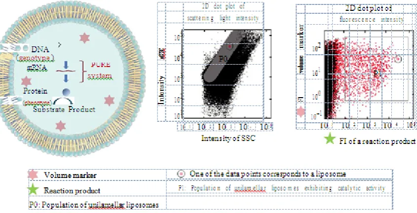
Figure 5: Characterization of in-liposome protein synthesis using a FACS. DNA, PURE
system, the fluorogenic substrate, and fluorescent volume marker are encapsulated in a
giant unilamellar liposome (left). The subpopulation of unilamellar liposomes is
represented by P0 in a 2D dot plot of scattering light intensity (middle).
The catalytic activity of enzymes expressed in liposomes is confirmed in P1 region of 2D dot
plot of fluorescence intensity obtained by FACS measurement (right) underlying this
technique is that a mutant gene library is compartmentalized as a single molecule into
cell-sized liposomes (giant liposomes) and a protein variant is synthecell-sized from the encapsulated
mutant gene through the PURE system in each liposome. Liposome-based IVC has two
primary advantages over emulsion-based IVC. Unlike W/O emulsion droplets, liposomes are
directly loaded onto a FACS apparatus when they are analyzed for gene screening (no
Anjum et al. World Journal of Pharmaceutical Research
expressed in a liposome is quantitatively evaluated using a FACS when a giant unilamellar
liposome is utilized for enzyme synthesis and catalytic activity expression. A FACS
measurement collects signals from two or more different fluorescence colours (a fluorescent
volume marker and fluorogenic substrate) from individual liposomes simultaneously and
quantitates the liposome size and reaction product concentration, both of which are necessary
for quantitative evaluation of the catalytic activity. In addition, membrane proteins can be
inserted into the phospholipid bilayer membrane of a liposome when giant unilamellar
liposome is utilized for membrane protein synthesis. Membrane protein incorporated into
lipid bilayer membrane is a prerequisite for quantitative evaluation of membrane protein
function and subsequent genetic screening.
We first performed a pilot experiment for liposome-based IVC and demonstrated that the
technique is promising for genetic screens.[24] Two GFP variants, GFPuv2 and GFPuv5, were
used in the pilot experiment, and they were encoded in the pETG2tag and pETG5tag vectors,
respectively. GFPuv5 emits a fluorescent signal eight times higher than GFPuv2 when
excited at 488 nm. A mixture of the pETG2tag and pETG5tag DNA at a molar ratio of
0.85: 0.15 was compartmentalized into giant liposomes with the PURE system and a
fluorescent volume marker. Giant liposomes were prepared by FDEL method (Section 3).
After incubation for GFP synthesis, the liposomes were measured for fluorescent signals
from the translated GFP as well as volume marker and sorted using the higher fluorescent
intensity of GFP and a certain liposome size. The pETG5tag was enriched over 10-fold from
the initial genetic mixture when the liposomes were collected from two liposome
subpopulations; one subpopulation ranged from 1.4fL to 6.7fL and the other ranged from
6.7fL to 13fL. Therefore, the genotype- (GFP gene-) phenotype (GFP) link was securely
constructed in individual liposomes, which encapsulated a single copy of DNA. Thus, the
pilot experiment successfully showed that GFP genes encapsulated in a liposome can be
screened for the fluorescence intensity from GFP emission.
However, we anticipated the following technical issue, which can be caused by multiple
compartments and lamella in giant liposomes prepared by the FDA method.[34] The issue is
the underestimation of catalytic activity where a gene is expressed only in a subset of the
multiple compartments in a giant liposome. This yields an inaccurate evaluation of catalytic
activity in individual liposomes and lower enrichment in the gene of interest. In addition,
Anjum et al. World Journal of Pharmaceutical Research
technical hurdle for detection of a functional membrane protein. To solve these problems, we
used a giant unilamellar liposome for liposome-based IVC. A giant unilamellar liposome was
prepared using the inverted-emulsion method (Section 3.2). We constructed a genetic
screening system composed of in vitro protein synthesis encapsulated within a giant
unilamellar liposome and a FACS (Figure 6). A mock genetic library for β-glucuronidase
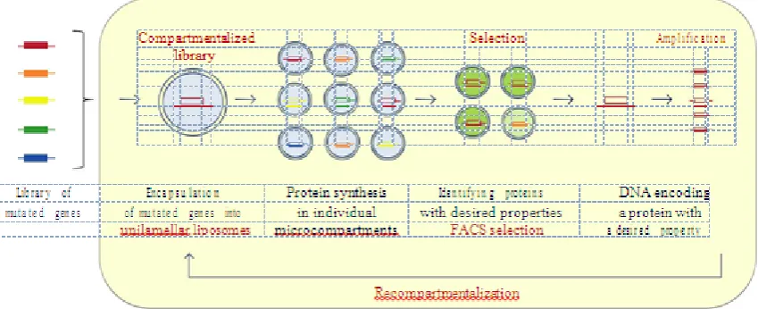
Figure 6: the Flowchart for protein screening using liposome-based IVC. Library of
mutated genes is compartmentalized with the PURE system and other reagents into the
liposome. Each protein variant is synthesized from a single copy of the gene in each
liposome. Liposomes encapsulating the gene of interest are screened using a FACS.
DNA extracted from the liposomes is amplified to be transferred to the next round of
gene screening.
(GUS) was compartmentalized into liposomes as a single molecule. The liposomes that
exhibited green fluorescence from hydrolysis of the fluorogenic substrate through the
synthesized GUS were sorted from the subpopulation of giant unilamellar liposomes using a
FACS. More than a 10-fold enrichment of the GUS gene with a higher catalytic activity was
generated when a single copy of the GUS gene was encapsulated in each liposome.
Quantitative analysis of the enrichment factors and their liposome size dependencies showed
that the experimentally generated and theoretical values agreed. Using this method, the genes
encoding active GUS were then enriched from a gene library of randomly mutated GUS
genes. Only three rounds of screening were required, which was also consistent with our
theoretical estimation. The consistency between the theoretical and experimental values
generated using our screening system indicates that the screening system operates as
[image:14.595.87.512.193.366.2]Anjum et al. World Journal of Pharmaceutical Research
6.2. Protein Evolution Directed by Compartment Size. Here,the directed evolution of protein
is discussed through the effect of compartment size on protein function. Nature contains
living prokaryotes with cell sizes that range from 0.02fL to 400Fl.[45] The lower limit of the cell size is determined by the catalytic efficiency of enzymes, protein synthesis machinery,
and machinery to cope with sudden environmental changes.[45] Under this theory, smaller cells could be generated if the enzyme catalytic efficiency was greater. Naturally occurring
proteins have evolved in the cell through Darwinian selection. However, directed evolution of
protein has never been discussed regarding compartment size because conventional
microcompartments (emulsions) have been unsuitable for this purpose. Of the gene screening
techniques for directed evolution of proteins, liposome-based IVC is the most promising
technique for studying how compartment size influences protein evolution because the
internal aqueous phase volume of the liposome is accurately evaluated by FACS
measurement. A molecular evolution system using liposome-based IVC is an experimental
approach for simulating the evolutionary process of protein function in a certain cell size.
We propose a molecular evolution system for evaluating the effect of compartment size on
protein evolution. The system comprises a giant unilamellar liposome, GUS, and a FACS.
Giant unilamellar liposomes are polydisperse in size ranging from 0.5fL to 250fL or larger,
which includes the cell size discussed. GUS is a tetrameric enzyme, and GUS tetramer
formation is a rate-limiting step in catalytic activity expression.[46] Kinetic analysis of GUS
tetramer formation in emulsion droplets showed that tetramer formation is susceptible to
compartment size when GUS is synthesized from a single gene in a W/O emulsion droplet.[47]
Monomeric GUS is prone to assemble in a smaller compartment because tetramer formation
is the rate-limiting step. In our molecular evolution system, a library of randomly mutated
GUS genes and the PURE system are compartmentalized in giant unilamellar liposomes.
GUS variants are synthesized in individual liposomes. Liposomes exhibiting GUS catalytic
activity are sorted from the subpopulation defined by a certain liposome size (100fL) and
green fluorescence intensity above the threshold value. We predict that GUS variants prone to
assemble in a larger compartment (100fL) will be generated after iterative rounds of genetic
screening. Our genetic screening experiment is in progress and will continue until it generates
a gene encoding active GUS variants, which are fit to a certain liposome size.
6.3. Adaptation of Membrane Protein Function to a Liposome Environment via Directed
Anjum et al. World Journal of Pharmaceutical Research
transport, signal transduction, and cell-cell contact. With recent progress in minimal cell
research using liposomes and an IVTT, experimental methods for including membrane
proteins are under development. Giant unilamellar liposomes are an ideal cell-mimetic
environment because the lipid composition can be optimized for reconstitution of membrane
proteins. Although certain water-soluble proteins have been synthesized using an IVTT inside
liposomes (Section 4), thus far only a few membrane proteins have been synthesized inside
liposomes and reconstituted into a lipid bilayer membrane. For a synthesis of membrane
proteins inside liposomes, a few groups have succeeded in synthesizing and characterizing α
-hemolysin for membrane permeation of nutrient molecules in giant unilamellar liposomes.[18]
as well as in-glycerol-3-phosphate acyltransferase (GPAT) and lysophosphatidic acid
acyltransferase (LPAAT) for lipid synthesis in liposomes.[48] We believe that through
liposome-based IVC development, in the vitro molecular evolution of membrane proteins
become possible. Advantages of using liposome-based IVC on the molecular evolution of
membrane proteins are expected as follows. (1) Various kinds of membrane proteins can be
engineered irrespective of their toxicity threatening cells' lives, (2) functions of membrane
proteins can be evaluated under various reaction conditions and also in membranes with
various lipid compositions. Thus, research on membrane proteins is entering a new stage for
applications aimed at complex molecular machines, such as biosensors for a monitoring
device, biochips for diagnosis, and biointerfaces for computing.
7. CONCLUSIONS
In this paper, we reviewed a novel in vitro genetic screening system comprising
liposome-based IVC and a FACS. Liposome-liposome-based IVC is a new technique developed to link genotype
and phenotype. This technique utilizes a giant unilamellar liposome and a PURE system. A
library of mutant genes and a PURE system are compartmentalized into giant unilamellar
liposomes for in vitro protein synthesis. A protein variant (phenotype) translated from a
single DNA is colocalized with the DNA (genotype) inside a liposome. Using a FACS for
high-throughput screening, liposomes encapsulating the gene of interest are sorted from a
large population of liposomes using fluorescent signals generated from the expression of a
protein function. The genes of interest are enriched through iterative rounds of genetic
screening.
With the gene screening system, genetic diversity at approximately 107 can be screened in a
Anjum et al. World Journal of Pharmaceutical Research
can screen various proteins including enzymes and membrane proteins. A large population of liposomes of different sizes (from 0.5fL to 250fL) facilitates the search for a protein function
that has adapted to a certain compartment size. In addition, the semipermeable character of
the liposomal membrane facilitates external feeding of a liposome compartment using
additional solutes. If the protein function of interest is coupled to the external solute, then the
protein screen is controlled by the timing of feeding and/or solute quantity.
Liposome-based IVC was successfully proven effective for screening a protein function
under simple conditions. However, in nature, proteins must have evolved to adapt to more
complex and dynamic environments where many biochemical reactions are coupled and
organized to control cell behaviour. This suggests that a reaction system comprising many
proteins and enzymes can evolve to perform more efficiently and more productively.
Liposome-based IVC will be a useful method for simulating versatile conditions by
assembling the necessary components into a liposome reactor. It is expected that such
liposome reactors containing a coupled reaction system have high potential as biochemical
sensors for monitoring chemicals (e.g., carcinogens, toxins, and environmental hormones)
and microreactors to produce biologically active substances for daily use (e.g., anticancer
drugs and antibiotics) with high efficiency and selectivity.
REFERENCES
1. H. Leemhuis, R. M. Kelly, and L. Dijkhuizen, "Directed evolution of enzymes: library
screening strategies," IUBMB Life, 2009; 61(3): 222–228.
2. F. H. Arnold, L. Giver, A. Gershenson, H. Zhao, and K. Miyazaki, “Directed evolution of
mesophilic enzymes into their thermophilic counterparts,” Annals of the New York
Academy of Sciences, 1999; 870: 400–403.
3. D. Lipovsek, M. Mena, S. M. Lippow, S. Basu, and B. M. Baynes, “Library construction
for protein engineering,” Protein Engineering and Design, 2010; 83–108.
4. S. Becker, H. Hubenreich,¨ A. Vogel et al., “Single-cell high-throughput screening to
identify enantioselective hydrolytic enzymes,” Angewandte Chemie, 2008; 47(27):
5085–5088.
5. D. Lipovsek,ˇ E. Antipov, K. A. Armstrong et al., "Selection of horseradish peroxidase
variants with enhanced enantioselectivity by yeast surface display," Chemistry and
Anjum et al. World Journal of Pharmaceutical Research
6. P. Amstutz, P. Forrer, C. Zahnd, and A. Pluckthun,¨ “In vitro display technologies: novel
developments and applications, ”Current Opinion in Biotechnology, 2001; 12(4):
400–405.
7. H. Leemhuis, V. Stein, A. D. Griffiths, and F. Hollfelder, "New genotype-phenotype
linkages for directed evolution of functional proteins," Current Opinion in Structural
Biology, 2005; 15(4): 472–478.
8. J. Hanes and A. Pluckthun,¨ “In vitro selection and evolution of functional proteins by
using ribosome display,” Proceedings of the National Academy of Sciences of the United
States of America, 1997; 94(10): 4937–4942.
9. R. Odegrip, D. Coomber, B. Eldridge et al., “CIS display: in vitro selection of peptides
from libraries of protein-DNA complexes," Proceedings of the National Academy of
Sciences of theUnited States of America, 2004; 101(9): 2806–2810.
10.R. W. Roberts and J. W. Szostak, “RNA-peptide fusions for the in vitro selection of
peptides and proteins,” Proceedings of the National Academy of Sciences of the United
States of America,1997; 94(23): 12297–12302.
11.V. Taly, B. T. Kelly, and A. D. Griffiths, "Droplets as microreactors for high-throughput
biology," Chem Bio Chem, 2007; 8(3): 263–272.
12.D. S. Tawfik and A. D. Griffiths, "Man-made cell-like compartments for molecular
evolution," Nature Biotechnology, 1998; 16(7): 652–656.
13.D. Griffiths and D. S. Tawfik, “Directed evolution of an extremely fast phosphotriesterase
by in vitro compartmentalization," EMBO Journal, 2003; 22(1): 24–35.
14.K. Bernath, M. Hai, E. Mastrobattista, A. D. Griffiths, S. Magdassi, and D. S. Tawfik, “In