www.wjpr.net Vol 4, Issue 06, 2015.
1245
A STUDY ON HEPATOPROTECTIVE ACTIVITY OF STEM
EXTRACTS OF
CUSCUTA REFLEXA
(ROXB) ON RANITIDINE
INDUCED HEPATOTOXICITY IN RATS
Nishant Singh Katiyar*1, Amrit Pal Singh1 and N Venkat Rao2
1
Sanjay College of Pharmacy, Mathura, Uttar Pradesh- 281406, India.
2
Dept of Pharmacology, V.L.College of Pharmacy, Raichur, Karnataka-584103, India.
ABSTRACT
The study was designed to evaluate the hepatoprotective activity of aqueous and alcoholic extracts of stem of Cuscuta Reflexa (Convolvulaceae) in acute experimental liver injury induced by ranitidine. The effects observed were compared with a known hepatoprotective agent, silymarin. In the acute liver damage induced by ranitidine, Cuscuta Reflexa stem extracts (100, 200 & 400mg/Kg, po) signeficantly reduced the elevated serum level of Alanine aminotransferase (ALT), Aspartate aminotransferase (AST), Serum alkaline phosphatase (ALP) & Serum bilirubin (BIL). Cuscuta Reflexa (Convolvulaceae) is a plant with a varity a of ethnic medicinal uses along with antioxidant activity. Hence it was planed to evaluate the hepatoprotective activity with alcoholic (AESCR) and aqueous (AQESCR) extracts of stem of C. reflexa.
In LD50 studies for AESCR and AQESCR up to the maximum dose level of 2000 g/kg dose
no mortality was observed in any of the animals, indicating the practically nontoxic. Hepatoprotective activity of both the extracts was studied against ranitidine induced hepatotoxicity in rats. The higher & medium dose of the extracts (400mg/Kg & 200mg/Kg, po) prevented the increase in liver weight when compared to hepatoxin treated control. Histological examination of the liver tissues supported the hepatoprotection.
AESCR and AQESCR showed a significant hepatoprotective effect against ranitidine induced hepatic damage. The medium and high doses of AESCR and AQESCR (200 and 400 mg/kg ) treated groups showd better hepatoprotective activity when compared to standard drug silymarin (25 mg/kg p.o.) treated group.
Volume 4, Issue 6, 1245-1256. Research Article ISSN 2277– 7105
Article Received on 22 March 2015,
Revised on 13 April 2015, Accepted on 08 May 2015
*Correspondence for
Author
Nishant Singh Katiyar
Sanjay College of
Pharmacy, Mathura, Uttar
www.wjpr.net Vol 4, Issue 06, 2015.
1246 KEYWORDS: Cuscuta reflexa, stem extracts, ranitidine, silymarin, hepatoprotective activity
INTRODUCTION
Hepatotoxicity may be defined as the effect of any agent on liver results in a deviation from normal function, morphology and implies chemical/drug/microbial-driven liver-damage.[1] Ranitidine hydrochloride is a potent H2- receptors antagonist, and is used for the treatment of
peptic ulcer diseases.[2] The drug has been shown to be safe at the therapeutic doses.[3] However, isolated cases of acute hepatitis due to therapy with ranitidine have been documented.[4-7] Liver injury induced by ranitidine was manifested in terms of moderate elevation of serum aminotransferases, hepatic infilteration by both lymphocytes eosinophils, and slight focal hepatocellular necrosis. The drug also caused liver cholestasis associated with increased plasma bilirubin and alkaline phosphatase.[7-8]
Cuscuta.reflexa is a leafless, delicate yellow coloured total stem parasite, belonging to the
plant family Convolvulaceae. The tiny white flowers appear in bunches. The fruits are pea shaped and seeds are black in colour.[9] It is found throughout India. The plant is acrid, bitter, astringent to the bowels, aphrodisiac, alternative, tonic and useful in diseases of the eye and of the heart, in biliousness, and in “kapha‟‟.
The herb has a bitter sharp taste; used as expectorant, carminative, tonic, anthelmintic, diuretic, blood purifier and lessens inflammation. It is also useful in jaundice, pain in the muscles and joints, headache, paralysis and also in lumbago.
It was reported that decoction prepared with stem is useful in constipation, flatulence, liver complaints and bilious affections.[10-11]
The seeds have a bitter bad taste, sedative, emmenagogue, diuretic; useful in diseases of the liver and the spleen, quartan fever, chronic fevers, griping, hiccough, purify the blood and cleanse the bowels; the infusion is given in opthalmia, the decoction in biliousness as a purgative (Unani).
The plant is purgative, it is used externally against itch and internally to protract fevers. The stems are specially useful in bilious disorders.[9-11]
www.wjpr.net Vol 4, Issue 06, 2015.
1247 other allopathic medication is available for the treatment of liver disorders. Some of the plants reported for their hepatoprotective activity are Andrographis paniculata[12], Calotropis procer[13], Fumaria indic [14], Luffa acutangula[15], Boerhavia diffuse[16] etc.
From the literature it was found that C. reflexa has also been traditionally indicated for treatment of hepatic disorders. Hence stem extracts of this plant was select for the study of hepatoprotective activity in ranitidine induced hepatotoxic rats.
MATERIALS AND METHODS Plant material
Stem of C. reflexa collected in the month of May and were identified by a botanist Prof. V. Hemanth Kumar, V.L. College of Pharmacy, Raichur and dried in shade at room temperature then subjected to size reduction to a fine powder with the help of mixer grinder.
Chemicals
Ranitidine and Silymarin are gift samples from Glaxo Smithkline Ltd, Mumbai, India and Micro Labs- Bangalore respectively. Thiopental sodium was purchased from Neon Laboratories Ltd., Mumbai, India. The following biochemical kits ALT, AST, ALP and BIL were purchased from Erba Diagnostics Mannheim GmbH, Germany.
Animals
Albino rats (Wistar strain) of either sex weighing between 150-200 g and Albino mice 16-25g were procured from National Centre for Laboratory Animal sciences, C/0 Sri.Venkateswara Enterprises, Bengaluru for experimental purpose. Then the animals were acclimatized for 7 days under standard husbandry condition. i.e.
Room temperature - 26 ± 20 C Relative humidity - 45-55% Light/ dark cycle - 12:12 h
www.wjpr.net Vol 4, Issue 06, 2015.
1248 Preparation of extracts
Preparation of alcoholic extract
The stem powder was packed in a soxhlet apparatus and extracted with 95% alcohol for 18 h. Appearance of colourless solvent in the siphon tube was taken as the termination of extraction. The extract was then transferred into the previously weighed empty beaker and evaporated to a thick paste on the water bath, maintained at 50oC to get alcoholic extract. The extract was finally air dried thoroughly to remove all traces of the solvent and the percentage yield was calculated.[17]
Preparation of aqueous extract
About 100 g of powder was taken in a round bottom flask (2000 ml) and macerated with 500 ml of distilled water with 10 ml of chloroform (preservative) for 7 days with occasional shaking for every hour in a closed vessel. Then the marc was removed by filtering the extract and then it was concentrated on a water bath maintained at 50oC.[17]
These two extracts were stored in airtight containers in a refrigerator below 10oC. The two extracts were examined for their colour and consistency. Their percentage yield was calculated with reference to air-dried powder sample used for the extraction.
Toxicity studies
The acute toxicity of C.reflexa was determined by using albino mice of either sex (16-20 g), maintained under standard husbandry conditions. The animals were fasted for 3 h prior to the experiment and were administered with single dose of individual extracts of C. reflexa and observed for the mortality upto 48 h study period (Short term toxicity). Based on the short-term toxicity profile, the next dose of the individual extracts was deshort-termined as per OECD guidelines No. 425. From the LD50 doses 1/20,1/10 and 1/5 doses were selected and
considered as low, medium and high dose respectively.[18]
Ranitidine induced hepatotoxicity[19-21]
www.wjpr.net Vol 4, Issue 06, 2015.
1249 and high) of AQESCR, for 21 days, animals of groups B, C, D, E, F, G, H and I were intoxicated with ranitidine (50 mg/kg, i.m) daily once for 21 days.
Assessment of hepatoprotective activity
On the 21th day after recording thiopentone induced sleeping time in all animals, all groups were anaesthetized and blood was collected from the retro-orbital puncture. Serum was separated after coagulating at 37°C for 30 min and centrifuging at 2000 rpm for 15 min, and estimated for alanine aminotransferase (ALT), aspartate aminotransferase (AST), serum alkaline phosphatase(ALP) and serum bilirubin (BIL).
The hepatoprotective activity was confirmed through histopathological studies on liver of rats. After collection of blood for biochemical estimation, the rats were sacrificed and the livers were carefully dissected out, cleaned of extraneous tissue, and fixed in 10% formalin for 24 h. Then the paraffin sections were prepared (automatic tissue processor, Autotechnique) and cut into sections of 5 µm thickness, using a rotary microtome. The sections were stained with Haematoxylin-Eosin dye and studied for histopathological changes.[22]
Stastical analysis
All the recorded results are expressed as mean ± SEM from 6 animals. Statistical difference in mean was analyzed by using one-way ANOVA (analysis of variance) followed by Post hoc test (Dunnett‟s„t‟ test). P< 0.05*, 0.01** and 0.001*** were considered as statistically significant.
RESULT
In the present study the effect of the AESCR and AQESCR on normal liver functions, was found to be non-toxic in nature. Ranitidine intoxication in normal rats elevated the levels of ALT, AST, ALP and BIL significantly, indicating acute centrilobular necrosis. The rats treated with AESCR and AQESCR showed a significant reduction in the biochemical parameters elevated by Ranitidine (Table1.)
www.wjpr.net Vol 4, Issue 06, 2015.
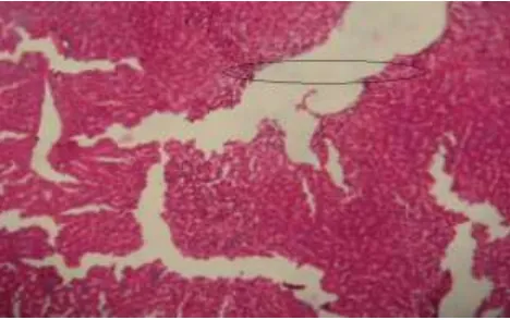
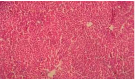
1250 Histopathological examination of liver sections of control group (Fig 1) showed normal cellular architecture with distinct hepatic cells, sinusoidal spaces and central vein. In the liver sections of the rats intoxicated with ranitidine (Fig 2), there is erosion of endothelium cells of blood vessels, disarrangement and degeneration of normal hepatic cells, dilation of blood sinusoids and increased number of binucleated cells. Also, inflammatory reactions around the blood vessels as reflected by the presence of infiltration of lymphocytes and increased activation of Kupffer cells were observed.
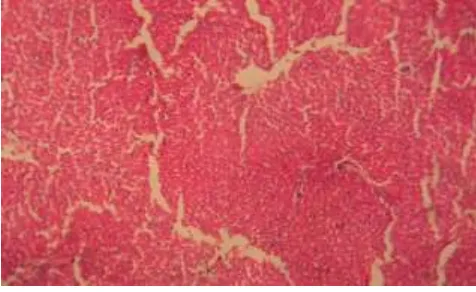
The liver sections of the rats treated with silymarin and intoxicated with ranitidine (Fig 3) and rats treated with AESCR and AQESCR (low, medium and high doses) and intoxicated with ranitidine (Fig 4-9) showed less vacuole formation, reduced sinusoidal dilation, and less disarrangement and degeneration of hepatocytes, indicating marked regenerative activity. The intensity of centrilobular necrosis was less.
Table 1. Hepatoprotective effect of different extracts of c. Reflexa on ranitidine induced liver damage in rats.
Groups Treatment mg/kg L wt g/100g TST min ALT
U/L AST U/L
ALP U/L BIL U/L Normal (vehicle) 10ml/kg
p.o. 4.43±0.12 67.86±2.02 31.74±3.30 76.87±7.91 126.78±8.83 0.17±0.01
Toxicant (Ranitidine)
100mg/kg
s.c. 7.77±0.15 194.73±3.13 116.01±12.97 185.05±18.57 253.71±16.84 2.42±0.43
Standard (silymarin) 25 mg/kg p.o. 5.35±0.11 ** 77.33 ±1.28 ** 47.18±4.18 ** 110.61±15.18 ** 186.18±10.04 ** 0.51±0.05 **
AESCR 100 7.24±0.14ns 185.83±1.49ns 111.80±2.41ns 183.43±
1.59ns
235.66±
5.98ns 1.87±0.05
ns
AESCR 200 6.46±0.14
** 147.83±2.09 ** 80.79±1.30 ** 153.56± 4.64
ns 197.40± 4.72
**
0.69±0.04 **
AESCR 400 5.84±0.15
** 117.00±1.83 ** 61.16±1.25 ** 131.55±2.33 ** 141.65±14.88 ** 0.55±0.04 **
AQESCR 100 6.90±0.34
* 169.16±2.27 ** 74.21±1.50 ** 181.28±3.58
ns 182.73± 2.05
**
1.75±0.18 *
AQESCR 200 6.40±0.16
** 146.5±6.62 ** 70.22±1.06 ** 139.46±1.34 ** 174.83±4.79 ** 0.75±0.05 **
AQESCR 400 5.75±0.28
** 110.83±4.39 ** 59.97±1.28 ** 126.28±1.90 ** 143.20±3.67 ** 0.53±0.02 ** n = 6, Significant at P< 0.05*, 0.01** and 0.001***, ns = not significant
AESCR- alcoholic extract of stem of C. reflexa, AQESCR- aqueous extract of stem of C.
reflexa,
www.wjpr.net Vol 4, Issue 06, 2015.
[image:7.595.181.417.72.205.2]1251 Fig 1. Histology of normal hepatic tissue
[image:7.595.179.418.247.379.2]
Fig 2. Ranitidine induced damage in Hepatictissue
Fig 3. Effect of Silymarin on ranitidine induced hepatic damage
[image:7.595.181.416.422.555.2] [image:7.595.180.414.597.744.2]www.wjpr.net Vol 4, Issue 06, 2015.
[image:8.595.181.416.71.208.2] [image:8.595.180.416.249.376.2]1252 Fig 5. Effect of AESCR (Med) dose on ranitidine induced hepatic damage
Fig 6. Effect of AESCR (High) dose on ranitidine induced hepatic damage
Fig 7. Effect of AQESCR (Low) dose on ranitidine induced hepatic damage
[image:8.595.184.416.418.550.2] [image:8.595.182.415.587.726.2]www.wjpr.net Vol 4, Issue 06, 2015.
[image:9.595.178.416.72.218.2]1253 Fig 9. Effect of AQESCR (High) dose on ranitidine induced hepatic damage
DISCUSSION
Liver diseases are among the most serious ailments and may are classified as acute or chronic hepatitis (inflammatory liver diseases), hepatosis (non-inflammatory diseases) and cirrhosis (fibrosis of liver). They are mainly caused by toxic chemicals, alcohol or drugs like paracetamol, anti-tubercular, anticancer agents etc. Most of the hepatotoxic chemicals damage liver cells either by lipid peroxidation or by other oxidative stress mechanisms induced cellular damage.
Liver is susceptible to injury from drugs and other chemical substances, 75% of blood coming to liver arrives directly from gastrointestinal organs and spleen via portal vein which brings xenobiotics and drugs in concentrated form. Several mechanisms are responsible for inducing hepatic injury or worsening the damage process. Many chemicals damage mitochondria and intracellular organelle that produce energy and their dysfunction releases excessive amount of oxidants, which in turn injure hepatic cells. Activation of some enzymes in the Cytochrome P-450 system such as CYP-2E1 also leads to oxidative stress and injury to hepatocytes, bile duct cells causing accumulation of bile acid inside liver and which in turn promotes further liver damage.
Liver plays an important role in regulation of physiological processes, involved in several vital functions such as metabolism, secretion and storage and also detoxifies a variety of
drugs and xenobiotics and bile secreted by the liver has an important role in digestion.
www.wjpr.net Vol 4, Issue 06, 2015.
1254 pronounced centrilobular and bridging necrosis. In the parenchyma there was focal liver cell necrosis with some accumulation of histocytic elements and slight steatosis and cholestasis. Portal tract shows fibrosis, bile duct proliferation and infiltrate consisting of lymphocytes, plasma cells, polymorphs and eosinophils. Liver injury is manifested in terms of increase in levels of serum aminotransferases, modest hepatic infiltration by both lymphocytes and eosinophils and slight focal hepatocellular necrosis also causes liver cholestasis associated with increased plasma bilirubin and alkaline phosphatase level.[20-21]
In chronic drug induced hepatotoxicity models, administration of thiopentone sodium results with an increased duration of sleeping time, as liver is the primary site for the metabolism of xenobiotics like barbiturates and functional damage to liver requires longer time to inactivate thiopentone resulting with an increased duration of action of this drug. Pretreatment with AESCR and AQESCR have decreased the thiopentone induced sleeping time as compared to toxic control indicating their protection of liver function against drug induced toxicity in rats.
Chronic administration of drugs (thioacetamide, ranitidine and paracetamol) to rats increased the levels of marker enzymes like ALT, AST and ALP as these are stored in the liver cells and increase in the levels of these marker enzymes in serum indicate damage to the liver cells. Pretreatment with AESCR and AQESCR decreased the levels of ALT, AST, ALP and BIL levels, an indication for the hepatoprotective activity of these extracts against drug induced hepatotoxicity.
The elevated levels of these parameters were significantly reduced by the treatment of C. reflexa stem extracts. It can be concluded from this investigation that stem of C. reflexa possess hepatoprotective activity.
CONCLUSION
www.wjpr.net Vol 4, Issue 06, 2015.
1255 It is found that the AQESCR is more potent than AESCR and that is confirmed by the functional and biochemical parameters followed by comparison of histological changes in liver.
Phytochemical constituents like alkaloids, flavonoids, glycosides and sterols are already reported for their hepatoprotective activity.[23] These phytoconstituents are present in both the extracts hence the hepatoprotective effect.
ACKNOWLEDGEMENT
The authors thank the authorities of V. L College of Pharmacy, Raichur, Karnataka, India for providing necessary facilities.
CONFLICT OF INTEREST: Declared none.
REFERENCES
1. Evelyn C.P. Anatomy and Physiology for Nurses, 16th edn, New Delhi , Jaypee Brothers Medical Publishers (P) Ltd., 1993; 241-246.
2. Zeldis J B, Friedman L S & Isselbacher K J, Ranitidine, A new H 2-Receptor antagonist, N Engl J Med., 1983; 309: 1368.
3. Mills J G, Koch K M, Webster C, Sirgo M A, Fidzgerald K & Wood J R, Safety of ranitidine in over a decade of use, Aliment Pharmacol Ther., 1997; 11: 129.
4. Barr G D & Piper D W, Possible ranitidine hepatitis, Med J Aust., 1981; 2: 421.
5. Black M, Scott W E & Kanter R, Possible ranitidine hepatotoxicity, Ann Intern Med., 1984; 101: 208.
6. Souza Lima M A, Ranitidine and the liver, Ann Intern Med., 1985; 102: 273.
7. Ramrakhiani S, Brunt E M & Bacon B R, Possible cholestatic injury from ranitidine with a review of the literature, Am J Gastroenterol., 1998; 93: 822.
8. Bredfeldt J E & Von Huene C, Ranitidine, acetaminophen and hepatotoxicity, Ann Intern Med., 1984; 101: 719.
9. Kurian JC. Plants that heals, 1st edn, Pune, Oriental Watchman Publishing House., 2007; 2: 53.
10.Kirtikar KR and Basu BD, Indian medicinal plants, 2nd edn, Delhi, Periodical Experts Book Agency, 1999; 1741-1742.
www.wjpr.net Vol 4, Issue 06, 2015.
1256 12.Neha T, Raval UM, “Hepatoprotective and toxicological evaluation of Andrographis
paniculata on severe liver damage” Indian J Pharmacol., 2000; 32: 288-293.
13.Viswanath KM, Quereshi AA , Setty SR, “Hepatoprotective activity of flowers of Calotropis procera in Paracetamol induced hepatic injury in rats”, Indian J Pharma Sci.,
2005; 44: 261.
14.Nimbakar SR, Juvekar AR, Jogalekar SN. “Hepatoprotective activity of Fumaria indica in hepatotoxicity induced by anti-tubercular drugs treatment”. Indian Drugs, 2000; 37(11): 537-542.
15.Muna Abid, Shivraj Gouda T, Venkat Rao N, “Hepatoprotective activity of fruit extracts of Luffa acutangula Roxb.Var. Indian Drugs., 2005; 40: 260.
16.Krupavaram K, Rao NV. “A Study on hepatoprotective activity of root extracts of B.diffusa” Indian Drugs, 2005; 40: 260.
17.Kokate CK. Practical pharmacognosy, 4th edn., Vallabh Prakashan Delhi, 1994; 110-111. 18.OECD 2001 guidelines on acute oral toxicity. Environmental health and safety
monograph series on testing and adjustment NO.425.
19.Valois M, Copper MA, Shear NH, An organized approach to drug induced hepatitis, Can J Clin Pharmacol., 2003;10: 59-62.
20.Faried AE, El Sayed AH, Biochemical and histological studies on H2-receptor antagonist
ranitidine-induced hepatotoxicity in rats. Indian J Exp Biol., 2005; 43: 782-785.
21.Vipul G, Patel Nilesh, Venkat Rao N, Nandakumar K, Gouda TS, Md. Shalam. Hepatoprotective activity of alcoholic and aqueous extracts of leaves of Tylophora indica in rats. Indian J Pharmacol., 2007; 39(1): 43-47.
22.Galigher AE, Kozloff EN. Essentials of practical microtechnique. 2nd ed., Lea and Febiger Philadelphia, 1971; 77.