Received 4 Jan 2016|Accepted 21 Mar 2016|Published 6 May 2016
The PDGF-BB-SOX7 axis-modulated IL-33 in
pericytes and stromal cells promotes metastasis
through tumour-associated macrophages
Yunlong Yang
1,*, Patrik Andersson
1,*, Kayoko Hosaka
1, Yin Zhang
1, Renhai Cao
1, Hideki Iwamoto
1, Xiaojuan Yang
1,
Masaki Nakamura
1, Jian Wang
1, Rujie Zhuang
2, Hiromasa Morikawa
3, Yuan Xue
1,4, Harald Braun
5,6,
Rudi Beyaert
5,6, Nilesh Samani
7, Susumu Nakae
8, Emily Hams
9, Steen Dissing
10, Padraic G. Fallon
9, Robert Langer
4&
Yihai Cao
1,7,11Signalling molecules and pathways that mediate crosstalk between various tumour cellular compartments in cancer metastasis remain largely unknown. We report a mechanism of the interaction between perivascular cells and tumour-associated macrophages (TAMs) in promoting metastasis through the IL-33–ST2-dependent pathway in xenograft mouse models of cancer. IL-33 is the highest upregulated gene through activation of SOX7 transcription factor in PDGF-BB-stimulated pericytes. Gain- and loss-of-function experiments validate that IL-33 promotes metastasis through recruitment of TAMs. Pharmacological inhibition of the IL-33–ST2 signalling by a soluble ST2 significantly inhibits TAMs and metastasis. Genetic deletion of host IL-33 in mice also blocks PDGF-BB-induced TAM recruitment and metastasis. These findings shed light on the role of tumour stroma in promoting metastasis and have therapeutic implications for cancer therapy.
DOI: 10.1038/ncomms11385 OPEN
1Department of Microbiology, Tumor and Cell Biology, Karolinska Institute, 171 77 Stockholm, Sweden.2The TCM Hospital of Zhejiang Province, Hangzhou,
Zhejiang 310006, China.3Unit of Computational Medicine, Department of Medicine, Center for Molecular Medicine, Karolinska Institute, 171 77 Stockholm,
Sweden.4Institute for Medical Engineering and Science, Massachusetts Institute of Technology, Cambridge, Massachusetts 02139, USA.5Department of
Biomedical Molecular Biology, Ghent University, B-9052 Ghent, Belgium.6Unit of Molecular Signal Transduction in Inflammation, Inflammation Research
Center VIB, B-9052 Ghent, Belgium.7Department of Cardiovascular Sciences, University of Leicester and NIHR Leicester Cardiovascular Biomedical Research
Unit, Glenfield Hospital, Leicester LE3 9QP, UK.8Laboratory of Systems Biology, Center for Experimental Medicine and Systems Biology, The Institute of
Medical Science, The University of Tokyo, Tokyo 108–8639, Japan.9School of Medicine, Trinity Biomedical Sciences Institute, Trinity College Dublin, College
Green, Dublin 2, Ireland.10Department of Cellular and Molecular Medicine, Panum Institute, University of Copenhagen, 2200N Copenhagen, Denmark.
11Department of Medicine and Health Sciences, Linko¨ping University, 581 83 Linko¨ping, Sweden. * These authors contributed equally to this work.
C
ancer metastasis is a complex process that involves in sophisticated interactions between malignant and hostcells1,2. Cancer cells often produce signalling molecules to
manipulate host cells in the local microenvironment to facilitate their invasion, dissemination and metastasis. The PDGF-PDGFR signalling often becomes activated in the tumour
micro-environment3–5 and endothelial cells in angiogenic vessels are
an important source for the production of PDGF-BB6, a
pluripotent member in the PDGF family. In epithelial cell- and other cell-originated cancer types, PDGF-BB primarily targets stromal fibroblasts and perivascular cells including pericytes and
vascular smooth muscle cells7. PDGF-BB stimulates the
proliferation and migration of perivascular cells through
activation of PDGFRbalthough interaction with PDGFRa also
occurs in fibroblasts5,7. Although it is well known that PDGF-BB
modulates vascular remodelling and maturation by recruiting pericytes and vascular smooth muscle cells onto angiogenic vessels, activation of these perivascular cells in the tumour microenvironment in cancer invasion and metastasis is poorly understood.
Tumour tissues often contain an exceptionally high number of inflammatory cells, which significantly alter tumour growth,
angiogenesis, metastasis and drug responses8,9. Inflammatory
cytokines including GM-CSF, TNF-a, IL-1b, IL-6 and various
chemokines are actively involved in recruitment of inflammatory
cells in tumours10,11. However, our current understanding of
recruitment of tumour-associated macrophages (TAMs) and their roles in cancer invasion and metastasis are far from complete. IL-33 as a relatively new cytokine belongs to IL-1 family and it can be produced by a broad range of cell types including fibroblasts, osteoblasts, endothelial cells, epithelial cells and
adipocytes12–15. IL-33 exerts its biological functions through
binding and activation of its receptor ST2, a member in the Toll-like receptor superfamily. IL-33 is known to regulate Th2
immune responses12. However, the role of IL-33 in tumour
inflammation and metastasis is unknown. A recent study shows that in a mouse breast cancer model, injection of IL-33 protein
stimulates primary tumour growth and metastasis16.
In the present study, we show that IL-33 is the most upregulated gene in PDGF-BB-stimulated pericytes and SOX7 transcription factor mediates PDGF-BB-induced IL-33 expres-sion. Gain-of-function and loss-of-function experiments demon-strate that pericyte- and stromal cell-derived IL-33 is a crucial cytokine for recruitment of TAMs in the tumour microenviron-ment. Importantly, in several human and mouse graft tumour models, we provide compelling evidence to demonstrate that pericyte- and stromal cell-derived IL-33-activated TAMs are
crucial for cancer metastasis. Finally, inin vivo tumour models,
we show that IL-33-activated TAMs mediate PDGF-BB-induced cancer metastasis. These findings shed new mechanistic lights on the crosstalk between various host cellular compartments and PDGF-BB-stimulated pericytes in promoting cancer metastasis. Functional blocking of the PDGF-BB-IL-33-TAM axis is an important approach for cancer therapy.
Results
PDGF-BB-PDGFRb signalling indirectly recruits TAMs. To
investigate the role of PDGF-BB in the recruitment of TAMs, we screened a panel of human tumour cell lines that spontaneously express PDGF-BB. We have found that human A431 squamous carcinoma cell line expressed a high level of endogenous
PDGF-BB protein (50 pg ml1) (Fig. 1a). The A431 xenograft tumour
contained a high number of Iba1þTAMs (Fig. 1b). Interestingly,
downregulation of PDGF-BB by Pdgfb-specific shRNA, which
effectively inhibited the Pdgfb mRNA level (Supplementary
Fig. 1a), markedly ablated TAMs in tumour tissues (Fig. 1b), suggesting that PDGF-BB was primarily responsible for TAM recruitment in this human xenograft model. To further validate these findings, we performed gain-of-function experiments in which mouse Lewis lung carcinoma (LLC) and T241
fibro-sarcoma were transfected with Pdgfb-retrovirus to stably express
PDGF-BB (Supplementary Fig. 1b and c). ShRNA knockdown of
Pdgfb significantly inhibited A431 tumour growth (Supplementary Fig. 1d), whereas PDGF-BB expression promoted tumour growth in T241 and LLC tumours (Supplementary Fig. 1e and f). Notably, FACS and immunohistochemical analyses showed that PDGF-BB-LLC and T241 tumours contained
sig-nificantly higher numbers of F4/80þ and Iba1þ TAMs as
compared with their respective vector-transfected tumours (Fig. 1c,d). Of note, Iba1 and F4/80 double immunostaining showed completely overlapping positive signals (Supplementary Fig. 1g), indicating that both markers detect the total macrophage population in tumour tissues. These findings demonstrate that PDGF-BB recruits TAMs in human and mouse cell line-derived graft tumour models.
To define PDGFRs that are responsible for TAM recruitment, we used various PDGFR inhibitors. Imatinib, a pan PDGFR
tyrosine kinase inhibitor17, significantly inhibited TAM
recruitment in A431, LLC and T241 tumours (Fig. 1e), suggesting that PDGFRs mediate PDGF-BB-induced TAM
infiltration. To distinguish PDGFRa and PDGFRbsignalling in
TAM recruitment, anti-mouse PDGFRa- and PDGFRb-specific
neutralizing antibodies (PDGFR blockades) were used for the
treatment of PDGF-BBþ T241 tumours. Interestingly, PDGFRb, but
not PDGFRa, blockade, markedly inhibited PDGF-BB-induced TAM
infiltration (Fig. 1e). These findings indicate that PDGFRb is the
receptor that mediates PDGF-BB-induced TAM recruitment. We next investigated the direct versus indirect role of PDGF-BB in the recruitment of TAMs. Surprisingly, co-localization of
PDGFRa and PDGFRb in T241 tumours by their specific
antibodies showed that TAMs lacked PDGFR expression (Fig. 1f), suggesting an indirect role of PDGF-BB in the
recruitment of TAMs. Consistent with this notion, PDGFRb
was primarily localized in non-TAM cells including NG2þ
pericytes and aSMAþ smooth muscle cells
(SMCs)/myofibro-blasts (Fig. 1g). These findings were further quantitatively validated by PCR with reverse transcription (RT–PCR), quanti-tative PCR (qPCR) and staining of various cell lines showing that
stromal fibroblasts and pericytes expressed high levels of Pdgfrb
mRNA, whereas mouse Raw macrophage-like cell line and
isolated TAMs completely lacked Pdgfrb mRNA expression
(Fig. 1h and Supplementary Fig. 1h). These findings further support our notion that PDGF-BB recruits TAMs in various tumour models through an indirect mechanism.
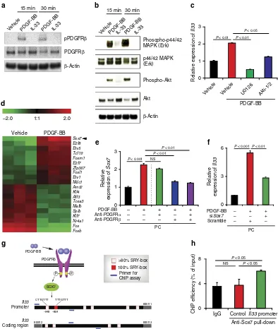
PDGF-BB induces pericyte- and fibroblast-derived IL-33. To
identify possible genes that mediate PDGF-BB-induced TAM recruitment, we performed a genome-wide expression microarray
analysis in PDGF-BB-stimulated pericytes. Surprisingly,Il33 was
the most upregulated gene product with more than an eight-fold increase among all the genes in the genome (Fig. 2a), and was the top one of the upregulated inflammatory cytokines (Fig. 2b). The PDGF-BB-induced IL-33 expression was further validated by
qPCR, which revealed more than a five-fold increase of Il33
mRNA expression (Fig. 2c). In contrast, PDGF-AA, a ligand that
only binds to PDGFRa, lacked ability to induceIl33 expression
(Fig. 2c), indicating that PDGFRbis responsible for
PDGF-BB-induced Il33 expression. In addition to pericytes, stimulation of
PDGFRbþstromal fibroblasts with PDGF-BB also led to marked
IL-33 protein expression from PDGF-BB-stimulated pericytes and stromal fibroblasts. Again, the IL-33 protein levels in PDGF-BB-stimulated pericytes and fibroblasts were significantly higher than those in non-stimulated cells (Supplementary Fig. 2a).
To validate these findingsin vivo, we analysed IL-33 protein
expression in PDGF-BBþ T241 tumours and found a marked
increase of IL-33 expression as compared with vector tumours (Fig. 2e). IL-33 protein expression levels in A431 tumour grafts
were markedly decreased by thePdgfb-specific shRNA (Fig. 2e).
We provided further in vivoevidence by delivery of adenoviral
Pdgfb(Adv-Pdgfb) into tumour-free mice. Again, delivery of
Adv-Pdgfbsignificantly induced IL-33 expression in the hepatic tissue
(Fig. 2f). Collectively, thesein vitroandin vivofindings provide
compelling evidence that PDGF-BB markedly induces IL-33
expression in PDGFRbþ perivascular cells and stromal
fibroblasts.
To further validate the pericytes and stromal cells as the major
source of IL-33 production in in vivo tumours, we isolated
different cell types from the tumour microenvironment. We
confirmed that the PDGFRbþ cell population including stromal
fibroblasts and perivascular cells were the important cells for the production of IL-33 in response to PDGF-BB (Fig. 2g).
Furthermore, NG2þ pericytes in PDGF-BB tumours produced
high levels of IL-33 as compared with those isolated from the
vector control tumours (Fig. 2g). In contrast, CD31þ vascular
endothelial cells did not significantly contribute to BB-induced IL-33 expression in tumours since IL-33 levels in PDGF-BB positive population was not increased (Fig. 2g). Similarly, tumour cells produced negligible levels of IL-33 in PDGF-BB-positive and -negative tumour cells, which remained unchanged. Taken together, these findings demonstrate that pericytes and tumour stromal cells are the primary source of IL-33 in the tumour microenvironment.
We treated PDGF-BB-stimulated pericytes with PDGFRaand
PDGFRb blockades to monitor IL-33 expression in vitro.
PDGFRb, but not PDGFRa, specific blockade significantly
inhibited PDGF-BB-induced IL-33 expression in pericytes
(Fig. 2h). The combination of PDGFRband PDGFRablockades
did not produce any additive effects. Similar to PDGFRb
blockade, imatinib also produced a markedly inhibitory effect
on IL-33 expression (Fig. 2h). Likewise, PDGFRbblockade also
significantly inhibited PDGF-BB-induced IL-33 expression in stromal fibroblasts (Supplementary Fig. 2b).
Role of PDGF-BB signalling pathways in IL-33 production.
Signalling pathway analysis showed that PDGF-BB induced
activation of PDGFRbby phosphorylation (Fig. 3a) and IL-33 has
no impact on activation of PDGFRbin pericytes. In concordance
with the activation of PDGFRb, downstream signalling
compo-nents including MAP kinase (Erk) and Akt also became activated in PDGF-BB-stimulated pericytes (Fig. 3b). Signalling network
analysis from cBioPortal18 showed that Akt and MAPK were
correlated with PDGF-BB expression (Supplementary Fig. 3a). Consistently, MAPK and Akt-specific inhibitors significantly and
effectively inhibited Il33 mRNA expression levels in
PDGF-BB-stimulated pericytes (Fig. 3c and Supplementary Fig. 3b). These findings show that PDGF-BB induces IL-33 expression in
pericytes through activation of the PDGFRbsignalling pathway.
SOX7 mediates PDGF-BB-induced IL-33 expression. We next
investigated potential mechanisms by which PDGF-BB induces
IL-33 expression in PDGFRbþ pericytes and fibroblasts.
Gen-ome-wide microarray analysis of PDGF-BB-stimulated pericytes revealed that SOX7 was the most upregulated transcription factor (about six-fold; Fig. 3d), which was ranked as the top three most
upregulated gene products in the genome (Fig. 2a and Supplementary Fig. 3c). The qPCR analysis further validated the
increased expression level of Sox7 mRNA in
PDGF-BB-stimu-lated pericytes (Fig. 3e). Notably, PDGFRb-specific blockade
significantly attenuated PDGF-BB-stimulated expression of Sox7,
whereas PDGFRa-specific blockade had no effect onSox7mRNA
expression (Fig. 3e). These findings suggest that PDGFRb
potentially mediates PDGF-BB-induced Sox7 expression. To decipher the functional relation between SOX7 and IL-33 expression, PDGF-BB-stimulated pericytes were treated with
Sox7-siRNA. Knockdown of SOX7 significantly impaired
PDGF-BB-induced IL-33 expression (Fig. 3f), which was correlated to the knockdown efficiency (Supplementary Fig. 3d). Similarly,
Sox7-siRNA knockdown also markedly reduced IL-33 production
in PDGF-BB-stimulated stromal fibroblasts (Supplementary Fig. 3e). To provide further supportive evidence of transcriptional regulation of IL-33 expression by SOX7, we analysed mouse sequences of the IL-33 promoter region and discovered a canonical SOX7-binding SRY box and five non-canonical binding sites (Fig. 3g). Chromatin immunoprecipitation (ChIP) assay
using the Il33 promoter fragment containing the canonical
binding site demonstrated that SOX7 directly bound to theIl33
promoter (Fig. 3h). However, it is possible that the non-canonical SOX7 binding sites might also mediate direct binding of SOX7. These data show that PDGF-BB induces IL-33 expression
through the PDGFRb-SOX7 signalling pathway.
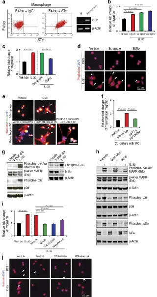
Signalling mechanisms of IL-33-induced Raw cell migration. As IL-33 was the most upregulated cytokine in PDGF-BB-stimulated pericytes, we investigated its functional impact on macrophages. FACS and RT–PCR analyses showed that Raw macrophage-like cell line expressed ST2 receptor that mediates biological functions of IL-33 (Fig. 4a). As a negative control, stromal fibroblasts lacked a detectable level of ST2 expression (Fig. 4a). Knowing that macrophages expressed ST2 receptor, we studied the functional impact of IL-33 on macrophages. Recombinant IL-33 stimulated Raw cell migration in a dose-dependent manner (Fig. 4b,c). In addition, IL-33 induced activated morphological changes of Raw cells that manifested an elongated cell shape (Fig. 4d).
Impor-tantly, theSt2-siRNA knockdown significantly ablated the
IL-33-induced Raw cell migration and morphological changes (Fig. 4c,d). To validate the biological effects of pericyte-derived
IL-33 in modulation of macrophage activitiesin vitro,
PDGF-BB-stimulated and non-PDGF-BB-stimulated pericytes were co-cultured with Raw cells. In this co-culture assay, PDGF-BB-stimulated pericytes induced elongated Raw cell morphological changes, which was neutralized by a soluble ST2 receptor (Fig. 4e). Similarly, PDGF-BB-primed pericytes also attract Raw cell motility in a co-culture system (Fig. 4f).
Consistent with the above-mentioned biological functions, IL-33 stimulation of Raw cells induced marked activation of MAPK, which became hyper-phosphorylated (Fig. 4g). In addition, IL-33 stimulated phosphorylation of p38 that is known in the regulation
of cellular actin reorganization and cell morphological changes19
(Fig. 4g). Notably, IL-33 stimulation led to potent activation of
IkBa, which became highly phosphorylated (Fig. 4g). However,
Akt levels in IL-33-stimulated and non-stimulated cells remained unchanged (Supplementary Fig. 4a). To functionally link the ST2 receptor and IL-33-activated intracellular signalling components,
we used the St2-siRNA knockdown technique. IL-33-induced
MAPK, p38 and IkBaphosphorylation in Raw cells were largely
inhibited by theSt2-specific siRNA (Fig. 4h), indicating that IL-33
induces the ST2-dependent activation of these intracellular signalling components in macrophages. In contrast to IL-33,
supporting the conclusion that PDGF-BB does not directly act on macrophages.
To define the biological functions of the IL-33-ST2-activated downstream signalling molecules, we treated the IL-33-stimulated Raw cells with various inhibitors that blocked the activation of a
specific signalling component. As expected, the MAPK, p38 and
IkBa inhibitors20–22 effectively blocked IL-33-stimulated
phosphorylation of these intracellular signalling molecules (Supplementary Fig. 4b). Treatment with a known MAPK inhibitor (U0126) completely abolished IL-33-induced Raw cell
0 U251 Vehicle T241-PDGF-BB T241-Vector A549 A431 OVCAR8 CAKI-1 MDA-MB-231 50 100 0 10 20 30 Expression of
hPDGF-BB (pg ml
–1)
0 10 20 30 P < 0.05
Number of F4/80
+
cells per field
Number of F4/80
+
cells per field
P < 0.001
P < 0.001
P < 0.001
P < 0.001
P < 0.001
A431 Scramble control Pdgfb shRNA Iba1 / GFP Iba1 / GFP Iba1 / GFP F4/80 / PDGFR α F4/80 / PDGFR β NG2 / PDGFR β α SMA / PDGFR β 0 5 10 15 A431 Pdgfb shRNA A431 Scramble control
P < 0.01
Iba1
+ area per
field (×10
3 μ
m
2 )
Iba1
+ area per
field (×10
3μ
m
2)
Iba1
+ area per
field (×10
3μ
m
2)
Iba1
+ area per
field (×10
3μ
m
2)
Iba1
+ area per
field (×10
3μ
m
2)
Iba1
+ area per
field (×10 3μ m 2) 0 1 2 - - -
P < 0.001
P < 0.001
P < 0.001
P < 0.001
NS
Relative expression of
Pdgfrb
MacrophageIsolated TAM T241 PC
SF LLC
P < 0.01
LLC Vector PDGF-BB Vector PDGF-BB T241 0 40 80 LLC-Vector LLC-PDGF-BB LLC-Vector LLC-PDGF-BB T241-Vector T241-PDGF-BB T241-Vector T241-PDGF-BB
P < 0.001
P < 0.001
0 20 40 A431 Vehicle Vehicle Iba1 / DAPI Iba1 / DAPI Iba1 / DAPI
Imatinib A431 LLC-PDGF-BB T241-PDGF-BB
T241-PDGF-BB T241-Vector
Imatinib
Imatinib Anti-PDGFRα Anti-PDGFRβ
LLC-PDGF-BB T241-PDGF-BB SF PDGFRβ β-Actin PC Macrophage 0 10 20 Vehicle Imatinib LLC-PDGF-BB 0 5 10 15 Vehicle Imatinib
P < 0.01
A431 0 5 10 15 T241-PDGF-BB
P < 0.01
P < 0.01
P < 0.01 NS - - - Vehicle Imatinib Anti-PDGFR α Anti-PDGFR β 0 5 10 15 T241-Vector T241-PDGF-BB F4/80
+ cells (%)
F4/80 GFP 104 103 R4 R4 102 101 100 104 103 102 101 100 100 101 102 103 104 100 101 102 103 104
P < 0.05
50 μm
50 μm
50 μm
50 μm 50 μm
50 μm
25 μm
25 μm
a b
c d
e f
g
migration and cell shape changes (Fig. 4i,j). These findings reconcile with the known functions of MAPK signalling. Similarly, p38 inhibitor also effectively inhibited IL-33-induced migration and cell shape changes of Raw cells (Fig. 4i,j). The
treatment of IL-33-stimulated Raw cells with an NF-kB inhibitor
also significantly attenuated IL-33-induced Raw cell migration and cell shape changes (Fig. 4i,j). These findings demonstrate that IL-33 displays direct effects on Raw cell migration and activation
through MAPK, p38 and IkBasignalling pathways.
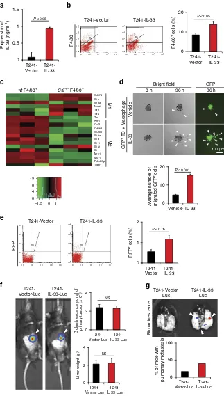
IL-33 increases TAMs in tumours. We performedin vivo
gain-of-function experiments that allowed studying the effect of IL-33 on tumour growth, TAM recruitment and tumour invasion and metastasis. Genetic propagation of T241 tumours with IL-33 by lentiviral approach led to overexpression of IL-33 protein in tumour tissues (Fig. 5a). Despite a high level of IL-33 expression,
IL-33-overexpressing tumours grew at similar rates in vitroand
in vivo as vector-transfected control tumours (Supplementary Fig. 5a and b). However, FACS analysis showed that the number
of F4/80þ TAMs in IL-33-tumours was markedly increased as
compared with that of vector control tumours (Fig. 5b). To further define the TAM phenotypes in tumours, we
implanted IL-33 positive tumours in wtand St2/ knockout
mice23 and isolated TAMs from these tumours. The isolated
primary TAMs fromwtandSt2/backgrounds were subjected
to genome-wide affymetrix analysis. Intriguingly, classical M2
markers including Cd206(Mrc1),Cd163, Pdl2(Pdcd1lg2),Ccr3,
Arg1and many others were all markedly downregulated in TAMs
isolated from the St2/ background as compared with those
isolated from tumours grown in wt mice (Fig. 5c and
Supplementary Fig. 5c).
We further investigated subpopulations of TAMs using M2 markers by FACS analysis in vector- and IL-33- overexpressed tumours. For defining the M2 population of macrophages, three independent cell surface markers including CD206, CCR3 and PDL2 were used in our FACS analysis. Altogether, three
independent analyses show that F4/80þCD206þ, F4/80þ
CCR3þ and F4/80þPDL2þ M2 subpopulations of macrophages
were significantly increased (Supplementary Fig. 5d–f). However,
the F4/80þCD206 subpopulation was also significantly
increased.
IL-33-primed macrophages promote metastasis. One of the
important characteristics of M2 macrophages is tumour promo-tion through various processes like metastasis. Since our study indicated increased tumour metastasis linked to IL-33, we investigated further the link between IL-33-induced TAMs and tumour metastasis. TAMs have been described to facilitate
tumour cell invasion, intravasation and dissemination1,24. To
functionally link IL-33-primed TAMs with cancer invasion, we
performedin vitromatrigel cancer invasion experiments in which
tumour cells and macrophages were co-embedded in matrigel as spheroids. IL-33-treated and non-treated macrophages were
mixed with GFPþ LLC tumour cells and spreading of GFPþ
cells was quantitatively measured. IL-33-stimulated macrophages, but not non-stimulated cells significantly promoted cancer cell
invasion in this in vitroinvasion assay (Fig. 5d). These findings
show that IL-33-primed macrophages promote cancer cell invasion and possibly metastasis.
We next analysed circulating tumour cells (CTCs) in IL-33 and vector tumour-bearing mice. Interestingly, a significantly higher number of CTCs were found in IL-33-tumour-bearing mice as compared with vector tumour-bearing mice (Fig. 5e). To provide further evidence of IL-33-induced metastasis, we developed an independent metastasis model in which luciferase-expressing primary tumours were implanted in the liver of each mouse. Although no differences of primary tumour growth were observed (Fig. 5f), a higher number of IL-33-tumour-bearing
mice developed luciferaseþ pulmonary metastasis as compared
with the vector control group (Fig. 5g). These findings were consistent with increased CTCs in IL-33-tumour-bearing mice and showed that IL-33 induced tumour cell intravasation and metastasis without affecting primary tumour growth.
The IL-33-ST2-TAM axis-dependent metastasis. To
mechan-istically link TAMs and cancer metastasis of IL-33-tumours, we
used clodronate liposomes25 as macrophage-ablating agent to
deplete TAMs. Expectedly, clodronate effectively ablated TAM numbers in both vector and IL-33-tumours (Fig. 6a). In contrast, the treatment of tumour-bearing mice with the control liposome did not significantly affect macrophage numbers in IL-33 tumours as compared with controls. Approximately 50% of
IL-33 tumour-bearing mice possessed visible pulmonary
metastatic nodules on the surface of their lungs at week 6 after removal of primary tumours (Fig. 6b). Conversely, the vector tumour control group had a lower rate of pulmonary metastasis. The lung metastatic lesions were validated by detection of red fluorescent protein (RFP; Fig. 6b). The depletion of TAMs by clodronate markedly decreased the metastatic incidences in the lungs of IL-33 tumour-bearing mice, whereas the low metastatic rate in vector-tumour bearing mice remained unchanged. These findings show that IL-33-promoted pulmonary metastasis is dependent on TAMs and the low metastatic incidence in control tumour-bearing mice might be mediated through a TAM-independent mechanism.
To further investigate the TAM-mediated metastatic potentials,
we used a zebrafish metastasis model26–28that allowed detection
Figure 1 | PDGF-BB induces PDGFRb-dependent macrophage recruitment in tumour cell line grafts tumours of human and mouse origin.(a) Expression
levels of PDGF-BB in conditioned medium of various human tumour cell lines (n¼3 samples per group). (b) Iba1þmacrophages (red) in human scrambled
shRNA-A431 andPdgfbshRNA-A431 squamous carcinomas cell line grafts. Arrowheads indicate tumour-infiltrating macrophages. Scale bar, 50mm. Iba1þ
TAMs were quantified as areas (n¼8 random fields per group). (c) Upper panels: FACS analysis of F4/80þ TAMs in vector-T241 and PDGF-BB-T241
fibrosarcoma cell line grafts and quantification of percentages of F4/80þmacrophages (n¼6 samples per group). Lower panels: quantification of F4/80þ
TAMs of immunohistochemical micrographs (n¼8 random fields per group). (d) Iba1þmacrophages (red) in T241 and PDGF-BB-T241, and
vector-and PDGF-BB-LLC tumours. The tumour cells express GFP (green). Arrowheads indicate tumour-infiltrated macrophages. Scale bar, 50mm. Iba1þTAMs
were quantified as areas (n¼8 random fields per group). (e) Iba1þmacrophages (green) in vehicle- or imatinib-treated A431 and PDGF-BB-LLC tumours,
and in vehicle-, imatinib-, anti-PDGFRa- or anti-PDGFRb-treated PDGF-BB-T241 tumours. Tissue sections were counter-stained with DAPI (blue).
Arrowheads indicate tumour-infiltrated macrophages. Scale bar, 50mm. Iba1þ TAMs were quantified from eight random fields per group. (f) PDGFRb
(green) and F4/80 (red), or PDGFRa(green) and F4/80 (red) double immunostaining of A431, PDGF-BB-T241 and PDGF-BB-LLC tumours. White
arrowheads indicate F4/80þ macrophages and yellow arrowheads point to PDGFRaþor PDGFRbþ cells. Scale bar, 25mm. (g)aSMA (green) and
PDGFRb(red), or NG2 (green) and PDGFRb(red) double immunostaining of vector- and PDGF-BB-T241 tumours. Arrowheads indicate double-positive
signals (yellow). Scale bar, 25mm. (h) RT–PCR and qPCR analyses ofPdgfrbin various cell types. Beta-actin was used as a standard loading (mean±s.e.m.,
of the interactions between malignant cells and macrophages at the single-cell level. This zebrafish metastasis model also permits kinetic monitoring of tumour cell invasion and metastasis in the
living fish body. Moreover, the availability of certain genetic
strains such as transgenic Fli1:EGFP zebrafish29,30 allows us
to study the event of tumour cell intravasation with or without 0
0.5 1 1.5
0 0.5 1 1.5
0 1 2 3 4 5
- - - - - -
Relative expression of
Il33
T241-Vector
T241-Vector
CD31+ PDGFRβ+ NG2+
T241-PDGF-BB
T241-PDGF-BB
CD31+ PDGFRβ+ NG2+
P< 0.001
P< 0.001
250 μm
0 3 6
Adv-Gfp
Adv-Pdgfb
Relative
expression of
Il33
Relative
expression of
Il33
P < 0.05
Expression of IL-33 (ng ml
–1)
Expression of IL-33 (ng ml
–1)
P < 0.05 P < 0.05
T241-Vector
A431-Scramble
control
A431-Pdgfb
shRNA
T241-PDGF-BB
P < 0.001
P < 0.001 NS
0 3 6
Vehicle
PDGF-AAPDGF-BB
Vehicle PDGF-BB
Relative expression of
Il33
Relative expression of
Il33
Vehicle PDGF-BB
–2.0 1:1 2.0
Il33 Mcpt8 Sox7 Sprr1a Lrp8 Kif14 Spc25 Il13ra2 Pbk Mfap3l Smoc2 Fosb Fmod Gdpd2 Ogn Cxcl13 Omd Aspn Slc10a6 Prss35
Vehicle PDGF-BB
–2.0 1:1 2.0
Il33 Spp1 Mif Il16 Cxcl10 Vegfa Cx3c11 Il1r1 Ccl7 Il5 Il7 Il8rb Il15 Tnfsf13 Il10rb Cxcl12 Cxcl15 Tnfsf13b Il1rn Cxcl13
0 2 4
SF
- - -
0 1 2 3 4 5
PDGF-BB –
Anti-PDGFRα –
Anti-PDGFRβ –
Imatinib –
+ – – –
+ – – –
+ – + –
+ + + –
+ – – + +
+ – –
- - -
P < 0.001
P < 0.01 NS
P < 0.01
P < 0.001
PC PC
Vehicle
Liver GFP
Adv-Gfp
a b c
d
e f
[image:6.595.66.534.51.575.2]g h
Figure 2 | PDGF-BB-PDGFRb-signalling induces IL-33 expression.(a) Heatmap of top 10 most upregulated and downregulated genes by genome-wide
expression profiling of PDGF-BB-stimulated lung pericytes culturedin vitro. (b) Heatmap of top 10 most upregulated and downregulated
inflammation-related signalling molecules by genome-wide expression profiling of PDGF-BB-stimulated lung pericytes culturedin vitro. (c) qPCR quantification ofIl33
mRNA expression levels in PDGF-AA- or PDGF-BB-stimulated lung pericytes culturedin vitro. (PC;n¼6 samples per group). NS, not significant. (d) qPCR
quantification ofIl33mRNA expression levels in PDGF-BB-stimulated bone marrow stromal fibroblasts culturedin vitro(SF;n¼6 samples per group).
(e) Quantification of mouse IL-33 protein levels of vector- and PDGF-BB-T241 tumours, and scrambled andPdgfbshRNA-transfected A431 tumours (n¼6
samples per group). (f) qPCR quantification ofIl33mRNA of Adv-Gfp- and Adv-Pdgfb-infected liver tissues (n¼6 samples per group). Adv-Gfp-infected
hepatocytes were visualized by a fluorescent microscope. (g) qPCR quantification ofIl33mRNA expression levels in CD31þ, PDGFRbþand NG2þ cell
populations isolated from vector and PDGF-BB T241 tumours (n¼6 samples per group). Vector and PDGF-BB T241 tumour cells served as controls.
(h) qPCR analysis ofIl33mRNA of vehicle-, anti-PDGFRa-, anti-PDGFRb- or imatinib-treated PDGF-BB-stimulated or non-stimulated lung pericytes
co-injection of macrophages. Interestingly, the implantation of IL-33 tumour cells alone in the perivitelline space did not significantly display high dissemination (Supplementary Fig. 6a and b). However, co-implantation of IL-33 tumour cells and macrophages resulted in massive tumour cell dissemination from the primary sites and distal metastasis. Interestingly, a substantial number of IL-33 metastatic tumour cells in distal regions of the
zebrafish body including the head and truck regions were coupled with co-injected macrophages, suggesting that tumour cells hijacked IL-33 stimulated macrophages for intravasation and dissemination. The injected macrophages in vector control tumours also significantly, albeit modestly, promoted tumour cell dissemination (Supplementary Fig. 6a and b). In another experimental setting, macrophages were stimulated with IL-33
0 4 8
IgG Control
Anti-Sox7 pull-down
Il33 promoter
ChIP efficiency (% of input)
P < 0.05
P < 0.05 NS 0
1 2 3
Vehicle PDGF-BB
–2.0 1:1 2.0
Sox7 E2f8 Etv5 Tcf19 Foxm1 E2f7 Zfp367 Fosl1 Etv1 Mdc1 Arnt2 Klf4 Aff3 Tcea3 Mafb Spib Klf7 Nr4a1 Fos Fosb
0 1 2 3
PDGF-BB –
Anti-PDGFRα –
Anti-PDGFRβ –
+ – –
+ + –
+ + + + – +
P < 0.001
P < 0.01
P < 0.01
Relative
expression of
Sox7
NS
- - -
PC
Relative
expression of
Il33
0 3 6
PDGF-BB –
siSox7 –
+ –
+ +
Scramble – + –
P < 0.001 P < 0.01
PC
Vehicle Vehicle U0126 Akti-1/2
P < 0.05
P < 0.01
P < 0.01
Relative expression of
Il33
PDGF-BB 15 min
pPDGFRβ
PDGFRβ
β-Actin
30 min 15 min
Phosopho-p44/42 MAPK (Erk)
p44/42 MAPK (Erk)
Phospho-Akt
Akt
β-Actin
30 min
Vehicle
Vehicle PDGF-BB
PDGF-BB IL-33
IL-33 PDGF-BB
PDGF-BB IL-33
IL-33
PDGF-BB
SOX7
Promoter
Coding region
II33
II33
PDGFRβ
p p
CTTGTTT TTTGTTT
Exon 1
Exon 3 Exon 2
–948 –854
>80% SRY-box
100% SRY-box Primer for ChIP assay a
d
g h
f e
[image:7.595.100.494.48.514.2]b c
Figure 3 | SOX7 transcription factor mediates the PDGF-BB-PDGFRb-induced IL-33 expression.(a) Western immunoblotting analysis of
phosphorylation of PDGFRbof vehicle-, PDGF-BB- and IL-33-treated lung pericytes culturedin vitro. Beta-actin indicates loading levels. (b) Western
immunoblotting analysis of Erk phosphorylation, Akt phosphorylation of vehicle-, PDGF-BB- and IL-33-treated pericytes. Beta-actin indicates loading levels.
(c) qPCR analysis ofIl33mRNA of vehicle-, U0126- or Akti-1/2-treated PDGF-BB-stimulated or non-stimulated lung pericytes culturedin vitro. (PC;n¼6
samples per group). NS, not significant. (d) Heatmap profiling of transcription factor gene expression of vehicle- and PDGF-BB-stimulated lung pericytes
culturedin vitro. (e) qPCR analysis ofSox7mRNA expression levels of anti-PDGFRa- or anti-PDGFRb-treated lung pericytes that received
PDGF-BB-stimulation (n¼6 samples per group). Vehicle-treated pericytes served as controls (n¼6 samples per group). NS, not significant; PC, pericyte. (f) qPCR
analysis ofIl33mRNA expression levels of vehicle- or PDGF-BB-stimulated lung pericytes that were transfected with scrambled orSox7siRNA (n¼6
samples per group). PC, pericyte. (g) Schematic diagram of IL-33 expression in pericytes regulated by the PDGF-BB-PDGFRbsignalling through SOX7
transcriptional regulation. PDGF-BB-activated PDGFRbinduces SOX7 that targets the SRY boxes located in theIl33promoter. (h) ChIP assay of SOX7
binding to theIl33gene promoter. Non-immune IgG andIl33coding region served as controls (n¼6 samples per group) (mean±s.e.m., NS, not significant,
protein in vitro before implantation with tumour cells in vivo. Again, the IL-33-educated macrophages significantly increased tumour cell invasion in this zebrafish model (Supplementary Fig. 6c and d), indicating that IL-33-activated macrophages play a critical role in cancer metastasis.
To encircle the functional loop between the
PDGF-BB-PDGFRb and IL-33-ST2 signalling pathways in the
TAM-associated cancer invasion and metastasis, we took both pharmacological and genetic approaches to execute the IL-33
loss-of-function experiments in PDGF-BB tumours. For
Macrophage
ST2
β-Actin
0 1 2
Relative fold change
of migration
Vehicle
IL-33 1 ng ml–1
10 ng ml–1
100 ng ml–1 P < 0.001
P < 0.001 P < 0.001
Relative fold change
of migration
- - -
IL-33 IL-33
Vehicle
Vehicle U0126 SB 203580 Withaferin A
Vehicle U0126
SB 203580Withaferin A P < 0.001
P < 0.001 P < 0.001
P < 0.001
0 1 2
Vehicle
Vehicle
Vehicle primed PC PDGF-BB primed PC
PDGF-BB primed PC + soluble ST2
IL-33
IL-33
Scramble
SiSt2
Relative fold change
of migration
IL-33
- - -
P < 0.001 P < 0.001
0 2 4
Relative fold change
of macrophage migration
Co-culture with PC Vehicle
PDGF-BB sST2 + PDGF-BB P < 0.001 P < 0.001
PC
PC
PC
50 μm
50 μm
50 μm
104
103
102
101
100
100
101
102
103
104
104
103
102
101
100
100
101
102
103
104
F4/80 + lgG F4/80 + ST2
ST2
F4/80
SF Macrophage
2
1
0
IL-33
PBS
Vehicle
Vehicle Scramble SiSt
2
Scramble SiSt2
Macrophage
Macrophage + PC
Phalloidin
/
F4/80
/
DAPI
Phalloidin
/
DAPI
Phalloidin
/
DAPI
PBS
lL-33
VehiclePDGF-BBIL-33 VehiclePDGF-BBIL-33
Phospho- p44/42 Phospho- lκBα
Phospho- lκBα
lκBα
β-Actin
β-Actin
lκBα
MAPK (Erk)
p44/42 MAPK (Erk)
Phospho- p38
p38
β-Actin
– + – + – + lL-33
Phospho- p44/42
Phospho- p38
p38 MAPK (Erk)
p44/42 MAPK (Erk)
β-Actin
β-Actin
a
c
e
g
i
j
h f d
pharmacological blocking of IL-33 functions, we treated PDGF-BB and vector tumour-bearing mice with a soluble ST2 receptor, which have been used to effectively block IL-33 functions in other
experimental settings31. Notably, treatment of PDGF-BB tumours
with the soluble ST2 completely blocked the PDGF-BB-elevated
Iba1þ TAMs that returned to the vehicle-treated control level
(Fig. 6c). Similarly, PDGF-BB-expressing LLC tumours grown in
Il33/ mice also showed significantly decreased infiltration of
TAMs that reached to a similar level of vector tumour grown in
wt mice (Fig. 6c). Furthermore, similar reduction of TAMs in
PDGF-BB tumours was also seen in St2/ deficient mice
(Fig. 6c). Collectively, these data show that the IL-33–ST2 signalling pathway mediates PDGF-BB-recruited TAMs in the tumour microenvironment.
Consistent with reduction of TAMs, treatment of PDGF-BB tumours with the soluble ST2 blocked PDGF-BB-promoted pulmonary metastasis (Fig. 6d). To further validate these findings,
we studied PDGF-BB-promoted cancer metastasis in Il33/
mice32. Primary PDGF-BB tumour growth was not altered inwt
andIl33/ mice (Supplementary Fig. 6e). However, PDGF-BB
tumour-bearing Il33/ mice showed attenuated metastasis as
compared with PDGF-BB tumour-bearingwtmice (Fig. 6d). The
presence of pulmonary metastatic lesions was further validated by GFP positivity. These findings provide compelling evidence that the IL-33–ST2 signalling pathway mediates PDGF-BB-triggered cancer metastasis.
Endogenous IL-33 recruits TAMs and promotes metastasis. To
relate our findings to pathophysiological relevance, we analysed IL-33 expression levels in various tumour tissues. We found that the Panc02 pancreatic xenograft tumour expressed endogenous
IL-33 at a high level (46,000 pg ml1) as compared with other
tumour types (Fig. 7a). Surprisingly, the analysis of IL-33
expression in cultured Panc02 cellsin vitroshowed only a modest
expression level (o500 pg ml1), although this level was higher
than other cultured tumour cells (Fig. 7a). High expression of IL-33in vivotumour tissues but notin vitrocultured Panc02 tumour cells indicated that host cellular components in tumour tissues contributed to IL-33 expression. We therefore analysed tumour tissues and found that Panc02 tumour tissues contained an extremely high proportion of the stromal component that con-stituted the majority of the tumour tissues (Fig. 7b). In contrast, other tumour tissues including those of T241 fibrosarcoma and LLC lung cancer possessed only little stromal components (Fig. 7b). These findings are in general agreements with pan-creatic cancers that contain high stromal cellular components,
which are correlated with an invasive phenotype33.
We localized PDGFRb expression in various tumour
xeno-grafts and found that the Panc02 tumour expressed PDGFRbat a
high level as compared with other tumour types (Fig. 7b).
Moreover, PDGFRb expression was restricted to stromal
fibroblast components and Panc02 tumour cells in vitro have
barely detectable levels of PDGFRb expression (Supplementary
Fig. 7a), supporting the non-tumour cell expression of PDGFRb.
Phosphorylation analysis showed that a substantial proportion of
PDGFRb molecules were phosphorylated in Panc02 tumour
tissue (Fig. 7c). PDGF-BB is a known and potent ligand for the
activation of PDGFRb34. However, PDGF-BB was barely
detectable in Panc02 tumour cells (Supplementary Fig. 7b),
suggesting an alternative mechanism for the PDGFRbactivation,
probably through a receptor autophosphorylation mechanism
owing to the formation of receptor dimers or oligomers35–37.
Consistent with the mouse IL-33 fibrosarcoma model, Panc02 tumours also contained an exceptionally high number of TAMs as compared with other tumour types (Fig. 7b), validating the causational relation between IL-33 and TAM recruitment.
In a subcutaneous xenograft model, Panc02 tumour-bearing mice manifested haematogenous metastasis in several organs including lung and liver (Fig. 7d). Notably, liver metastasis was the dominant route for Panc02 tumour spreading, whereas
pulmonary metastatic nodules were occasionally
detectable (Fig. 7d). These findings demonstrate that the Panc02 pancreatic tumour is a highly invasive and metastatic cancer type.
TAM-dependent metastasis of high IL-33 tumours. To define
the causational relation between TAMs and Panc02 metastasis, Panc02 tumour-bearing mice were treated with clodronate lipo-somes to deplete TAMs. Expectedly, clodronate treatment sig-nificantly ablated the total number of TAMs in Panc02 tumour
tissues (Fig. 7e). Similarly, Panc02 tumours grown in Il33/
mice contained a significantly less number of TAMs as compared
with those tumours grown in wt mice as measured by
immu-nohistochemistry and FACS (Fig. 7e and Supplementary Fig. 7c). Consistently, significant reduction of IL-33 expression in Panc02
tumours was detected in Il33/ mice (Supplementary Fig. 7d),
supporting the fact that host cellular components are main sources of IL-33 production. Importantly, both pulmonary and liver metastases were markedly inhibited in clodronate-treated and
Il33/ deficient tumour-bearing mice (Fig. 7f). Finally, multiple
data set network analyses of human tissues from Genemania38
showed that expression of Pdgfrb and Il33 are positively
co-localized (Supplementary Fig. 7e), supporting the existence of a regulatory pathway in humans as seen in mice. These data further strengthen our conclusions that IL-33-induced TAMs are largely responsible for metastasis.
We next analysed gene expression profiles of isolated TAMs
from Panc02 tumours grown in wt and St2/ mice.
Interestingly,Cd206(Mrc1) was among the top 10 downregulated
Figure 4 | IL-33 stimulates Raw cell migration through activation of the ST2-intracellular signalling pathways.(a) Left panels: FACS analysis of
F4/80þST2þ mouse Raw macrophage cell line (n¼6 samples per group). Non-immune IgG served as a negative control. Right panel:St2mRNA
expression in macrophages and stromal fibroblasts. (b) Dose-dependent stimulation of Raw cell migration by IL-33 (n¼6 samples per group).
Vehicle-treated macrophages served as controls. (c) Inhibition of IL-33-induced Raw cell migration by a siRNA specifically targetingSt2(n¼6 samples per group).
Scrambled siRNA serves as a control. (d) Inhibition of IL-33-induced morphological changes of Raw cells by a siRNA specifically targetingSt2. Scrambled
siRNA serves as a control. Arrowheads indicate filopodia sprouts of the IL-33-activated macrophages. Scale bar, 50mm. (e) Cell morphologies of Raw cells
co-cultured 48 h with PDGF-BB- or buffer-stimulated lung pericytes. F4/80 were shown in green (yellow overlapped with phalloidin red). Soluble ST2 was
added to block IL-33 function (200 ng ml1). PC, pericyte. Scale bar, 50mm. Arrowheads indicate filopodia sprouts of the IL-33-activated macrophages.
Recombinant IL-33-stimulated mouse Raw cells served as a positive control. (f) Migration of Raw cells co-cultured with PDGF-BB- or buffer-stimulated lung
pericytes in the presence of a soluble ST2 or vehicle (n¼6 samples per group). (g) IL-33 induces phosphorylation of MAPK and p38 at 10 min, and IkBaat
5 min in mouse Raw cells. Beta-actin indicates the loading level in each lane. (h) SiRNA specifically targetingSt2inhibited IL-33-induced phosphorylation of
MAPK, p38 and IkBain Raw cells. Beta-actin indicates the loading level in each lane. (i) Inhibition of IL-33-induced mouse Raw cell migration by MAPK, p38
and IkBaspecific inhibitors (n¼6 samples per group). Vehicle-treated cells served as controls. (j) Inhibition of IL-33-induced Raw cell shape changes by
MAPK, p38 and IkBaspecific inhibitors (n¼6 samples per group). Vehicle-treated cells served as controls. Arrowheads indicate filopodia sprouts of the
0 10 20
0 2 4
Liver weight (g)
Bioluminescence
NS
0 2 4
T241-Vector-Luc
T241-IL-33-Luc
T241-Vector-Luc
T241-IL-33-Luc
T241-Vector-Luc T241-IL-33-Luc
Bioluminescence signal of primary tumour (×10
7)
NS
0 1 2
100 μm
0 0.5 1 1.5
T241-Vector T241-IL-33
T241-Vector
T241-IL-33
T241-Vector
T241-IL-33
T241-Vector
T241-IL-33
Expression of IL-33 (ng ml
–1
)
F4/80
RFP
P < 0.05
0 50 100
% of mice with
pulmonary metastasis
RFP
+ cells (%)
P < 0.05
0 10 20
F4/80
+cells (%)
P < 0.05
Average number of migrated GFP
+ cells
wt F4/80+ St2–/–F4/80+
M1
0 h 36 h 36 h
M2
GFP
+ TC + Macrophage
IL-33
Vehicle
Bright field GFP
Vehicle
T241-Vector
T241-Vector-Luc T241-Vector-Luc
T241-IL-33 -Luc
T241-IL-33-Luc
T241-IL-33
IL-33
P < 0.001
Cd274 II1b II23a Nos2 Tlr2 Tlr4 Tnf Arg1 Ccr3 Cd163 Chi3I3 II12a II1a II1r1 II1r2 II6 Mrc1 Msr1 Pdcd1lg2 Tgfb1
12
8 4
0
–1.5 0 1
100
100
101
R3 R3
R2 R2
101
102
102
103
103
104 100 101 102 103 104
104
100
100
101
101
102
102
103
103
104
104
100
100
101
101
102
102
103
103
104
104
100
101
102
103
104
a b
c d
e
[image:10.595.135.458.48.617.2]f g
Figure 5 | IL-33 induces infiltration of M2-like TAMs and metastasis.(a) IL-33 expression levels in vector- and IL-33-T241 tumour xenografts (n¼6
samples per group). (b) FACS analysis of the total F4/80þmacrophages in vector- and IL-33-T241 tumour tissues (n¼5 samples per group). (c) Heatmap
of M1 and M2 related genes by genome-wide expression profiling of F4/80þcells isolated from Panc02 tumour grafts implanted inwtandSt2/mice
(n¼3 samples per group). (d)In vitromatrigel invasion of GFPþLLC tumours in the presence of IL-33 or vehicle-stimulated macrophages (n¼6 samples
per group). Arrowheads point to spread GFPþtumour cells. Scale bar, 100mm. (e) FACS analysis and quantification of RFPþcirculating tumour cells in the
peripheral blood of vector- and IL-33-T241 tumour-bearing mice (n¼5 samples per group) at the time point of the average tumour size of 1.5 cm3.
(f) Bioluminescent imaging of tumour-bearing mice implanted in livers with luciferaseþvector- and IL-33-T241 tumours. Arrowheads point to luciferaseþ
tumours. Quantifications of bioluminescence signals and liver weights (n¼5 samples per group). NS, not significant. (g) Bioluminescent imaging of lungs
of luciferaseþ vector- and IL-33-T241 tumour-bearing mice. Arrowheads point to luciferaseþmetastatic nodules. Quantifications of luciferaseþ
genes, indicating loss of the M2 phenotype of macrophages in
St2/ TAMs (Supplementary Fig. 8a). Macrophage
metastasis-related genes including matrix-degradation proteases, angiogenic
factors and direct tumour invasion effectors were analysed39.
Interestingly, among 56 metastasis-related gene products, many
matrix metalloproteinases (MMPs) including Mmp2, Mmp9,
Mmp11, Mmp15, Mmp19 and Mmp28 are among the top 10
downregulated genes inSt2/ TAMs (Fig. 8a). These findings
suggest that TAMs possibly promote cancer invasion through the production of MMPs. In addition, we also performed genome-wide gene expression analysis of cytokines, chemokines and their receptors. However, both upregulation and downregulation of
these cytokines were found (Fig. 8b). Notably, Ccl2 expression
was not altered between St2þ/þ andSt2/ TAMs. Similarly,
Ccl2expression is not decreased in Panc02 tumours implanted in
St2/ andIl33/ mice compared with those inwtmice as
validated by qPCR (Supplementary Fig. 8b). Also, chemokine
receptors were not among top regulated genes betweenSt2þ/þ
and St2/ TAMs. These data suggest that the CCL2-CCR
signalling is less likely involved in mediating TAM-induced metastasis in our model system. However, we cannot completely exclude the possibility of involvement of chemokines and their receptors in recruiting TAMs.
Discussion
The current work was initiated by our original surprising finding that PDGF-BB-expressing tumours contained a high number of TAMs that lacked PDGFR expression. We therefore asked a crucial mechanistic question: Through what mechanism does PDGF-BB recruit macrophages? The fact that monocytes and macrophages lack detectable PDGFR expression implies the existence of an indirect mechanism that underlies macrophage recruitment by PDGF-BB in the tumour microenvironment. To uncover this indirect mechanism, we first analysed
possible receptor types that mediate PDGF-BB-induced
TAM recruitment and demonstrated that PDGFRb, but
PDGFRa, is the crucial receptor mediating PDGF-BB-recruited
TAMs. PDGFRbis primarily expressed in perivascular cells and
stromal fibroblasts as epithelial LLC cancer cells completely lack
PDGFRb expression7. Thus, perivascular cells and stromal
fibroblasts should be the primary cell types responsible for TAM recruitment.
Pericytes as the main perivascular cell type often exist in tumour
microvasculatures5,7,40 and their functions in tumour growth,
invasion and metastasis are largely unknown. Coverage of microvessels with pericytes increases maturation and stability of
tumour vessels that would potentially support tumour growth41.
Conversely, pericyte coverage of tumour vessels may prevent intravasation of tumour cells into the circulation and thus
decreases metastatic potentials42. Therefore, mechanisms of
tumour vasculature-associated pericytes in tumour growth and metastasis may be complex and somehow paradoxical. In general, molecular mechanisms of pericyte-derived signalling molecules in modulation of the tumour microenvironment are overlooked in the field of cancer research. To date, most studies focus on characterization of signalling molecules that affect pericyte
proliferation, migration and morphological changes41. Unlike
most other studies, we have taken a genome-wide approach to define pericyte-derived signalling molecules that affect behaviour and functions of other cellular components in the tumour microenvironment. One of the most striking discoveries of our present study is that IL-33 is the most upregulated gene product in the whole genome of PDGF-BB-stimulated pericytes. This is an unexpected discovery because PDGF-BB is known to stimulate pericyte proliferation and migration. Thus, gene products involving
in cell division, motility and cytoskeleton reorganization would be expected to be within the top-listed genes of PDGF-BB-stimulated pericytes. Further, the PDGF-BB-induced IL-33 expression is also observed in stromal fibroblasts, indicating the existence of a
generally regulatory mechanism of the PDGF-BB-PDGFRb-IL-33
axis. In contrast to pericytes and stromal fibroblasts, vascular endothelial cells isolated from PDGF-BB-positive tumours did not show elevated expression levels of IL-33. However, endothelial cells have been described as the major cellular source of IL-33 in chronically inflamed tissues under other pathological conditions
such as rheumatoid arthritis and Crohn’s disease43. Perhaps, the
cellular sources of IL-33 are different under different
pathophysiological conditions. We provide mechanistic data to
demonstrate that the PDGF-BB-PDGFRb signalling pathway
modulates the IL-33 promoter activity through the SOX7-mediated transcriptional regulation. This seems to be a
generalized regulatory mechanism existing in PDGFRbþ cells.
The exceptionally high level of IL-33 in PDGF-BB-stimulated pericytes suggests the existence of a novel functional pathway since IL-33 is a relatively newly identified cytokine. Despite its known
functions in the regulation of immune responses12,14,23,32, the role
of IL-33 on monocytes/macrophages is relatively unexplored. Particularly, the IL-33-triggered signalling in the tumour microenvironment in relation to inflammation-associated tumour invasion is unknown.
We showed that monocytes and macrophages express ST2 receptor, which becomes activated in response to IL-33 stimulation. The interaction between IL-33 and ST2 is functionally meaningful as downstream signalling components such as MAP kinase become activated, leading to macrophage migration. IL-33-induced migratory effect could be important for the recruitment of TAMs in tumours from peripheral tissues such as those in surrounding tissues and peripheral blood. TAMs showed a M2-like phenotype characterized
by expression ofCd206(Mrc1),Cd163,Pdl2(Pdcd1lg2),Chi3i3, Arg1,
as well as tumour-promoting molecules involved in invasion and
metastasis likeMMPs.
The next question is what IL-33-stimulated TAMs do for tumour growth and invasion. To address this important functional issue, we have taken both gain-of-function and loss-of-function approaches. Overexpression of IL-33 in tumours has no impact on primary tumour growth. However, IL-33 triggers extensive haematogenous metastasis, which is dependent on TAMs. These findings are in general agreement with TAM
functions, especially the CD206þ M2 macrophages44 that
facilitate tumour invasion and metastasis. Although TAMs might affect several steps of the metastatic cascade, the IL-33-stimulated TAMs are likely to increase intravasation of tumour cells into the circulation. At this early stage of metastasis, TAMs may guide tumour cells to transmigrate through the vessel wall by interacting with the endothelial cells. Again, this is another example how tumour cells manipulate various host cells for invasion and metastasis. In contrast to our findings, a recent study shows that systemic injection of IL-33 stimulates primary
tumour growth and metastasis in a mouse tumour model16. At
this time of writing, the difference between our findings and this study is unclear. It is plausible that systemic delivery of IL-33 protein in mice as shown in that study could elicit a broad immune response that favour tumour growth. Consequently, IL-33-accelerated tumour growth rates are also coupled to increased metastasis. Thus, in that study, it is unclear whether IL-33-associated metastasis is owing to large tumour sizes or other mechanisms. Two published studies also show that IL-33 exhibits antitumour activity through modulation of cytolytic T cells and
NK cells45,46. In addition, ‘alarmin’ IL-33 may also act as an
immunoadjuvant to inhibit tumour growth47. Although these
tumour growth, our data show that IL-33 promotes metastasis through a distinct mechanism by which metastasis occurs through a primary tumour size-independent mechanism.
Taken together, our present work not only defines a novel mechanistic pathway of host cells in the tumour microenvironment
that controls cancer metastasis, but also indicates that TAMs are the primary cell types that governs the metastatic process (Fig. 8c).
Targeting the PDGF-BB-PDGFRb-IL-33–ST2 signalling axis in the
stromal compartment would provide a novel therapeutic option for the treatment of cancer and metastasis.
T241-PDGF-BB LLC-PDGF-BB
T
T
T
T
Control liposomes
T241-Vector
T241-Vector
T241-Vector
T241-Vector T241-lL-33
T241-lL-33
T241-lL-33
T241-lL-33 Clodronate liposomes
T
T
Control liposomes Clodronate liposomes
Clodronate liposomes Control liposomes 0 20 40
T241-Vector
T241-IL-33
T241-Vector
T241-IL-33
0 50 100
% of mice with pulmonary
metastasis
Clodronate liposomes Control
liposomes T241-Vector
T241-IL-33
T241-Vector
T241-IL-33
Iba1
+ area per field (×10 4μ
m
2)
P < 0.01 P < 0.001
P < 0.001
NS
Vector
Vehicle + wt Vehicle + wt Soluble ST2 + wt Vehicle + St–/–
PDGF-BB
T241
Vector
Vehicle Soluble ST2
wt wt
wt ll33–/–
ll33–/–
PDGF-BB
LLC
Lung
Lung
H&E
GFP
0 50 100
% of mice with pulmonary metastasis
Vehicle
Soluble ST2 wt
Il33
–/–
T241-PDGF-BB LLC-PDGF-BB
- - -
0 40 80
LLC-Vector
LLC-PDGF-BBLLC-PDGF-BB
wt Il33–/–
0 20 40
wt St2–/–
T241-Vector T241-PDGF-BB
Soluble ST2 Vehicle
Vehicle Vehicle
P < 0.001
P < 0.001 P < 0.001
P < 0.001
P< 0.001
50 μm
50 μm
50 μm
50 μm
50 μm
50 μm 50 μm 5 mm
5 mm
RFP
H&E
lba1
/
GFP
lba1
/
GFP
lba1
/
GFP
Iba1
+ area
per field (×10
4μ
m
2)
Iba1
+ area
per field (×10
4μ
m
2)
a
b
c
Methods
Cell culture.PDGF-BB- and vector-transfected T241 fibrosarcoma and LLC stable cell lines were established by a Murine Stem Cell Virus (MSCV) vector containing enhanced green fluorescent protein (GFP). The human A431 epidemoid carcinoma cell line was kindly provided by Dr Keiko Funa from the Gothenburg University, Sweden. The shRNA-Pdgfb-containing lentivirus (HSH012856, GeneCopoeia) was amplified in 293 T cells. The infected EGFPþA431 cells were sorted by FACS and shRNA efficiency was detected by qPCR. Murine pancreatic cancer cell line Panc02 was kindly provided by Dr Maximilian Schnurr from University of Munich, Germany. The S17 stromal cells were cultured in a Minimum Essential Medium Alpha Medium supplemented with 10% fetal bovine serum (FBS)48. Mouse monocyte/macrophage RAW 264.7 cell line was kindly provided by Dr Martin Rottenberg from the Karolinska Institutet, Sweden. FACS sorting was used to isolate primary NG2þ pericytes (NG2 antibody: Catalogue (Cat.) No. AB5320,
Millipore) from mouse healthy lung tissues and F4/80þTAMs (F4/80 antibody: Cat No.123122, Biolegend) from mouse tumours. All other cell lines were grown and maintained in Dulbecco’s modified Eagle’s medium (DMEM) supplemented with 10% FBS. All the cell lines were not authenticated after purchase or transferred from other laboratories. We routinely tested mycoplasma contaminations in all our cell lines and they were negative.
Chromatin immunoprecipitation assay.Mouse primary pericytes were used for the ChIP assay, which was performed according to the manufacturer’s standard protocol using an EZ-ChIP kit (Cat. No. 17-371, Millipore). In brief, the cells were fixed with 4% paraformaldehyde (PFA) before sonication with an agarose-blocking buffer, followed by incubation overnight with 2.5mg of a non-immune sheep IgG (Cat. No. 12-515, Millipore) or an anti-SOX7 antibody49(kindly provided by Dr Valerie Kouskoff, Cancer Research UK Manchester Institute, United Kingdom) per immunoprecipitation reaction. To quantitatively analyse relative levels of precipitated chromatin, quantitative PCR was used with primers directed against specific fragments of interested genes. The SRY-box containing promoter fragment of mIL-33 was amplified using the following primers: forward 50-TGCAAGAAGG
CAAATGCTAC-30; and reverse 50-ATAGCTGACCTGCCTCCCTAC-30. To
amplify a control fragment lacking the SRY-box consensus sequence within the coding region of IL-33, the following primers were used: Forward 50-CACTGATCT
GGAAACTCGCAAC-30; and reverse 50-TTATAGCCTGGTCCTTCATCTC-30.
Fragment amplification in total input was used to adjust the enriched values after immunoprecipitation.
Animals.All animal studies were approved by the North Stockholm Animal Ethical Committee. Female C57BL/6 and immunodeficient CB17/Icr-Prkdcscid/ IcrCrl (SCID) mice were provided by the breeding unit of the animal facility of Department of Microbiology, Tumor and Cell Biology, Karolinska Institute, Swe-den. The C57BL/6-Il33/mice were generated by Dr Susumu Nakae (University of Tokyo, Japan). The C57BL/6-T1/ST2/mice were generated as described23. Zebrafish of the Tg(fli1:EGFP)y1 (ZFIN, Eugene) was used for metastasis assay as described29,30.
Xenograft tumour models and metastasis.Female 4- to 8-week-old C57/B6 or SCID mice were used. For most experiments, 1106cells per 0.1 ml tumour cells were subcutaneously injected into the middle region of the dorsal back of each mouse. In a subset of experiments, tumour cells were stably transfected with a luciferase-expressing lentiviral vector. After creating an incision 0.5106cells per 0.03 ml tumour cells were injected into the left liver lobe of each mouse. After tumour cell implantation, the incision was sutured. For subcutaneous tumour implantation experiments, the tumour size was measured every other day using a caliper and the tumour volumes were calculated by a standard formula: Volume¼LengthWidthWidth0.52 (ref. 50). For liver tumour
implantation, the tumour sizes were monitored with an IVIS Spectrum CT system (PerkinElmer). Briefly, tumour-bearing mice were injected with D-luciferin
(150 mg kg1, PerkinElmer). Luminescence positive signals were detected by IVIS Spectrum CT system after 10–20 min injection (PerkinElmer). Subcutaneous pri-mary tumours were surgically removed at the approximate size of 1.5 cm3. The mice were observed for 4–6 weeks for development of metastases. Once the mice were killed, the organs including liver and lungs were removed and surface metastases were photographed. Metastatic lesions were detected by haematoxylin and eosin (H&E) histological analysis and fluorescent microscopy.
Drug treatment.For depletion of macrophages, 100ml control or clodronate liposomes (dichloromethylene bisphosphonate; ClodronateLiposomes, The Nether-lands) were intravenously injected every 4 days starting from 3 days before tumour implantation and continued until primary tumour removal. The mice were kept for an additional 4–6 weeks for the detection of metastases. For IL-33 neutralization
in vivo, tumour-bearing mice were daily treated by subcutaneous injection with phosphate-buffered saline (PBS) or a soluble ST2 (sST2, 0.1 mg per mouse)31starting from 1 day before tumour cell injection. After surgical removal of primary tumours, the treatment was terminated and the mice continued for metastasis experiments. For specifically neutralizing PDGFRs, an anti-PDGFRa(PDGFRablockade, IH3, ImClone Inc.) or an anti-PDGFRb(PDGFRbblockade 2C5, ImClone Inc.) was intraperitoneally injected (0.8 mg per mouse) twice per week for 2 weeks. The tumours were collected for further experimentation. For imatinib (LC Laboratories, Woburn, MA, USA) treatment, the mice were orally administrated with imatinib (50 mg kg1daily). For metastasis experiments, imatinib treatment was terminated after primary tumour removal and the experiments were continued for 4–6 weeks. A lethal dose of CO2was used to kill the animals.
Whole-mount staining.The whole-mount protocols in our laboratory were used25,51,52. Briefly, fresh tumour tissues were fixed with 4% PFA at 4°C overnight and the fixed tissues were cut into small pieces and digested with proteinase K (20 mM) in a Tris buffer, permeabilized with 100% methanol, washed and blocked overnight with 3% milk in 0.3% Triton X-100 in PBS. Primary antibodies against Iba1 (rabbit, Cat. No. 019-19741, WAKO), F4/80 (Rat, Cat. No. MCA497G, AbD Serotec), F4/80 (Rabbit, Cat. No. NBP2-12506, Novus Biologicals) and Ki67 (Rat, Cat. No. 14-5698-82, eBioscience) were incubated overnight at 4°C, followed by washing, blocking with 3% milk and incubation with fluorescent-conjugated secondary antibodies for 2 h at room temperature. Additional washing was performed before mounting. The stained tissues were mounted with Vectashield mounting medium (Cat. No. H-1000, Vector Laboratories). Fluorescent signals were examined with a confocal microscope (Zeiss LSM510 Confocal, or Nikon C1 Confocal microscope) and quantitative analysis was performed with a Photoshop (CS5) software.
Affymetrix gene-array analysis.The primary pericytes isolated from the lung tissues were treated with or without 100 ng ml1human PDGF-BB for 5 days and RNA samples were prepared using an RNAeasy kit (Qiagen) and hybridized using Affymetrix 1.0 ST Gene arrays. The sample preparation and analysis method for microarrays of PDGF-BB-treated pericytes is described as follows53. Triplicates of each sample were used for gene expression analysis. Normalization and analysis for differentially expressed genes are performed using robust multi-array analysis and significance analysis of microarrays (SAM) via R statistical software packages, oligo and samr. Heatmaps were presented for up- and downregulation of gene expression using the Multiple Experiment Viewer system (version 4.7). Survival data and gene expression data of uterine carcinosarcoma patients and kidney renal papillary cell carcinoma patients from The Cancer Genome Atlas (TCGA) is analysed for IL-33-high (above mean) and IL-33-low (below mean) groups. For breast cancer, the top 25% IL-33-high and lowest 25% IL-33-low groups were analysed. The statistical difference was analysed using Kaplan–Meier survival method followed by log-rank test.
Figure 6 | IL-33 mediates PDGF-BB-stimulated cancer metastasis through a TAM-dependent mechanism.(a) Clodronate effectively inhibited Iba1þ
macrophage infiltration (green) in IL-33-T241 tumours. Tumour cells were labelled with RFP (red). Arrowheads indicate TAMs. Quantification of Iba1þ
macrophage in clodronate-treated and non-treated vector- and IL-33-T241 tumours (n¼8 random fields per group). (b) Lung metastasis in
clodronate-treated and non-clodronate-treated vector- and IL-33-T241 tumour-bearing mice. Arrowheads indicate lung surface metastatic nodules. Dashed line marks the border
between the RFPþmetastatic nodule and surrounding lung tissues. T, tumour. Quantification of percentage of animals with pulmonary metastasis on the
surface of lungs (n¼10–16 mice per group). (c) Detection of Iba1þmacrophages (red) and tumour cells (green) in vehicle- and soluble ST2-treated
PDGF-BB-T241 tumours implanted inwtmice. Iba1þ macrophages (red) were also detected in PDGF-BB-T241 tumours implanted inSt2/mice. Vector-T241
tumour serves as a control. The detection of Iba1þ macrophages (red) and tumour cells (green) in PDGF-BB-LLC tumours implanted inwtandIl33/
mice. Vector-LLC tumour serves as a control. Arrowheads indicate Iba1þ macrophages. Quantification of Iba1þ macrophages (n¼8 random fields per
group). (d) Pulmonary metastasis in vehicle- and soluble ST2-treated PDGF-BB-T241 tumour-bearing mice. Pulmonary metastasis in PDGF-BB-LLC
tumour-bearingwtandIl33/ mice. Metastases were detected by gross examination of lung surface, fluorescent detection for GFPþ signals and
histological staining with H&E. Arrowheads indicate lung and liver surface metastases in tumour-bearing mice. Dashed lines encircle the borders between
tumour nodules and surrounding tissues. T, tumour. Quantification of lung surface metastases in tumour-bearing mice (n¼8–10 mice per group;