Case Report
Dynamization of the locking compression plate for
treating tibia nonunion: a case report
Junguo Wu*, Lei Zhou*, Jian Chen, Yueming Yu, Tieqi Zhang, Minghai Wang
Department of Orthopedics, The Fifth People’s Hospital of Shanghai, Fudan University, Shanghai, China. *Equal
contributors.
Received March 16, 2017; Accepted July 28, 2017; Epub September 15, 2017; Published September 30, 2017
Abstract: There have been a large number of clinical studies to approve the application of locking compression plate (LCP) is effective in treatment of comminuted fracture of long bones. However, orthopedists also find stiffness of the LCP construct lead to nonunion by preventing micromotion at the fracture site. Distinctly, treatment of bone nonunion is challenging as a more effective operation mode is always a necessity. The dynamization surgery, which is often applied to intramedullary nails to generate micromotion, may be one possible solution for fracture nonunion that caused by the extremely rigid LCP. We herein introduce a splintered tibiofibular fracture case treated with the LCP but developed into bone nonunion. The patient was treated with dynamization surgery, namely, by taking out three locking screws closest to the fracture site, and then achieved successful bone healing one year later. In this regard, we aimed at understanding the principle of the LCP more in-depth to correctly utilize it for treating compli-cated comminuted fractures.
Keywords: Dynamization, locking compression plate, bone nonunion
Instruction
Among various techniques that have been applied to restore the integrity of bone after fracture, the LCP is one of the most active ways for immobilizing bone fracture [1]. In terms of the definition of the LCP, it’s a biological plate functioned as an alternative internal fixator, which consists of the conventional hole and the threaded hole [2]. It has been recommended as a favorable instrument in treating complex frac-tures, but incidents of nonunion or refracture occurs when the LCP construct is fixed very rigid [3]. Kubiak et al. reported that despite “the good feeling” for its ease of use and firm fixation in operation, the locked plate may fail to obtain bone union as a result of poor new bone quality and nonunion would occur conse-quently [4]. We hereby applied the concept of dynamization of the plate-bone structure in clinical practice to make it less stiff to stimulate bridging callus formation.
Case report
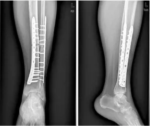
On examination of his legs, the physical exam indicated no neurological findings or impaired blood vessels. X-ray plain film showed closed multiple tibiofibular fractures (AO-OTA 42C23) on his left lower limb region (Figure 1). Besides, the patient had a smoking history for over 30 years with one pack of cigarettes a day, without other chronic diseases or relevant surgical his-tory. After being admitted, the patient was given calcaneal traction immediately. He was taken to operating room a week after injury for limited open reduction and percutaneous LCP fixation to restore tibiofibular fractures. Successfully, postoperative plain radiography showed good alignment and reduction (Figure 2). The patient was evaluated regularly with radiography in postoperative follow-up.
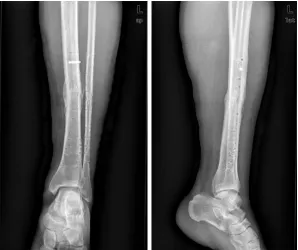
carefully and gaining pa- tient’s informed consent, we practiced dynamization to the LCP (Figure 5), in other words, three locking screws were removed from the center of the LCP [5, 6]. Meanwhile, autologous iliac graft was transplanted to the fracture site for the optimal therapeutic effect. The principle of dynamiza-tion was to enhance micro-motion in fracture site so as to stimulate callus for-mation through a given load. Six weeks after dyna- mization, the broken end displayed callus growth. Radiographs taken three months after dynamization displayed solid fracture healing and bridging callus formed across the gap (Figure 6), which means the callus kept increasing and remodeling. So we sug-gested the patient started walk with weight again. We removed the internal fixa-tion on account of patient’s demand since the fracture was healed a year and a half later, but one locking screw broke and remained (refer to the Figure 7 for details). Up to now, the patient walked with weight painlessly and refracture has not happened again. Discussion
[image:2.612.89.387.71.340.2]The LCP therapy and intra-medullary nail therapy are two major treatment of comminuted fracture of long bones currently. Dy- namization of Intramedul- lary nail can speed up the process of bone healing in most cases, while the dy- namization of LCP was not reported until 2011 by a
Figure 1. Left tibiofibular fracture (AO-OTA 42C23), on September 4th, 2012.
[image:2.612.89.387.387.676.2]Korean scholar [7]. But poor reduction of frac-ture and removed lag screws instead of not locking screws for dynamization were two major problems in the report, and there were also some relevant querying reports later [6]. There are few relevant reports of malunion after the LCP treatment published because the internal fixation was in frequenly taken out in other countries, while in China, patients usually ask
ture at the same site tend to occur within a short period of time. It is known that fracture site need comparable stress stimulation at dif-ferent stages during the healing process. Stress stimulation comes from interfragmen-tary movement. As the vital biomechanical and biological factor, interfragmentary movement depends on the rigidity of the fixation, load of the operated leg due to gravity and muscles for removing the internal fixation. That’s why the inci-dent of refracture is much more common in China [8]. With the popularization of the LCP treatment in the past decades, the efficacy of LCP in treatment of com-minuted fracture of long bones has been recogniz- ed, despite some failure cases [9].
It is secondary bone heal-ing mostly when applyheal-ing LCP to treat comminuted fracture of long bones, whi- ch takes a long course. At the early stage, the broken end is relatively stable be-cause stress is bore by the LCP, and primary callus is forming at the fracture end through cell proliferation and differentiation at the same time. Afterwards, wo- ven bone and lamellar bone formed at the middle and later stages through matrix mineralization and deposi-tion, finally, union will be achieved by means of con-tinuously molding and re- building. But at later stage, stress shielding as a result of rigid LCP construct may lead to low bone strength, even result in fatigue rup-ture or loosening of inter- nal fixation, which was rare when treated with ordinary compression plate previ-ously. Meanwhile, if the LCP is removed all at once after fracture union,
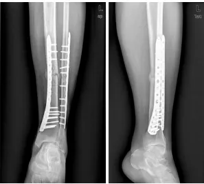
[image:3.612.90.388.71.322.2]refrac-Figure 3. Distal tibia fracture line disappeared and fibular fracture line was vague about one year after the surgery, on October 14th, 2013.
[image:3.612.89.388.371.569.2]forces, and the number of load cycles [10]. It’s worth thinking that whether the LCP shall be taken out gradually in order to train the fracture end step by step for further molding until the patient meet the requirement of bearing lo- ads in daily life completely. According to Perren’s strain theory, bone fracture would have healed in secondary intention under a deforma-tion of 2% to 10% [11]. The broken end would grow into bone malunion or even no- nunion as a result of insta-bility if the deformation af- ter internal fixation is over 10%. For such cases, treat-ments are usually replacing or adding internal fixations (intramedullary nail, lock-ing plate, blocklock-ing nail, etc.) to make the broken end become stable. There are also cases with so stable construction of the LCP that make the deformation less than 2%, indicating insufficient stress stimula-tion at the broken end and a tendency to develop into bone atrophy and non-union. This kind of case is treated with replacing inter-nal fixation conventiointer-nally. Above all, it would speed up the process of fracture healing as well as decrease failure of internal fixation and occurrence of refrac-ture with the help of pro-gressive stress training due to timely dynamization to stiff LCP construction at the later stage of fracture union [12].
[image:4.612.90.389.71.351.2]Accordingly, we propose a new concept of dynamiza-tion of the LCP, which is
Figure 5. Dynamization surgery: removed 3 locking screws from the center on November 7th, 2013.
[image:4.612.90.387.408.675.2]mainly applicable to patients who display non-union or delayed non-union in response to absolute stability after using the LCP to treat comminut-ed fracture of long bones. Actually, dynamiza-tion is a derivative concept after the extensive clinical application that base on Wolff’s law of bone growth and remodeling, Perren’s strain theory, as well as bridging working principle of the LCP [13-16]. It mainly has two advantages: firstly, reducing the economic burden comes from replacement of internal fixation; and sec-ondly, avoiding the possibility of refracture because of insufficient osseous strength at the early phase after removing internal fixation. As a result, patients would have one more choice except replacing internal fixation for nonunion if this concept applied to clinical practice. Stress shielding because of the over stiff LCP construction was considered the reason of the patient’s bone nonunion, in other words, low stress stimulation prevented the newly regen-erated bone transformed from fibrous cartilage into primary callus. So we tried to practice dynamization to the LCP so as to make micro-motion in the construction. And the break of the locking screw was considered fatigue frac-ture due to the last second locking screw at the proximal end endured most stress after the
None.
Address correspondence to: Minghai Wang, De- partment of Orthopedics, The Fifth People’s Hospital of Shanghai, Fudan University, Shanghai, China. E-mail: [email protected]
References
[1] Schutz M, Muller M, Krettek C, Hontzsch D, Re-gazzoni P, Ganz R and Haas N. Minimally inva-sive fracture stabilization of distal femoral fractures with the LISS: a prospective multi-center study. Results of a clinical study with special emphasis on difficult cases. Injury 2001; 32 Suppl 3: Sc48-54.
[2] Miller DL and Goswami T. A review of locking compression plate biomechanics and their ad-vantages as internal fixators in fracture heal-ing. Clin Biomech (Bristol, Avon) 2007; 22: 1049-1062.
[3] Strauss EJ, Schwarzkopf R, Kummer F and Egol KA. The current status of locked plating: the good, the bad, and the ugly. J Orthop Trau-ma 2008; 22: 479-486.
[4] Kubiak EN, Fulkerson E, Strauss E and Egol KA. The evolution of locked plates. J Bone Joint Surg Am 2006; 88 Suppl 4: 189-200.
[5] Perren SM, Fernandez A and Regazzoni P. Un-derstanding fracture healing biomechanics based on the “strain” concept and its clinical
dynamization surgery, com-bined with the requirement of longer working distance of the LCP with the incre- ased stress after the pa- tient walked with weight. The other possibility is the steel plate was too short. The patient in the case achieved bone union after dynamization therapy. How- ever, this is just a case and the patient had received autogenous bone graft. It required further verificat- ion by large sample clinical study and fundamental re- search to determine whe- ther dynamization surgery plays the leading role in fracture healing.
[image:5.612.90.388.71.321.2]Disclosure of conflict of interest
applications. Acta Chir Orthop Traumatol Cech 2015; 82: 253-260.
[6] Goyal T, Nag HL and Tripathy SK. Dynamization of locked plating on distal femur fracture. Arch Orthop Trauma Surg 2011; 131: 1331-1332. [7] Oh JK, Hwang JH, Lee SJ and Kim JI.
Dynamiza-tion of locked plating on distal femur fracture. Arch Orthop Trauma Surg 2011; 131: 535-539.
[8] Miller SD and Katcherian DA. Refracture after removal of a condylar plate from the distal third of the femur. J Bone Joint Surg Am 1991; 73: 949-950.
[9] Button G, Wolinsky P and Hak D. Failure of less invasive stabilization system plates in the dis-tal femur: a report of four cases. J Orthop Trau-ma 2004; 18: 565-570.
[10] Claes L, Blakytny R, Gockelmann M, Schoen M, Ignatius A and Willie B. Early dynamization by reduced fixation stiffness does not improve fracture healing in a rat femoral osteotomy model. J Orthop Res 2009; 27: 22-27.
[11] Perren SM. Evolution of the internal fixation of long bone fractures. The scientific basis of bio-logical internal fixation: choosing a new bal-ance between stability and biology. J Bone Joint Surg Br 2002; 84: 1093-1110.
[12] Wagner M. General principles for the clinical use of the LCP. Injury 2003; 34 Suppl 2: B31-42.
[13] Wolff J. Das Gesetz der Transformation der Knochen A. Dtsch Med Wochenschr 1892; 19: 1222-1224.
[14] Claes LE, Heigele CA, Neidlinger-Wilke C, Kas-par D, Seidl W, Margevicius KJ and Augat P. Ef-fects of mechanical factors on the fracture healing process. Clin Orthop Relat Res 1998; S132-147.
[15] Kenwright J, Richardson JB, Goodship AE, Ev-ans M, Kelly DJ, Spriggins AJ, Newman JH, Bur-rough SJ, Harris JD and Rowley DI. Effect of controlled axial micromovement on healing of tibial fractures. Lancet 1986; 2: 1185-1187. [16] Egol KA, Kubiak EN, Fulkerson E, Kummer FJ