Original Article
Efficacy of neoadjuvant chemotherapy in breast
cancer: arterial infusion chemotherapy
vs intravenous chemotherapy
Duanming Du, Leichang Jiang, Shuibo Qiu, Ruming Zhou
Department of Interventional Therapy, Shenzhen Second People’s Hospital, The First Affiliated Hospital of Shenzhen University, Shenzhen, China
Received March 11, 2016; Accepted May 26, 2016; Epub July 15, 2016; Published July 30, 2016
Abstract: Objective: This study aimed to compare the efficacy of arterial infusion chemotherapy with that of intra -venous chemotherapy as neoadjuvant chemotherapy in breast cancer patients. Methods: A total of 92 patients were recruited and received doxorubicin dominant chemotherapy. Patients were randomized into arterial infusion chemotherapy group (n=44; chemoembolization was performed if necessary; group A) and intravenous chemo-therapy group (n=48; group V). After chemochemo-therapy, surgical interventions were employed. The adverse effects
were evaluated, and the time interval between chemotherapy and surgery was determined. The therapeutic efficacy
and long survival rate were compared between them. Results: The remission rate (complete remission and partial
remission) in group A (93.18%) was significantly higher than in group V (62.5%). In group V, the incidence of adverse effects (bone marrow suppression, gastrointestinal reactions and alopecia) was significantly higher than in group A. The mean time interval between chemotherapy and surgery in group A was significantly shorter than in group V (25.00±5.34 days vs 56.00±15.65 days; P<0.05). The survival rate within first 5 years was comparable between
groups, but the 10-year survival rate in group A was slightly higher than in group V. Conclusion: Arterial infusion chemotherapy may serve as an effective strategy for the neoadjuvant chemotherapy of breast cancer.
Keywords: Breast cancer, neoadjuvant chemotherapy, arterial infusion chemotherapy, intravenous chemotherapy
Introduction
Breast cancer is a common malignancy in women and causes over a half-million deaths each year worldwide [1]. The latest world can-cer statistics available from the International Agency for Research on Cancer (IARC) showed that 1,677,000 women were diagnosed with breast cancer and 577,000 women died in 2012. Since 2008, breast cancer incidence has increased by over 20% and breast cancer deaths have risen by 14% [1]. Moreover, most countries with the highest breast cancer mor-tality rate are low- to middle-income countries (LMICs) and breast cancer in LMICs often pres-ents when locally advanced breast cancer (LABC) [2] that can be easily appreciated at physical exam but is still limited to the breast and draining lymph nodes, without clinical evi-dence of metastatic spread. Despite being con-fined to the breast and regional nodes, locally
advanced stage often heralds the rapid onset of metastatic disease, explaining high mortality rates.
inop-erable disease opinop-erable. In recent years, neo-adjuvant therapy has increasingly been used in patients with operable disease [4].
In this study, we compared the efficacy of arte -rial infusion chemotherapy with that of intrave-nous chemotherapy in breast cancer patients, and the long term survival rate and adverse effects were evaluated, aiming to find an opti -mal way by which the neoadjuvant therapy is conducted in breast cancer patients.
Materials and methods
General information
Women who were pathologically diagnosed with breast cancer were recruited between January 2003 and December 2006 from the Second People’s Hospital of Shenzhen City. These patients received neoadjuvant chemo-therapy. Of these patients, 44 received arterial infusion chemotherapy (A) and 48 underwent intravenous chemotherapy (V). The patients’ characteristics were comparable at baseline between two groups (P>0.05) and are shown in Table 1. Before therapy, routine blood test, urine and stool analyses, detection of liver and kidney functions, chest X ray, and electrocardi-ography were conducted to exclude organic lesions and patients had no contradictions to chemotherapy.
Treatment
Group A: Patients lied in a supine position and the right inguina region was sterilized, followed by local anesthesia with 1% lidocaine (10 ml). The right femoral artery was punctured with Seldinger method, and super-selective arteriog-raphy was performed at the internal mammary artery, lateral thoracic artery and subclavian artery of the affected breast. The blood supply, solid lesions and lymph node status were eval-uated. Chemotherapy with CEF or MEF proto-col: Mitomycin (MMC; 10 mg), cyclophospha-mide (CTX; 400 mg), epirubicin (EADM; 50 mg) and 5-fluorouracil (5-Fu; 1000 mg) were used
for chemotherapy. Arterial infusion chemother-apy was performed via above three arteries. When the affected breast was supplied mainly by the internal mammary artery and the lateral thoracic artery, 50% of chemotherapeutics was infused via major vessel, 30% via subclavian artery and 20% via secondary artery; when the cancer or lymph nodes were not obvious in arteriography, 50% of chemotherapeutics was infused via subclavian artery, 30% via internal mammary artery, and 20% via lateral thoracic artery. When the cancer was very obvious, the major vessel was occluded with 1000-1400 µm gelatin sponge. When infusion was con-ducted via the subclavian artery, inflatable cuff was used to maintain the pressure at 10-20 mmHg higher than systolic blood pressure, and the cuff was released 30 min later and then maintained for 5 min. When infusion was con-ducted via the internal mammary artery and the lateral thoracic artery, inflatable cuff was not used. Infusion was performed slowly with a micropump for 2-3 h.
Group V: Anthracyclines (epirubicin) dominant protocol was used. CEF or MEF protocol was used. The chemotherapeutics included MMC (8 mg/m2), CTX (400-600 mg/m2), EADM (40 mg/
m2), and 5-Fu (500-750 mg/m2). Intravenous
infusion was conducted once every 3-4 weeks for a total of 1-3 courses.
After chemotherapy, surgical interventions were performed with classical radical mastec-tomy, modified radical mastectomy or breast-conserving resection.
Angiography
After angiography, the blood supply to the breast cancer was evaluated by at least 2 expe-rienced physicians, and any discrepancy was resolved by discussion.
Determination of therapeutic efficacy
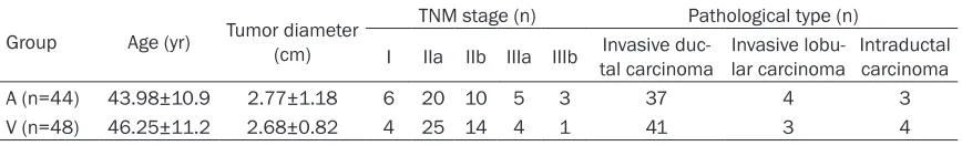
Before and after neoadjuvant chemotherapy, the cancer size was determined by the physi-Table 1. Characteristics of patients with breast cancer at baseline
Group Age (yr) Tumor diameter (cm)
TNM stage (n) Pathological type (n)
I IIa IIb IIIa IIIb Invasive duc-tal carcinoma Invasive lobu-lar carcinoma Intraductal carcinoma
A (n=44) 43.98±10.9 2.77±1.18 6 20 10 5 3 37 4 3
[image:2.612.90.524.85.151.2]cians for interventional therapy and surgeons together, and it was measured clinically. Acc- ording to the Response Evaluation Criteria in Solid Tumors (RECIST1.1), the therapeutic effi -cacy was classified as complete remission (CR), partial remission (PR), stable disease (SD) and progressive disease (PD). The overall efficacy was calculated as follow: Sum of patients with CR and PR/total patients ×100%. The adverse effects of chemotherapy were evaluated acc- ording to the classification criteria for acute and subacute adverse effects of anti-tumor drugs. Histological evaluation was conducted as follows: mild pathological change: pathologi-cal change was confined to <1/3, a large amount of residual cancer cells was observed, and a variety of lymph nodes were involved; significant pathological change: pathological change was confined to <1/2, residual cancer cells were still observable, and lymph node metastasis was observed; pathological com-plete remission: there were no invasive lesions in the primary lesions and lymph nodes collect-ed by surgery. The time interval between che-motherapy and surgery referred to the interval from day of initiation of first chemotherapy to the day of surgery.
Follow up
The patients’ information was collected by reviewing the medical record and via telephone. The date of the last hospital visit or hospitaliza-tion was used for patients lost to follow up. The main end point was the overall survival. Follow up was conducted for 6-130 months in these patients and the last follow up was performed on March 1, 2014.
Statistical analysis
Statistical analysis was performed with SPSS version 19.0. Quantitative data are expressed as mean ± standard deviation (_x±s) and com-pared with t test. Qualitative data are expressed as percentage and compared with chi square test. Survival analysis was conducted with Kaplan-Meier method and Log Rank (Mantel-Cox) test. A value of P<0.05 was considered statistically significant.
Results
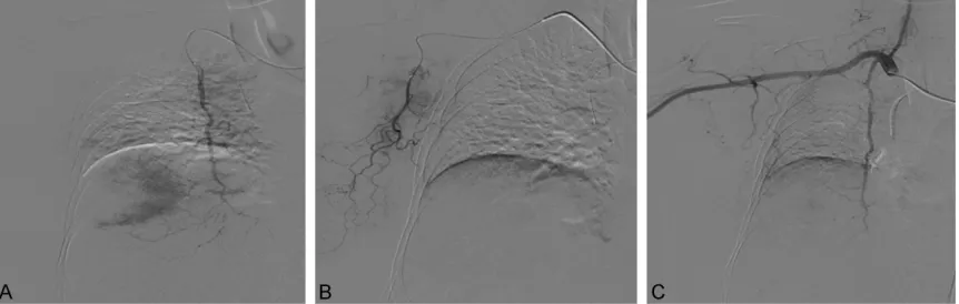
Findings from angiography
Angiography showed the blood vessels en- larged, were irregularly arranged and formed networks. In parenchymal phase, the cancer was not observed and spotty or mass-like. The axillary lymph nodes were significantly enlarged and irregular, and branches of blood vessels were obvious (Figure 1A and 1B). For breast cancer which was obvious in angiography, the major blood vessel was embolism with gelatin sponge after infusion. Re-examination by angi-ography is shown in Figure 1C.
Therapeutic efficacy
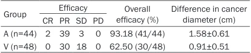
According to the RECIST1.1, CR and PR were found in 41 patients in group A with the overall efficacy of 93.81%, which was significantly higher than in group V (62.5%, n=30; P<0.05) (Table 2).
Adverse effects
[image:3.612.93.524.72.209.2]Besides adverse effects shown in Table 3, skin flushing was also observed in group A; blisters
or even ulcer were found in several patients in group V, but resolved after symptomatic thera-py; light chromatosis was found in several patients in group V. In group V, skin lesions were not observed. In group V, the incidence of bone marrow suppression, gastrointestinal reactions and alopecia was significantly higher than in group A (Table 3).
Time interval between chemotherapy and sur-gery
The mean time interval between chemotherapy and surgery was 25.00±5.34 days in group A, which was significantly shorter than in group V (56.00±15.65 days; P<0.05).
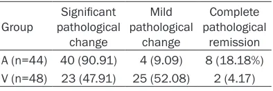
Histological examination
In group A, karyopyknosis, nuclear fragmenta-tion, necrosis and infiltration of inflammatory cells as well as cells with abnormal nucleus were observed in sections of 40 patients (40/44; 90.91%). In group V, these findings were observed in 23 patients (23/48; 47.92%). There was significant difference in the propor -tion of patients with above pathological chang-es between two groups (Figure 2) (P<0.05; Table 4).
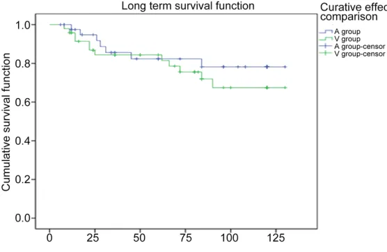
Long term survival
The 1-year, 3-year, 5-year and 10-year survival rate was 97.5%, 85.6%, 82.3% and 78.2%, respectively, in group A and 95.8%, 84.4%, 78.6% and 67.5%, respectively, in group V. Log Rank (Mantel-Cox) test showed the survival rate within first 5 years was comparable between groups, but the 10-year survival rate in group A was slightly higher than in group V (Figure 3).
Discussion
Breast cancer is the most commonly diagnosed cancer in women [1]. Surgery followed by adju-vant treatment has been the gold standard for
Traditional indications for neoadjuvant therapy in breast cancer include N2 stage-fixed or mat -ted lymph node on ipsilateral side, or clinically apparent ipsilateral internal mammary nodes in the absence of axillary node, making the clini-cal staging at least stage IIIA or above. Patients with stage IIIB disease with tumors invading the chest wall, skin or both, or with breast cancer of inflammatory nature, would be a good candi -date for neoadjuvant therapy [5]. Neoadjuvant therapy should also be considered for women with clinical stage IIA and IIB tumors with a larg-er tumor who wish to have breast-conslarg-erving operations and avoid mastectomy. Not in all, but in many patients, neoadjuvant therapy results in sufficient tumor response to make breast-conserving operations possible. Several studies in the early 2000 s showed that neoad-juvant chemotherapy successfully reduced both locoregional and in breast tumor recur-rence even in large T3 and T4 tumors [6, 7]. Neoadjuvant therapy has been evolving rapidly given this benefit [8].
[image:4.612.89.341.84.138.2]In 1968, Fisher et al [9] proposed the primary lesions of breast cancer were only the focal manifestation of the systematic disease. Neoadjuvant chemotherapy may benefit patients and has been one of standard and classic strategies in the therapy of cancers [10]. The proven benefits of NST justifying its routine clinical use include the following: it improves disease-free survival and overall sur-vival to the same extent as postoperative che-motherapy; it increases breast-conserving sur-gery (BCS) rates in patients with operable local-ly advanced breast cancer (clinical stages IIIA except of T3N1M0, IIIB, and IIIC); and it reduces the extent of resection in cancers >2 cm even if a patient is a candidate for BCS. The extent of residual cancer after NST is a powerful prog-nostic marker [8, 10]. In arterial infusion che-motherapy, chemotherapeutics are infused via the vessels supplying the cancer, which leads to a high concentration of chemotherapeutics Table 2. Therapeutic efficacy in tow groups
Group Efficacy efficacy (%)Overall Difference in cancer diameter (cm) CR PR SD PD
A (n=44) 2 39 3 0 93.18 (41/44) 1.58±0.61 V (n=48) 0 30 18 0 62.50 (30/48) 0.91±0.51
Note: Difference in cancer diameter refers to the difference between cancer sizes before and after chemotherapy; CR: complete remission; PR: partial remission; SD: stable disease; PD: progressive disease.
at the cancer and adjacent lymph nodes. The drugs that are not metabolized may enter the systemic vein and contact with the cancer again via circulation (second chemotherapy), increasing the therapeutic efficacy [11].
Feldman et al [12] for the first time described the blood supply to the breast cancer by angi-ography after brachial artery puncture. In re- cent years, Zhou et al and Zhang et al [13, 14] found that lateral thoracic artery was the major vessel supply the breast cancer, and the blood supplied via the lateral thoracic artery is more than that via the internal mammary artery. The supplied arteries forms a network in breast cancer, and each vessel also has a lot of col-lateral vessels which connect with each other. Thus, the extent of infusion should be expand-ed during the arterial infusion chemotherapy, and the chemotherapeutics are infused mainly via the internal mammary artery, lateral tho-racic artery and subclavian artery. The dose of chemotherapeutics is determined according to findings from arteriography.
Breast cancer cells are sensitive to chemother-apeutics, especially the anthracyclines and tax-anes. CMF protocol has been used as a gold standard in the therapy of breast cancer for more than 30 years [15]. In the present study, anthracycline (epirubicin) dominant protocol was used for chemotherapy (CEF or MEF proto-col). Our results showed the overall efficacy was as high as 93.08% in A group, which was significantly higher than in V group. In addition, in our study, more patients in A group devel-oped skin related adverse effects as compared to V group. This may be ascribed to the high concentration of chemotherapeutic in the can-cer and surrounding lymph nodes and the con-tact between drugs and breast cancer is pro-longed, leading to endangiitis, vascular thicken-ing and thrombosis as well as skin lesions. These adverse effects resolved after symptom-atic therapy.
[image:5.612.90.522.84.276.2]Liu et al [15] reported that the 5-year survival rate was 93.3% and 56% after arterial infusion chemotherapy and systemic chemotherapy, respectively. Zeng et al reported that the over-all effectiveness rate of arterial infusion che-motherapy and embolic therapy was 93.3% for advanced breast cancer, and the 5-year surviv-al rate was as high as 93.3%. In addition, Shimamoto et al and Miura et al [16, 17] pro-posed that arterial infusion chemotherapy could achieve better efficacy as compared to traditional venous chemotherapy: the short Table 3. Adverse effects in two groups (n; %)
Group Bone marrow suppression Gastrointestinal reactions Alopecia Peripheral sen-sory neuropathy Liver dys-function Grade I Grade II Grade III Grade IV Grade I Grade II Grade III
A (n=44) 4 (9.09) 1 (2.27) 5 (11.36) 2 (4.54) 10 (22.73) 8 (18.18) 0 12 (27.27) 6 (13.64) 2 (4.5%) V (n=48) 13 (27.08) 11 (22.92) 3 (6.25) 3 (6.25) 15 (31.25) 22 (45.83) 3 (6.25) 30 (62.50) 5 (10.42) 6 (12.5%)
P <0.05 <0.05 <0.05 >0.05 >0.05
Figure 2. A: Acidophilic change in cytoplasm of breast cancer cells; B: Enlargement and singularity of nucleuses of breast cancer cells; C: Vacuolation of breast cancer cells (HE*400).
Table 4. Histological examination of breast cancer in both groups (n, %)
Group pathological Significant change
Mild pathological
change
Complete pathological
[image:5.612.92.289.349.415.2]term effectiveness rate was 20%-30% higher than that of venous chemotherapy; the quality of life was significantly improved and long term survival time was prolonged. These findings were consistent with above mentioned. In the present study, results showed the effective-ness rate was 93.18% in A group, which was similar to previously reported. However, the effectiveness rate in V group was 62.50%, which was lower than previously reported. This might be ascribed to the small course of venous chemotherapy (1-3 courses; 4 weeks in each course). Patients in both groups were followed up for 6-130 months. The survival rate within first 5 years was comparable between two groups, but the 10-year survival rate in A group was higher than in V group.
Our study indicates that arterial infusion che-motherapy is an effective strategy for the neo-adjuvant chemotherapy of breast cancer. This neoadjuvant chemotherapy is able to reduce cancer size, which is helpful for the surgical interventions, reduces adverse effects, and shortens the time interval between chemother-apy and surgery. However, arterial infusion che-motherapy fails to significantly improve the long term survival rate in breast cancer patients. This is a retrospective study, and more pro-spective, randomized and controlled studies are required to confirm our findings.
Disclosure of conflict of interest
None.
Address correspondence to: Dr. Duanming Du, Department of Interventional Therapy, Shenzhen
ast cancer-strategies for developing nations. Front Oncol 2015; 5: 89.
[3] Frei E 3rd. Clinical cancer research: an embat-tled species. Cancer 1982; 50: 1979-1992. [4] Miller E, Lee HJ, Lulla A, Hernandez L, Gokare
P and Lim B. Current treatment of early breast cancer: adjuvant and neoadjuvant therapy. F1000 Res 2014; 3: 198.
[5] Deo SV, Bhutani M, Shukla NK, Raina V, Rath GK and Purkayasth J. Randomized trial com-paring neo-adjuvant versus adjuvant chemo-therapy in operable locally advanced breast cancer (T4b N0-2 M0). J Surg Oncol 2003; 84: 192-197.
[6] Sweeting RS, Klauber-Demore N, Meyers MO, Deal AM, Burrows EM, Drobish AA, Anders CK and Carey LA. Young women with locally ad-vanced breast cancer who achieve breast con-servation after neoadjuvant chemotherapy have a low local recurrence rate. Am Surg 2011; 77: 850-855.
[7] Akay CL, Meric-Bernstam F, Hunt KK, Grubbs EG, Bedrosian I, Tucker SL, Kuerer HM, Hoffman KE, Babiera GV, Strom EA, Buchholz TA and Mittendorf EA. Evaluation of the MD Anderson Prognostic Index for local-regional recurrence after breast conserving therapy in patients receiving neoadjuvant chemotherapy. Ann Surg Oncol 2012; 19: 901-907.
[8] Kaufmann M, von Minckwitz G, Mamounas EP, Cameron D, Carey LA, Cristofanilli M, Denkert C, Eiermann W, Gnant M, Harris JR, Karn T, Liedtke C, Mauri D, Rouzier R, Ruckhaeberle E, Semiglazov V, Symmans WF, Tutt A and Pusztai L. Recommendations from an international consensus conference on the current status and future of neoadjuvant systemic therapy in primary breast cancer. Ann Surg Oncol 2012; 19: 1508-1516.
[9] Fisher B, Ravdin RG, Ausman RK, Slack NH, Moore GE and Noer RJ. Surgical adjuvant
che-Figure 3. Long term survival rate in two groups.
Second People’s Hospital, The
First Affiliated Hospital of
Sh-enzhen University, ShSh-enzhen, China. E-mail: dmdu69@sina. com
References
[1] Ferlay J, Soerjomataram I, Dikshit R, Eser S, Mathers C, Rebelo M, Parkin DM, Forman D and Bray F. Cancer incidence and mor-tality worldwide: sources, methods and major pat-terns in GLOBOCAN 2012. Int J Cancer 2015; 136: E359-386.
[image:6.612.92.370.74.255.2]motherapy in cancer of the breast: results of a decade of cooperative investigation. Ann Surg 1968; 168: 337-356.
[10] Liu SV, Melstrom L, Yao K, Russell CA and Sener SF. Neoadjuvant therapy for breast can-cer. J Surg Oncol 2010; 101: 283-291. [11] Stephens FO. Intraarterial induction
chemo-therapy in locally advanced stage III breast cancer. Cancer 1990; 66: 645-650.
[12] Feldman F, Habif DV, Fleming RJ, Kanter IE and Seaman WB. Arteriography of the breast. Radiology 1967; 89: 1053-1061.
[13] Zhou T, Zhuang YQ, Qin ST, Xu B and Huang P.
Therapeutic efficacy of superselective chemo -embolization under digital subtraction angiog-raphy in locally advanced breast cancer. Guangdong Med J 2012; 33: 1993-1995. [14] Zhang JX, Liu YE and Tang WH. Preoperative
interventional regional chemotherapy versus systemic chemotherapy for advanced breast cancers: a comparison study. J Intervent Radiol 2013; 22: 587-590.
[15] Liu ZY and Zhang J. Comparison of clinical ef-fect of TE versus CEF regimens as neoadjuvant chemotherapy in the treatment of breast can-cer. Chin J Breast Dis (Elec Ver) 2008; 2: 30-39.
[16] Shimamoto H, Takizawa K, Ogawa Y, Yoshimatsu M, Yagihashi K, Okazaki H, Kanemaki Y, Nakajima Y, Ohta T, Ogata H and
Fukuda M. Clinical efficacy and value of redis -tributed subclavian arterial infusion chemo-therapy for locally advanced breast cancer. Jpn J Radiol 2011; 29: 236-243.