warwick.ac.uk/lib-publications
Original citation:
Dockree, Tamsin, Holland, Christopher J., Clement, Mathew, Ladell, Kristin, McLaren, James
E., van den Berg, Hugo A., Gostick, Emma, L. Miners, Kelly, Llewellyn-Lacey, Sian, S
Bridgeman, John, Man, Stephen, Bailey, Mick, Burrows, Scott R., Price, David A. and
Wooldridge, Linda. (2017) CD8+ T-cell specificity is compromised at a defined MHCI/CD8
affinity threshold. Immunology and Cell Biology, 95 . pp. 68-76.
Permanent WRAP URL:
http://wrap.warwick.ac.uk/84130
Copyright and reuse:
The Warwick Research Archive Portal (WRAP) makes this work of researchers of the
University of Warwick available open access under the following conditions.
This article is made available under the Creative Commons Attribution 4.0 International
license (CC BY 4.0) and may be reused according to the conditions of the license. For more
details see:
http://creativecommons.org/licenses/by/4.0/
A note on versions:
The version presented in WRAP is the published version, or, version of record, and may be
cited as it appears here.
ORIGINAL ARTICLE
CD8
+
T-cell speci
fi
city is compromised at a de
fi
ned
MHCI/CD8 af
fi
nity threshold
Tamsin Dockree
1, Christopher J Holland
2, Mathew Clement
1, Kristin Ladell
1, James E McLaren
1,
Hugo A van den Berg
3, Emma Gostick
1, Kelly L Miners
1, Sian Llewellyn-Lacey
1, John S Bridgeman
1,
Stephen Man
4, Mick Bailey
2, Scott R Burrows
5, David A Price
1,6and Linda Wooldridge
2The CD8 co-receptor engages peptide-major histocompatibility complex class I (pMHCI) molecules at a largely invariant site distinct from the T-cell receptor (TCR)-binding platform and enhances the sensitivity of antigen-driven activation to promote effective CD8+T-cell immunity. A small increase in the strength of the pMHCI/CD8 interaction (~1.5-fold) can disproportionately amplify this effect, boosting antigen sensitivity by up to two orders of magnitude. However, recognition specificity is lost
altogether with more substantial increases in pMHCI/CD8 affinity (~10-fold). In this study, we used a panel of MHCI mutants with altered CD8-binding properties to show that TCR-mediated antigen specificity is delimited by a pMHCI/CD8 affinity threshold. Ourfindings suggest that CD8 can be engineered within certain biophysical parameters to enhance the therapeutic efficacy of adoptive T-cell transfer irrespective of antigen specificity.
Immunology and Cell Biology(2017)95,68–76; doi:10.1038/icb.2016.85
CD8+ T cells recognize antigens in the form of short peptide fragments bound to major histocompatibility complex class I (MHCI) molecules on the target cell surface.1Specific engagement of peptide-MHCI (ppeptide-MHCI) complexes via the clonotypically expressedαβT-cell receptor (TCR) triggers a range of effector functions that play a critical role in protective immunity against intracellular infections and various malignancies. The ability to identify and eliminate cancerous cellsin vivois particularly intriguing2,3and promises novel therapies based on the immunobiology of CD8+ T cells. Indeed, adoptive transfer of in vitro-expanded CD8+ T cells can cause tumour regression in the clinical setting.4,5These seminal observations have sparked great interest in the use of cellular therapy to combat cancer.6,7 However, a number of obstacles preclude the widespread use of this approach. In biological terms, one key limitation relates to the naturally low affinity of self-derived antigen-specific TCRs,8,9 which constrains the functional properties of tumour-associated antigen-specific CD8+T-cell populations. This intrinsic problem stems from the negative selection of high-affinity autoreactive αβ TCR clonotypes during thymic education and most likely explains why it has proven difficult to develop cancer vaccines in the absence of a clear oncogenic microbial agent. Although high-affinity TCRs can be engineered to circumvent suboptimal antigen recognition, most notably via phage display technology,10,11the requirement to reiterate this process for each pMHCI specificity tailored to individual tumour proteomes is a major barrier to therapeutic applicability.
The surface-expressed CD8αβ glycoprotein (CD8 from here on) serves as a co-receptor for MHCI-restricted T cells.12CD8 binds to a largely invariant region of MHCI at a site distinct from the TCR-binding platform and acts to enhance T-cell antigen sensitivity by up to six orders of magnitude.12–14 This effect is mediated via several mechanisms, including: (i) promotion and stabilization of the TCR/pMHCI interaction at the cell surface;15–18 (ii) recruitment of signalling molecules to the intracellular side of the TCR/CD3ζ complex;19–22and (iii) localization of TCR/pMHCI complexes within specialized membrane microdomains enriched for early intracellular signal transduction molecules.23,24These properties can potentially be harnessed to modulate antigen-specific CD8+ T-cell immunity. It is notable in this regard that pMHCI/CD8 binding is characterized by very low solution affinities (average KD~ 145μM).25 Moreover, an incremental increase in the strength of this interaction (KD~ 98μM) can boost antigen sensitivity by up to 100-fold.17,26Such manipula-tions are globally applicable across TCR specificities due to the non-polymorphic nature of CD8, thereby providing a generic opportunity to enhance CD8+ T-cell reactivity for therapeutic purposes.27 How-ever, substantial increases in pMHCI/CD8 affinity can abrogate antigen specificity.28
In this study, we used a panel of MHCI mutants with altered CD8-binding properties to show that the specificity of peptide-dependent TCR recognition is maintained within a defined pMHCI/ CD8 affinity window. Collectively, the data provide biophysical
1Institute of Infection and Immunity, Cardiff University School of Medicine, Cardiff, UK;2Faculty of Health Sciences, University of Bristol, Bristol, UK;3Mathematics Institute,
University of Warwick, Coventry, UK;4Institute of Cancer and Genetics, Cardiff University School of Medicine, Cardiff, UK;5Cellular Immunology Laboratory, QIMR Berghofer
Medical Research Institute, Brisbane, QLD, Australia and6Human Immunology Section, Vaccine Research Center, National Institute of Allergy and Infectious Diseases, National
Institutes of Health, Bethesda, MD, USA
Correspondence: T Dockree, Institute of Infection and Immunity, Cardiff University School of Medicine, Cardiff CF14 4XN, UK. E-mail: [email protected]
guidelines for the rational design of high-affinity CD8 molecules to optimize the therapeutic efficacy of adoptive T-cell transfer.
RESULTS
Development of a novel MHCI mutant to probe the pMHCI/CD8 interaction
The pMHCI/CD8 interaction is characterized by very low solution binding affinities and extremely rapid kinetics.29–31Although some variation exists between different MHCI molecules due to polymorphisms that affect the CD8 binding site, the average pMHCI/CD8 interaction occurs with an equilibrium dissociation constant (KD) ~ 145μM (range=100–220μM).25,32 Substantially weaker pMHCI/CD8 solution binding affinities have been reported for human leukocyte antigen (HLA) A*6801, HLA B*4801 and HLA B*8101.22,25The introduction of a glutamine (Q) to glutamic acid (E) substitution at position 115 of the MHCI α2 domain increases the pMHCI/CD8 interaction by ~ 1.5-fold (KD~ 98μM) without impact-ing the TCR/pMHCI bindimpact-ing platform.26This mutation significantly enhances the sensitivity of pMHCI antigen recognition (up to 100-fold) without compromising TCR-mediated specificity. In contrast, a human to murine MHCIα3 domain switch increases the pMHCI/CD8 interaction by ~ 15-fold (KD~ 11μM) and bypasses the requirement for cognate TCR engagement.28
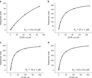
To determine the pMHCI/CD8 affinity at which antigen specificity is lost, we introduced an alanine (A) to valine (V) substitution at position 245 of A2/Kb (a fusion molecule comprising the α1/α2
[image:3.595.296.545.237.326.2]peptide-binding platform of HLA A*0201 and the α3 domain of H2-Kb) to generate the novel MHCI mutant A2/Kb A245V. Surface plasmon resonance analysis revealed that A2/Kb A245V binds CD8 with a KD of 27μM (Figures 1a and b), while the TCR/pMHCI interaction remains unchanged (Figures 1c and d). Combined with previously developed mutants, we then had an extended panel for functional analysis that incorporated MHCI molecules spanning a range of CD8 interaction affinities as follows: abrogated (A2 D227K/ T228A);21 weak (A2 A245V);22 wild type (A2); slightly enhanced (A2 Q115E);26 enhanced (A2/Kb A245V); and superenhanced (A2/ Kb).28Importantly, none of these mutations affect the integrity of TCR binding to pMHCI (Table 1; Figure 2a).
Figure 1A2/KbA245V exhibits enhanced affinity for CD8 without impacting the TCR/pMHCI interaction. Biotinylated A2 (a, c) or A2/KbA245V (b, d)
monomers refolded with wild typeβ2m and the heteroclitic peptide ELAGIGILTV were immobilized on a streptavidin-coated BIAcore chip. Serial dilutions of
[image:3.595.108.473.380.690.2]soluble human CD8αα (a, b) or MEL5 TCR (c, d) wereflowed over the chip to measure equilibrium binding by surface plasmon resonance. Data were analyzed using BIAevaluation 3.1, Microsoft Excel and Origin 6.1.
Table 1 CD8-binding affinity measurements for the MHCI molecules used in this study
Location of mutation Description of mutation pMHCI/CD8KD(μM)
MHCIα3 domain A2 D227K/T228A 410 000 (NDB)a
MHCIα3 domain A2 A245V 498a
Wild type No mutation 137±9.7a
MHCIα2 domain A2 Q115E 98±14.5a
MHCIα3 domain A2/KbA245V 27±1
MHCIα3 domain A2/Kb 11a
Abbreviations: MHC1, major histocompatibility complex class I; NDB, no detectable binding; pMHC1, peptide-MHC1.
aMeasurements reported previously for MHCI molecules refolded with wild type humanβ2m and the nonamer peptide LLFGYPVYV, an immunodominant epitope derived from the human T-cell lymphotropic virus type 1 Tax protein (residues 11–19).17
The pMHCI/CD8 interaction controls T-cell specificity T Dockreeet al
69
Increasing the strength of the pMHCI/CD8 interaction enhances pMHCI engagement at the cell surface
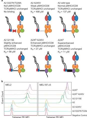
To investigate the relationship between pMHCI/CD8 affinity and pMHCI engagement at the cell surface, we generated fluorescent tetrameric complexes of A2 D227K/T228A, A2 A245V, A2, A2 Q115E, A2/Kb A245V and A2/Kb refolded with wild type β
2microglobulin (β2m) and the decamer peptide ELAGIGILTV, which is a heteroclitic variant of the Melan-A26-35epitope EAAGIGILTV. These pMHCI tetramers were used at standardized concentrations to stain two different ELAGIGILTV-specific CD8+ T-cell clones (MEL2 and MEL187.c5). Tetramer staining of MEL2 and MEL187.c5 was very
poor in the absence of an interaction with CD8 (A2 D227K/T228A) (Figure 2b). As the strength of the pMHCI/CD8 interaction increased, however, progressive increments in pMHCI tetramer staining were observed for both CD8+T-cell clones. Thus, pMHCI engagement at the cell surface is enhanced in the presence of stronger pMHCI/CD8 interactions.
pMHCI binding specificity is compromised at a defined pMHCI/ CD8 affinity threshold
Standard wild type pMHCI tetramers bind cell surface TCRs with exquisite specificity.33,34In contrast, nonspecific binding occurs in the A2 D227K/T228A
Null pMHCI/CD8 TCR/pMHCI unchanged No binding
A2 A245V Weak pMHCI/CD8 TCR/pMHCI unchanged KD = 498 µM
A2 wild type Normal pMHCI/CD8 TCR/pMHCI unchanged KD = 137 µM
A2 Q115E Slightly enhanced pMHCI/CD8
TCR/pMHCI unchanged KD = 98 µM
A2/Kb A245V
Enhanced pMHCI/CD8 TCR/pMHCI unchanged KD = 27 µM
A2/Kb
Superenhanced pMHCI/CD8
TCR/pMHCI unchanged KD = 11 µM
MEL2
A2/Kb
A2/Kb A245V
A2 Q115E
A2
A2 A245V
A2 D227K/T228A
Negative Control
MEL187.c5
Event Count
[image:4.595.127.475.70.540.2]Tetramer PE MFI Tetramer PE MFI
Figure 2Increasing the strength of the pMHCI/CD8 interaction enhances pMHCI binding at the cell surface. (a) Schematic representation of the six different MHCI mutants spanning a range of pMHCI/CD8 interaction affinities. None of the introduced mutations affect TCR/pMHCI binding. (b) 5 × 10⁴clonal MEL2
or MEL187.c5 CD8+T cells were stained with ViViD and the indicated ELAGIGILTV tetramer (A2 D227K/T228A, A2 A245V, A2, A2 Q115E, A2/KbA245V
or A2/Kb) at 25μg ml−1. Viable events are shown in concatenated histogram plots. Data were acquired using a FACSCantoIIflow cytometer and analyzed
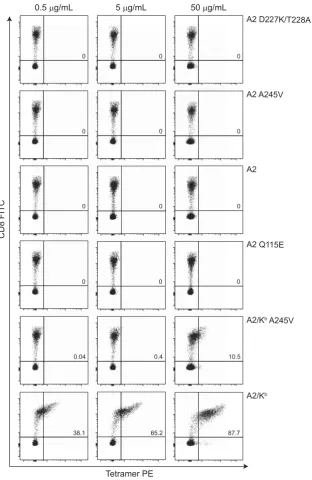
presence of a superenhanced pMHCI/CD8 interaction (KD~ 11μM).28 To define the pMHCI/CD8 affinity threshold at which pMHCI binding specificity is compromised, we stained healthy donor periph-eral blood mononuclear cells (PBMCs) with fluorescent tetrameric complexes of A2 D227K/T228A, A2 A245V, A2, A2 Q115E, A2/Kb A245V and A2/Kbrefolded with wild typeβ
2m and ELAGIGILTV. First, we stained A2– PBMCs. In the absence of alloreactivity, we would not expect these samples to harbour TCRs that recognize peptides in the context of A2. Any observable tetramer staining under these circumstances can therefore be attributed to peptide-independent recognition of pMHCI. No background staining was
detected when A2–PBMCs were stained with the A2 D227K/T228A, A2 A245V, A2 or A2 Q115E tetramers up to a concentration of 50μg ml−1(Figure 3). A similar pattern was observed with the A2/Kb A245V tetramer at 0.5 and 5μg ml−1. In line with a concentration-dependent effect, however, the same reagent displayed moderate background staining at 50μg ml−1. The A2/Kb tetramer was almost entirely nonspecific, as described in a previous report.28
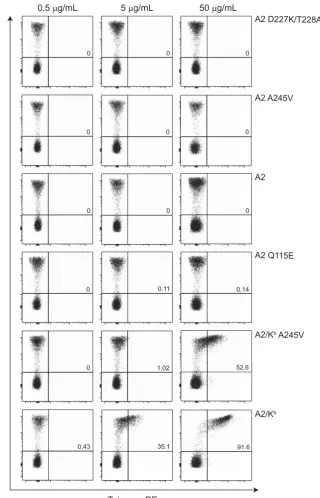
[image:5.595.135.448.205.686.2]Next, we repeated this analysis using A2+PBMCs, which frequently harbour TCRs specific for ELAGIGILTV. The clonotypic repertoire in these samples is also shaped by positive selection to ensure an intrinsic level of reactivity with A2. Staining specificity was maintained with the
Figure 3pMHCI binding specificity is compromised at a defined pMHCI/CD8 affinity threshold in A2–donors. 1 × 10⁶A2–PBMCs were stained with ViViD and the indicated ELAGIGILTV tetramer (A2 D227K/T228A, A2 A245V, A2, A2 Q115E, A2/KbA245V or A2/Kb) at 0.5, 5 or 50μg ml−1, followed by a
panel of lineage-specific monoclonal antibodies as described in the Methods section. Plots are gated on live, CD3+populations. Data were acquired using a
FACSCantoIIflow cytometer and analyzed with FlowJo software version 10.6. Values shown in the upper right quadrant indicate % tetramer+CD8+T cells.
The pMHCI/CD8 interaction controls T-cell specificity T Dockreeet al
71
A2 D227K/T228A, A2 A245V, A2 and A2 Q115E tetramers up to a concentration of 50μg ml−1 (Figure 4). Similarly, no background staining was detected with the A2/Kb A245V tetramer at 0.5 and 5μg ml−1. Reactivity was apparent with the same reagent at 50mg ml 1, however, exceeding the levels observed in comparable experiments with A2 PBMCs. The A2/Kbtetramer was again largely nonspecific, although this effect was not obvious at 0.5μg ml−1.
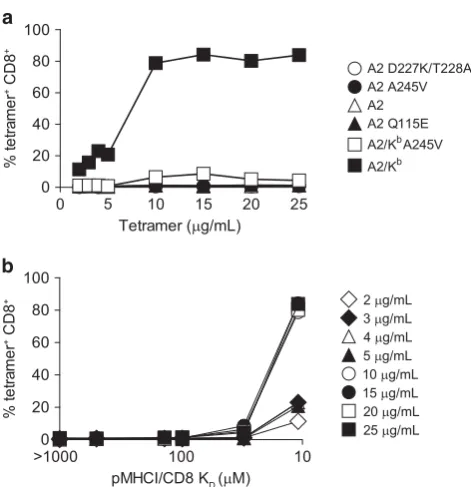
To consolidate thesefindings, we performed analogous experiments across a broader range of tetramer concentrations using PBMCs from a different A2+ donor (Figure 5a). Again, no loss of specificity was detected with the A2 D227K/T228A, A2 A245V, A2 or A2 Q115E
[image:6.595.149.470.207.706.2]tetramers up to a concentration of 25μg ml−1. The A2/Kb A245V tetramer was also highly specific at p5μg ml 1, but modest reactivity was observed with the same reagent at45μg ml 1. Considerable background staining was apparent with the A2/Kb tetramer. To clarify these data, we plotted nonspecific staining as a function of tetramer concentration versus pMHCI/CD8 affinity (Figure 5b) and used non-parametric tests to examine the impact of these variables on tetramer binding at the cell surface (Figure 6). Our analyses revealed that loss of tetramer specificity does not occur gradually with incremental increases in the strength of the pMHCI/ CD8 interaction. Instead, the specificity of pMHCI engagement is
Figure 4 pMHCI binding specificity is compromised at a defined pMHCI/CD8 affinity threshold in A2+donors. 1 × 10⁶A2+PBMCs were stained and analyzed
as described in the legend for Figure 3. Values shown in the upper right quadrant indicate % tetramer+CD8+T cells.
compromised beyond a certain pMHCI/CD8 affinity threshold, epitomized by the A2/KbA245V (K
D~ 27μM) and A2/Kb(KD~ 11μM) tetramers.
T-cell activation specificity is compromised at a defined pMHCI/ CD8 affinity threshold
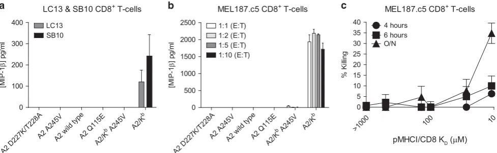
CD8+T-cell activation is exquisitely sensitive, requiringo10 pMHCI molecules for full calcium release and mature synapse formation.35As a consequence, effector functions can be elicited at cognate pMHCI concentrations well below those necessary for detectable tetramer binding.36To determine the pMHCI/CD8 affinity at which activation specificity is lost, we used a panel of Hmy.2 C1R (C1R) B cells transduced to express A2 D227K/T228A, A2 A245V, A2, A2 Q115E, A2/Kb A245V or A2/Kb at equivalent surface densities. Nonspecific activation as a function of pMHCI/CD8 affinity was initially tested using the LC13 and SB10 CD8+ T-cell clones, which are neither restricted by nor alloreactive against A2.37,38 After overnight stimulation, nonspecific macrophage inflammatory protein-1brelease was only observed in the presence of A2/KbC1R B cells (Figure 7a). Similar results were obtained with the A2-restricted CD8+T-cell clone MEL187.c5 (Figure 7b).
To confirm these findings with a different effector read-out, we used the same panel of C1R B cells in standard chromium release assays with the MEL187.c5 CD8+ T-cell clone to measure peptide-independent cytotoxicity (Figure 7c). The A2 D227K/T228A, A2 A245V, A2 and A2 Q115E C1R B-cell targets remained largely intact throughout the experiment. Similarly, there was no detectable short-term killing of A2/KbA245V C1R B cells. Marginal nonspecific lysis was apparent with the same targets after prolonged incubation, however, consistent with a subtle time-dependent effect triggering the release of cytolytic enzymes. The A2/KbC1R B-cell targets were killed in substantial numbers over time. Collectively, these data mirror the
corresponding tetramer staining patterns and indicate that CD8+ T-cell activation specificity is maintained below a defined pMHCI/ CD8 affinity threshold (KD~ 27μM).
DISCUSSION
Despite an extremely weak interaction with MHCI (average KD~ 145μM), the CD8 co-receptor mediates profound biological effects that enhance the sensitivity of TCR-driven activation in response to cognate antigen.12,39A small increment in pMHCI/CD8 affinity can further amplify the functional consequences of this interaction, increasing antigen sensitivity in responding CD8+T cells by up to 100-fold.26These observations suggest a possible translational role for affinity-enhanced CD8 molecules.27 For example, the introduction of such modified co-receptors together with tumour-specific TCRs may facilitate the activation of engineered T cells in the presence of naturally expressed cancer antigens, compensating both for low-affinity TCR/pMHCI interactions and low-density cognate pMHCI expression on the target cell surface. However, excessive increases in the strength of the pMHCI/CD8 interaction (KD~ 11μM) lead to nonspecific T-cell activation.28 It is therefore important to define the optimal affinity at which CD8 co-receptor engagement enhances pMHCI recognition without compromising the specificity of antigen-specific CD8+T cells.
In this study, we used a panel of MHCI molecules spanning a range of CD8-binding affinities to delineate the impact of variable pMHCI/ CD8 interactions on the specificity of TCR-mediated antigen recognition. Surface plasmon resonance studies confirmed that none of these mutations affect the TCR/pMHCI-binding platform. Tetrameric pMHCI complex engagement at the cell surface was enhanced in a stepwise manner with increasing pMHCI/CD8 affinities. In contrast, the specificity of pMHCI binding and T-cell activation was compromised at a defined pMHCI/CD8 affinity threshold (KD~ 27μM).
[image:7.595.41.277.63.307.2]Biophysical studies have shown that the murine pMHCI/CD8 interaction (average KD~ 49μM) is considerably stronger than the
Figure 5Detailed analysis of pMHCI binding specificity across a range of pMHCI/CD8 affinities in an A2+donor. (a) 1 × 10⁶A2+PBMCs were stained
and analyzed as described in the legend for Figure 3, with the exception that each tetramer was used at 2, 3, 4, 5, 10, 15, 20 or 25μg ml−1. (b)
The same data shown as % tetramer+ CD8+ T cells versus pMHCI/CD8
affinity.
>10000 1000
100 10
10
5 15 20 25 0
100
50
% tetramer
+ CD8
+
Tetramer (µg/mL)
pMHCI/CD8 K
D (µM)
Figure 6pMHCI binding specificity is a function of tetramer concentration and pMHCI/CD8 affinity. The percentage of tetramer+ CD8+T cells varies
with tetramer concentration (P=4.4 × 10⁻³; Friedman test). Modest to strong
evidence was found for individual MHCI mutants (A2 D227K/T228A:
P=1.6 × 10⁻²; A2 A245V: P=1.4 × 10⁻¹; A2: P=1.4 × 10⁻¹; A2 Q115E: P=1 × 10⁻²; A2/Kb A245V: P=5.4 × 10⁻²; A2/Kb: P=8.8 × 10⁻⁴;
Jonckheere–Terpstra test for increasing dependence on tetramer concentration). There was strong evidence for an effect of pMHCI/CD8 affinity on tetramer staining (P=3 × 10⁻⁷; Friedman test), although this was
not apparent when data for the two lowest KD values were excluded
(P=1.7 × 10⁻¹; Friedman test). Tetramer staining was strongly dependent on
theKDof the pMHCI/CD8 interaction (Po10–7; Jonckheere–Terpstra test for
increasing dependence on KD). The virtual absence of staining at pMHCI/
CD8 affinities427μMsuggests that a value within this order of magnitude behaves as a threshold.
The pMHCI/CD8 interaction controls T-cell specificity T Dockreeet al
73
[image:7.595.302.536.71.189.2]human pMHCI/CD8 interaction (average KD~ 145μM).21,25 This peculiar feature of mice may act to enhance T-cell cross-reactivity, allowing a size-limited repertoire to provide effective coverage against a common universe of pMHCI antigens.40It is also notable that the affinity of the murine pMHCI/CD8 interaction lies just below the specificity threshold defined in this study (KD~ 27μM). A conserved optimum may therefore dictate the evolutionary limits of co-receptor binding within a functional mammalian immune system.
The data presented here suggest the existence of an affinity window that potentially enables optimization of the pMHCI/CD8 interaction for therapeutic purposes without nonspecific T-cell activation. However, it is important to note that CD8+ T cells are naturally cross-reactive and that this phenomenon is controlled to some extent by the CD8 co-receptor.41–43It will therefore be important to examine this effect in more detail to avoid potentially dangerous off-target reactivity.44,45Nonetheless, the maintenance of CD8+T-cell specificity below a supranormal pMHCI/CD8 affinity threshold offers an exciting opportunity to enhance the therapeutic efficacy of adoptive cell transfer irrespective of antigen specificity.
METHODS
Cells
The following CD8+T-cell clones were used in this study: (i) MEL2 and
MEL187.c5, specific for the Melan-A-derived epitope ELAGIGILTV (residues
26–35) restricted by HLA A*0201 (A2); (ii) LC13, specific for the Epstein–Barr
virus EBNA3A-derived epitope FLRGRAYGL (residues 339–347) restricted by
HLA B*0801;37and (iii) SB10, specific for the cytomegalovirus pp65-derived
epitope CPSQEPMSIYVY (residues 103–114) restricted by HLA B*3508.38
Clones were maintained in RPMI 1640 containing 100 U ml−1 penicillin,
100 mg ml−1 streptomycin, 2 mM L-glutamine and 10% heat-inactivated
fetal calf serum (R10; all components from Life Technologies, Carlsbad, CA, USA), supplemented with 2.5% Cellkines (Helvetica Healthcare, Geneva,
Switzerland), 200 IU ml−1 interleukin-2 and 25 ng ml−1 interleukin-15
(both PeproTech, Rocky Hill, NJ, USA). Healthy donor PBMCs were isolated by standard density gradient centrifugation using Ficoll-Hypaque (GE Health-care, Chicago, IL, USA). C1R B cells expressing full-length A2 and variants
thereof were generated and maintained as described previously.26
pMHCI tetramer staining andflow cytometry
Soluble pMHCI tetramers were produced as described previously.17For A2
typing, 1 × 10⁶ PBMCs were stained withαA2-FITC (clone BB7.2; Serotec,
Oxford, UK) for 30 min at 4 °C. For pMHCI tetramer staining, 1 × 10⁶PBMCs
were resuspended in phosphate-buffered saline and stained with LIVE/DEAD Fixable Violet (ViViD; Life Technologies) for 5 min at room temperature. After washing in phosphate-buffered saline, cells were stained with tetramer-PE (A2 wild type and variants thereof) at the indicated concentrations for 20 min at 37 °C. The following mouse anti-human monoclonal antibodies were then
added for 20 min at 4 °C:αCD3-PerCP (clone SK7; BioLegend, San Diego, CA,
USA); αCD4-FITC (clone VIT4; Miltenyi Biotec, Bergisch Gladbach,
Ger-many); αCD8-APC (clone HIT8a; BD Pharmingen, San Diego, CA, USA);
αCD14-Pacific Blue (clone HCD14; BioLegend); and αCD19-Pacific Blue
(clone HIB19; BioLegend). Cells were washed twice in phosphate-buffered
saline after staining and 5 × 10⁴events per condition were acquired using a
FACSCantoIIflow cytometer (BD Biosciences, San Jose, CA, USA). Data were
analyzed with FlowJo software version 10.6 (TreeStar Inc., Ashland, OR, USA).
Macrophage inflammatory protein-1benzyme-linked immunosorbent assay
Clonal CD8+T-cells were incubated with C1R B cells expressing full-length A2
or variants thereof at different effector-to-target (E:T) ratios as indicated.
Supernatants were collected after 18 h and assayed for macrophage infl
amma-tory protein-1b by enzyme-linked immunosorbent assay according to the
manufacturer’s instructions (R&D Systems, Minneapolis, MN, USA).
Chromium release assay
Target C1R B cells (1 × 10⁶) were loaded with51Cr (30μCi) for 1 h and plated
in triplicate at 2 × 10³cells per well in R10. Clonal CD8+T cells were then
applied at an E:T ratio of 5:1 in afinal volume of 150μl. Target cells incubated alone were used to calculate spontaneous release. Total release was measured via the addition of Triton X-100 (Sigma-Aldrich, St Louis, MO, USA). Supernatants were collected after 4, 6 or 18 h at 37 °C and mixed with
OptiPhase Supermix Scintillation Cocktail (150μl per well; PerkinElmer Life
Sciences, Waltham, MA, USA).51Cr content was measured using a MicroBeta
Counter (PerkinElmer Life Sciences). Specific lysis (%) was calculated
accord-ing to the followaccord-ing formula: (experimental release−spontaneous release/total
[image:8.595.62.551.68.219.2]release−spontaneous release) × 100.
Figure 7 CD8+T-cell activation specificity is compromised at a defined pMHCI/CD8 affinity threshold. (a) 3 × 10⁴clonal SB10 or LC13 CD8+T cells were
incubated overnight with 6 × 10⁴C1R B cells expressing A2 D227K/T228A, A2 A245V, A2, A2 Q115E, A2/KbA245V or A2/Kb. Supernatants were collected
and assayed for macrophage inflammatory protein (MIP)-1b by enzyme-linked immunosorbent assay (ELISA). Data are shown corrected for background production of MIP-1b. (b) 3 × 10⁴clonal MEL187.c5 CD8+T cells were incubated overnight at the indicated E:T ratios with C1R B cells expressing A2
D227K/T228A, A2 A245V, A2, A2 Q115E, A2/KbA245V or A2/Kb. Supernatants were collected and assayed for macrophage inflammatory protein (MIP)-1b
by ELISA. Data are shown corrected for background production of MIP-1β. (c) 1 × 10⁴clonal MEL187.c5 CD8+T cells were incubated with 2 × 10³C1R B
cells expressing A2 D227K/T228A, A2 A245V, A2, A2 Q115E, A2/KbA245V or A2/Kbin standard chromium release assays as described in the Methods
Surface plasmon resonance
Soluble TCRs and CD8ααwere produced as described previously.22,46Binding
analysis was performed using a BIAcore 3000 (GE Healthcare) equipped with a CM5 sensor chip. Between 200 and 400 response units of biotinylated pMHCI were immobilized to streptavidin, which was chemically linked to the chip
surface. The pMHCI was injected at a slowflow rate (10μl min−1) to ensure
uniform distribution on the chip surface. Combined with the small amount of pMHCI bound to the chip surface, this reduced the likelihood of off-rate
limiting mass transfer effects. Soluble MEL5 TCR and CD8ααwere purified
and concentrated to 100 and 150μM, respectively, on the day of analysis to
reduce the likelihood of aggregation affecting the results. For equilibrium analysis, eight serial dilutions of analyte were carefully prepared in triplicate for each sample and injected over the relevant sensor chips at 25 °C. Soluble MEL5
TCR or CD8αα were injected over the chip surface at a flow rate of
30μl min−1. Results were analyzed using BIAevaluation 3.1 (GE Healthcare),
Microsoft Excel (Microsoft, Redmond, WA, USA) and Origin 6.1 (OriginLab,
Northampton, MA, USA). The equilibrium binding constant (KD) values were
calculated using a nonlinear curvefit (y=[P1x]/[P2+x]).
Statistical analysis
The dependence of nonspecific CD8+ T-cell staining intensity on tetramer
concentration and theKDof the pMHCI/CD8 interaction was assessed using
the Friedman test for one-way effects and the Jonckheere–Terpstra test for the
dependent variable increasing with the treatment variable.47
CONFLICT OF INTEREST
The authors declare no conflict of interest.
ACKNOWLEDGEMENTS
We thank Dr Anya Lissina for helpful discussions. TD is supported by a Wellcome Trust Research Training Fellowship (WT099067AIA). DAP is supported by a Wellcome Trust Senior Investigator Award (100326/Z/12/Z). LW was supported by a Wellcome Trust Intermediate Clinical Fellowship (WT079848MA). Additional funding was provided by a Wellcome Trust Entry Level Fellowship awarded to TD (WT096454AIA).
1 Rossjohn J, Gras S, Miles JJ, Turner SJ, Godfrey DI, McCluskey J. T cell antigen receptor recognition of antigen-presenting molecules.Annu Rev Immunol2015;33: 169–200.
2 Koebel CM, Vermi W, Swann JB, Zerafa N, Rodig SJ, Old LJ et al. Adaptive immunity maintains occult cancer in an equilibrium state. Nature 2007; 450: 903–907.
3 Boon T, van der Bruggen P. Human tumor antigens recognized by T lymphocytes. J Exp Med1996;183: 725–729.
4 Dudley ME, Wunderlich JR, Robbins PF, Yang JC, Hwu P, Schwartzentruber DJet al. Cancer regression and autoimmunity in patients after clonal repopulation with antitumor lymphocytes.Science2002;298: 850–854.
5 Rosenberg SA, Restifo NP, Yang JC, Morgan RA, Dudley ME. Adoptive cell transfer: a clinical path to effective cancer immunotherapy. Nat Rev Cancer 2008; 8: 299–308.
6 Dudley ME, Wunderlich JR, Shelton TE, Even J, Rosenberg SA. Generation of tumor-infiltrating lymphocyte cultures for use in adoptive transfer therapy for melanoma patients.J Immunother2003;26: 332–342.
7 Morris EC, Bendle GM, Stauss HJ. Prospects for immunotherapy of malignant disease. Clin Exp Immunol2003;131: 1–7.
8 Cole DK, Pumphrey NJ, Boulter JM, Sami M, Bell JI, Gostick Eet al.Human TCR-binding affinity is governed by MHC class restriction. J Immunol 2007; 178: 5727–5734.
9 Bridgeman JS, Sewell AK, Miles JJ, Price DA, Cole DK. Structural and biophysical determinants of ab T-cell antigen recognition. Immunology 2012; 135: 9–18.
10 Li Y, Moysey R, Molloy PE, Vuidepot AL, Mahon T, Baston Eet al.Directed evolution of human T-cell receptors with picomolar affinities by phage display.Nat Biotechnol 2005;23: 349–354.
11 Liddy N, Bossi G, Adams KJ, Lissina A, Mahon TM, Hassan NJet al.Monoclonal TCR-redirected tumor cell killing.Nat Med2012;18: 980–987.
12 Zamoyska R. CD4 and CD8: modulators of T-cell receptor recognition of antigen and of immune responses?Curr Opin Immunol1998;10: 82–87.
13 Dembic Z, Haas W, Zamoyska R, Parnes J, Steinmetz M, von Boehmer H. Transfection of the CD8 gene enhances T-cell recognition. Nature 1987; 326: 510–511.
14 Zamoyska R. The CD8 coreceptor revisited: one chain good, two chains better. Immunity1994;1: 243–246.
15 Luescher IF, Vivier E, Layer A, Mahiou J, Godeau F, Malissen Bet al.CD8 modulation of T-cell antigen receptor-ligand interactions on living cytotoxic T lymphocytes. Nature1995;373: 353–356.
16 Gakamsky DM, Luescher IF, Pramanik A, Kopito RB, Lemonnier F, Vogel Het al.CD8 kinetically promotes ligand binding to the T-cell antigen receptor.Biophys J2005;89: 2121–2133.
17 Wooldridge L, van den Berg HA, Glick M, Gostick E, Laugel B, Hutchinson SLet al. Interaction between the CD8 coreceptor and major histocompatibility complex class I stabilizes T cell receptor-antigen complexes at the cell surface.J Biol Chem2005;280: 27491–27501.
18 van den Berg HA, Wooldridge L, Laugel B, Sewell AK. Coreceptor CD8-driven modulation of T cell antigen receptor specificity. J Theor Biol 2007; 249: 395–408.
19 Veillette A, Bookman MA, Horak EM, Bolen JB. The CD4 and CD8 T cell surface antigens are associated with the internal membrane tyrosine-protein kinase p56lck. Cell1988;55: 301–308.
20 Barber EK, Dasgupta JD, Schlossman SF, Trevillyan JM, Rudd CE. The CD4 and CD8 antigens are coupled to a protein-tyrosine kinase (p56lck) that phosphorylates the CD3 complex.Proc Natl Acad Sci USA1989;86: 3277–3281.
21 Purbhoo MA, Boulter JM, Price DA, Vuidepot AL, Hourigan CS, Dunbar PRet al.The human CD8 coreceptor effects cytotoxic T cell activation and antigen sensitivity primarily by mediating complete phosphorylation of the T cell receptorz chain. J Biol Chem2001;276: 32786–32792.
22 Hutchinson SL, Wooldridge L, Tafuro S, Laugel B, Glick M, Boulter JMet al.The CD8 T cell coreceptor exhibits disproportionate biological activity at extremely low binding affinities.J Biol Chem2003;278: 24285–24293.
23 Arcaro A, Gregoire C, Boucheron N, Stotz S, Palmer E, Malissen Bet al.Essential role of CD8 palmitoylation in CD8 coreceptor function. J Immunol 2000; 165: 2068–2076.
24 Arcaro A, Gregoire C, Bakker TR, Baldi L, Jordan M, Goffin L et al. CD8b endows CD8 with efficient coreceptor function by coupling T cell receptor/CD3 to raft-associated CD8/p56(lck) complexes. J Exp Med 2001; 194: 1485–1495.
25 Cole DK, Laugel B, Clement M, Price DA, Wooldridge L, Sewell AK. The molecular determinants of CD8 co-receptor function. Immunology 2012; 137: 139–148.
26 Wooldridge L, Lissina A, Vernazza J, Gostick E, Laugel B, Hutchinson SLet al. Enhanced immunogenicity of CTL antigens through mutation of the CD8 binding MHC class I invariant region.Eur J Immunol2007;37: 1323–1333.
27 Devine L, Thakral D, Nag S, Dobbins J, Hodsdon ME, Kavathas PB. Mapping the binding site on CD8b for MHC class I reveals mutants with enhanced binding. J Immunol2006;177: 3930–3938.
28 Wooldridge L, Clement M, Lissina A, Edwards ES, Ladell K, Ekeruche Jet al.MHC class I molecules with superenhanced CD8 binding properties bypass the requirement for cognate TCR recognition and nonspecifically activate CTLs.J Immunol2010;184: 3357–3366.
29 Wyer JR, Willcox BE, Gao GF, Gerth UC, Davis SJ, Bell JIet al.T cell receptor and coreceptor CD8aa bind peptide-MHC independently and with distinct kinetics. Immunity1999;10: 219–225.
30 Gao GF, Jakobsen BK. Molecular interactions of coreceptor CD8 and MHC class I: the molecular basis for functional coordination with the T-cell receptor.Immunol Today 2000;21: 630–636.
31 Gao GF, Rao Z, Bell JI. Molecular coordination ofabT-cell receptors and coreceptors CD8 and CD4 in their recognition of peptide-MHC ligands.Trends Immunol2002;23: 408–413.
32 Gao GF, Willcox BE, Wyer JR, Boulter JM, O'Callaghan CA, Maenaka Ket al.Classical and nonclassical class I major histocompatibility complex molecules exhibit subtle conformational differences that affect binding to CD8aa.J Biol Chem2000;275: 15232–15238.
33 Altman JD, Moss PA, Goulder PJ, Barouch DH, McHeyzer-Williams MG, Bell JIet al. Phenotypic analysis of antigen-specific T lymphocytes.Science1996;274: 94–96. 34 Burrows SR, Kienzle N, Winterhalter A, Bharadwaj M, Altman JD, Brooks A.
Peptide-MHC class I tetrameric complexes display exquisite ligand specificity. J Immunol2000;165: 6229–6234.
35 Purbhoo MA, Irvine DJ, Huppa JB, Davis MM. T cell killing does not require the formation of a stable mature immunological synapse.Nat Immunol2004;5: 524–530.
36 Wooldridge L, Lissina A, Cole DK, van den Berg HA, Price DA, Sewell AK. Tricks with tetramers: how to get the most from multimeric peptide-MHC.Immunology2009;126: 147–164.
37 Argaet VP, Schmidt CW, Burrows SR, Silins SL, Kurilla MG, Doolan DLet al.Dominant selection of an invariant T cell antigen receptor in response to persistent infection by Epstein-Barr virus.J Exp Med1994;180: 2335–2340.
38 Wynn KK, Fulton Z, Cooper L, Silins SL, Gras S, Archbold JKet al.Impact of clonal competition for peptide-MHC complexes on the CD8+T-cell repertoire selection in a persistent viral infection.Blood2008;111: 4283–4292.
39 Holler PD, Kranz DM. Quantitative analysis of the contribution of TCR/pepMHC affinity and CD8 to T cell activation.Immunity2003;18: 255–264.
The pMHCI/CD8 interaction controls T-cell specificity T Dockreeet al
75
40 Mason D. A very high level of crossreactivity is an essential feature of the T-cell receptor. Immunol Today1998;19: 395–404.
41 Wooldridge L, Laugel B, Ekeruche J, Clement M, van den Berg HA, Price DAet al.CD8 controls T cell cross-reactivity.J Immunol2010;185: 4625–4632.
42 Wooldridge L, Ekeruche-Makinde J, van den Berg HA, Skowera A, Miles JJ, Tan MP et al.A single autoimmune T cell receptor recognizes more than a million different peptides.J Biol Chem2012;287: 1168–1177.
43 Wooldridge L. Individual MHCI-restricted T-cell receptors are characterized by a unique peptide recognition signature.Front Immunol2013;4: 199.
44 Linette GP, Stadtmauer EA, Maus MV, Rapoport AP, Levine BL, Emery L et al. Cardiovascular toxicity and titin cross-reactivity of affinity-enhanced T cells in myeloma and melanoma.Blood2013;122: 863–871.
45 Stauss HJ, Morris EC. Immunotherapy with gene-modified T cells: limiting side effects provides new challenges.Gene Ther2013;20: 1029–1032.
46 Cole DK, Yuan F, Rizkallah PJ, Miles JJ, Gostick E, Price DAet al.Germ line-governed recognition of a cancer epitope by an immunodominant human T-cell receptor. J Biol Chem2009;284: 27281–27289.
47 Sprent P, Smeeton NC. Applied Nonparametric Statistical Methods. Chapman & Hall/CRC: London, UK. 2007.
This work is licensed under a Creative Commons Attribution 4.0 International License. The images or other third party material in this article are included in the article’s Creative Commons license, unless indicated otherwise in the credit line; if the material is not included under the Creative Commons license, users will need to obtain permission from the license holder to reproduce the material. To view a copy of this license, visit http:// creativecommons.org/licenses/by/4.0/