warwick.ac.uk/lib-publications
Manuscript version: Author’s Accepted ManuscriptThe version presented in WRAP is the author’s accepted manuscript and may differ from the published version or Version of Record.
Persistent WRAP URL:
http://wrap.warwick.ac.uk/111068 How to cite:
Please refer to published version for the most recent bibliographic citation information. If a published version is known of, the repository item page linked to above, will contain details on accessing it.
Copyright and reuse:
The Warwick Research Archive Portal (WRAP) makes this work by researchers of the University of Warwick available open access under the following conditions.
Copyright © and all moral rights to the version of the paper presented here belong to the individual author(s) and/or other copyright owners. To the extent reasonable and
practicable the material made available in WRAP has been checked for eligibility before being made available.
Copies of full items can be used for personal research or study, educational, or not-for-profit purposes without prior permission or charge. Provided that the authors, title and full
bibliographic details are credited, a hyperlink and/or URL is given for the original metadata page and the content is not changed in any way.
Publisher’s statement:
Please refer to the repository item page, publisher’s statement section, for further information.
Head growth and intelligence from birth to adulthood in very preterm and term born
individuals
Authors: Julia Jaekel 1,2; Christian Sorg 3,4,5; Josef Baeuml 3,4,5; Peter Bartmann 6; Dieter Wolke 2,7
Affiliations: 1Department of Child and Family Studies, University of Tennessee, Knoxville, TN,
United States; 2Department of Psychology, University of Warwick, Coventry, United Kingdom;
3TUM-Neuroimaging Center and Departments of 4Neuroradiology and 5Psychiatry of Klinikum
rechts der Isar, Technische Universität München TUM, Munich, Germany; 6Department of
Neonatology, University Hospital Bonn, Bonn, Germany; 7Division of Mental Health and
Wellbeing, Warwick Medical School, University of Warwick, Coventry, United Kingdom
Corresponding author: Dieter Wolke, Department of Psychology, University of Warwick,
Coventry CV4 7AL, United Kingdom, work phone: +44 2476573217, fax: +44 2476524225,
D.Wolke@warwick.ac.uk
Total word count: 3,426
Abstract
Objective: To investigate the effects of infant and toddler head growth on intelligence scores
from early childhood to adulthood in very preterm (<32 weeks gestational age; VP) and/or very
low birth weight (<1500g; VLBW) and term born individuals.
Method: 203 VP/VLBW and 198 term comparisons were studied from birth to adulthood as part
of the prospective geographically defined Bavarian Longitudinal Study (BLS). Head
circumference was assessed at birth, 5, 20 months, and 4 years of age. Intelligence was assessed
with standardized tests in childhood (6 and 8 years: K-ABC) and at 26 years (Wechsler Adult
Intelligence Scale, WAIS). Structural equation modeling (SEM) was used to model the effect of
head growth on IQ.
Results: On average, VP/VLBW had lower head circumference at birth (27.61cm versus 35.11cm,
mean difference 7.49, 95% CI [7.09 – 7.90]) and lower adult intelligence scores (88.98 versus
102.54, mean difference 13.56 [10.59 - 16.53]) than term born comparison individuals. Head
circumference at birth (e.g., total effect β=.48, p<.001 for adult IQ) and head growth in
childhood predicted intelligence development from age 6 to 26 years in both VP/VLBW and
term born individuals (70% of variance in adult IQ explained by full model). Effects of gestation
and birth weight on intelligence were fully mediated by head circumference and growth.
Conclusions: This longitudinal investigation from birth to adulthood indicates head growth as a
proxy of brain development and intelligence. Repeated early head circumference assessment
adds valuable information when screening for long-term neurocognitive risk.
Key words: early childhood; gestational age; prospective longitudinal study; brain development;
Introduction
Very preterm (<32 weeks gestational age; VP) and very low birth weight (<1500g, VLBW) infants
grow up with life-long risks for neurocognitive impairment (Breeman, Jaekel, Baumann,
Bartmann, & Wolke, 2015; Eryigit Madzwamuse, Baumann, Jaekel, Bartmann, & Wolke, 2015).
Early identification allows timely provision of resources. Follow-up after VP/VLBW birth is a
general standard in many Western countries (Doyle et al., 2014). However, assessing cognitive
development through descriptive behavioral data is time consuming and expensive (Aslin &
Fiser; Hack et al., 2005). Repeated head circumference assessments may be a cost-effective
indicator of brain development (Bartholomeusz, Courchesne, & Karns, 2002; Cheong et al.,
2008; Garcia-Alix, Saenz-de Pipaon, Martinez, Salas-Hernandez, & Quero, 2004). Regional brain
and cortical growth is significantly associated with brain maturation (Makropoulos et al., 2016),
generally indicating that size matters.
Head circumference is measured in infancy in most countries, but its predictive validity remains
controversial (Wright & Emond, 2015). At term, preterm infants have smaller whole brain
volumes than term born infants (Ball et al., 2012; Makropoulos et al., 2016). Researchers argue
that head growth between birth and 2 years is critical for intellectual development (Räikkönen
et al., 2009) and studies of preterm populations have documented the value of head growth as
a predictor of long-term neurocognitive abilities (Sammallahti et al., 2017; Sammallahti et al.,
2014). Accelerated postnatal head growth suggests catch up after prenatal restraint (Cockerill,
Uthaya, Doré, & Modi, 2006), but may not compensate for poor earlier growth after infancy
(Gale, O'Callaghan, Bredow, & Martyn, 2006). Thus, it is important to investigate whether there
In clinical practice, individual head growth is usually documented by plotting raw head
circumference values on age-standardized growth chart curves, by converting raw values into
standard scores, or by categorizing scores (Fenton & Kim, 2013). This is done to (clinically)
identify individuals who grow at a substantially slower than average rate. Instead, growth over
time assessed with latent class growth curve analyses to identify how the growth rate across all
individuals (i.e., head growth) affects growth in another dimension (i.e., IQ). Structural equation
modeling (SEM) has been proposed as the most appropriate in the context of additional factors
(e.g., gestational age, birth weight) (Usami, Hayes, & McArdle, 2017).
Our aims were (1) to investigate VP/VLBW and term born individuals’ head growth from birth to
4 years and intelligence in childhood and adulthood, and (2) to determine the specific timing of
head growth that matters for intelligence development.
Method
Design
Participants were all VP and/or VLBW infants and an equally sized group of healthy term
comparisons born in a geographically defined area of South Bavaria (Germany) between January
1985 and March 1986 as part of the prospective whole population Bavarian Longitudinal Study
(BLS). The current study uses data collected at birth, term and at 5 and 20 months, and at 4, 6,
8, and 26 years of age.
Standard Protocol Approvals and Patient Consents. This research was completed in accordance
with the Helsinki Declaration. Original ethical approval was obtained from the University of
Munich Children’s Hospital and the Landesärztekammer Bayern. Ethical approval for the adult
Informed written consent was provided by parents within 48 hours of their child’s birth and all
participants gave fully informed written consent for the adult assessment.
Participants. This study assesses a whole population sample of 682 VP/VLBW individuals (Wolke
& Meyer, 1999). Of this cohort, 411 VP/VLBW were presumed alive, living in Germany, and
eligible for inclusion at 26 years of age, and 260 (63.3%) participated in the adult assessment
(see Appendix Figure 1). The BLS VP/VLBW participants did not differ from VP/VLBW adults who
dropped out in terms of GA, BW, duration of hospitalization, gender, maternal age, parental
marital status, and childhood cognitive scores, but had fewer prenatal complications and were
of higher socioeconomic status (SES) (Eryigit Madzwamuse et al., 2015).
Of 916 healthy term-born comparison infants at birth, 350 were selected and stratified to match
the VP/VLBW participants at the 6 years follow-up assessment. Of these, 308 individuals were
eligible for inclusion and 229 (74.4%) participated at 26 years (Eryigit Madzwamuse et al., 2015).
Only participants who had complete childhood and adulthood IQ data (203 VP/VLBW and 198
term comparisons) were included in the current analyses.
Head Circumference. Head circumference (cm) was assessed by trained research nurses during
clinical assessments at birth, 5, and 20 months, and at 4, 6, and 8 years of age. At 5 and 20
months, examination ages were corrected for gestational age at birth. HC was measured twice
at each assessment and the average score was recorded. Raw HC scores and standardized
scores are reported in Table 1. Raw scores are used in main analyses in order to investigate
separate contributions of gestation and birth weight in our statistical models.
Biological and Demographic Characteristics. Gestational age (weeks, range: VP/VLBW=25-36,
comparisons=2120-5050g) were coded from Bavarian perinatal survey forms at birth (Zander,
Holzmann, & Selbmann, 1989). Family SES at birth was assessed as a weighted composite score
of parents’ education and occupation (Bauer, 1988).
Neurocognitive Assessments. Cognitive development (i.e., intelligence) was assessed
longitudinally by psychologists with standardized tests in childhood and adulthood (Breeman et
al., 2015). At age 20 months, children were administered the Griffiths’ Mental Development
ScalesDevelopmental Quotient (DQ) items (Brandt, 1983). At 6 and 8 years, IQ was assessed
with the German version of the Kaufman Assessment Battery for Children (K-ABC) (Melchers &
Preuss, 1991). Reliability and construct validity of the K-ABC are high (e.g., 0.70 correlation with
the Wechsler Intelligence Scale for Children-Revised (WISC-R) total score) (Melchers & Preuss,
1991). At 26 years, age-normed Full-Scale IQ was assessed with a short German version of the
Wechsler Adult Intelligence Scale (WAIS III) (Von Aster, Neubauer, & Horn, 2006). In our models,
VP/VLBW individuals’ DQ and IQ data from different ages are directly compared to healthy term
born comparison individuals’ DQs and IQs at each assessment point.
Statistical Approach.Data were analyzed with SPSS 23 and Amos 24 (Armonk, NY: IBM Corp.).
Structural Equation Modeling (SEM) was used to simultaneously test direct and indirect
associations between head circumference and growth, and intelligence to 26 years of age.
Gestational age, birth weight, and family SES were included as additional predictors of interest.
In the initial, full model, all variables and the potential paths between each measure were
included (see Appendix Figure 2). Model fit values were then used to identify the best fitting
intelligence development. Final model results are presented combined for VP/VLBW and term
comparisons (Model 1), and as separate multiple-group models (Models 2 and 3).
Results
On average, VP/VLBW had lower head circumference at birth, lower head growth from 20
months to 4 years, and lower intelligence scores than term born comparison individuals (Table
1). However, VP/VLBW had higher head growth from birth to 5 months and, on average, from
birth to 20 monthsthan term born comparison infants, both assessments were scheduled
corrected for gestational age.
- Table 1 and Figure 1 about here -
The initial, full model did not have sufficient fit (χ2=80.44(df=16), p<0.001, CFI=.983,
RMSEA=1.00 (90% CI=.079 to .123), PCLOSE=.000; Appendix Figure 2) and was thus reduced to
the best fitting, most parsimonious Model 1 (Figure 1). In particular, there was no added value
in assessing head growth from birth to five months and then from five to 20 months separately,
thus growth from birth to 20 months was collapsed into one variable. Moreover, cognitive
development at 20 months was less accurately predicted by infants’ head growth than IQ at 6+
years. Model 1 showed that head circumference at birth and head growth in childhood
predicted intelligence development from age 6 to 26 years in both VP/VLBW and term born
individuals. Effects of gestation and birth weight on intelligence were found to be fully mediated
by head circumference and growth (i.e., indirect effects) while family SES directly predicted
intelligence. Table 2 shows detailed direct, indirect, and total effects for all variables in the
70% at 26 years of age. Overall model fit was excellent with χ2=19.85(df=16), p<0.227, CFI=.999,
RMSEA=.025 (90% CI=.000-.055), PCLOSE=.907.
- Table 2 about here -
Since part of the explained variance in adult IQ was carried by childhood IQ (i.e., longitudinal
stability of IQ), we also explored how much variance was explained if only the isolated effects of
HC at birth and head growth were used to predict adult IQ. A substantial R2=.24 were explained
just by HC at birth and early head growth in the total sample (R2=.22 in VP/VLBW and R2=.02 in
healthy term control individuals, respectively).
To test if the associations between head growth and intelligence were different among
VP/VLBW compared with term comparison individuals, we ran the same SEM separately as a
multiple-group model. As before, head circumference at birth, head growth from birth to 20
months, and from 20 months to 4 years predicted intelligence development from age 6 to 26
years in VP/VLBW and term born individuals, but the path coefficients were slightly different
(Figures 2 and 3).
- Figures 2 and 3 about here -
Model 2 shows that among VP/VLBW, head circumference at birth and head growth in early
childhood directly predicted intelligence at age 6, while there were only indirect effects on
intelligence at 8 and 26 years. The total variance explained in intelligence was 22% at 6 years,
Model 3 shows that among term comparisons, only head growth from birth to 20 months
directly predicted intelligence at age 26. The total variance explained in term comparison
individuals’ intelligence was lower with 9% at 6 years, 53% at 8 years, and 53% at 26 years.
As before, model fit was excellent with χ2=19.78(df=16), p<0.230, CFI=.999, RMSEA=.024 (90%
CI=.000-.053), PCLOSE=.924; and effects of gestation and birth weight on intelligence were fully
mediated by head circumference and growth while family SES directly predicted intelligence.
Discussion
This longitudinal investigation from birth to adulthood confirms that early head growth as a
proxy of brain development predicts later intelligencein VP/VLBW but also in term born
individuals. Larger head circumference at birth and higher head growth during the first four
years predicted IQfrom age 6 to 26 years in both VP/VLBW and term born individuals while
effects of gestation and birth weight on intelligence were fully mediated by head circumference
and growth. These findings add to previous evidence about the value of repeated head
circumference assessments in early childhood, in particular in VP/VLBW children, as a proxy of
brain volume and screening for later intellectual impairments (Kapellou et al., 2006).
Head growth is driven by brain growth and thus an indicator of brain development (Kiesler &
Ricer, 2003). Researchers have suggested that the same genetic factors have an effect on
physical (i.e., head) growth and cognitive development (Silventoinen, Iacono, Krueger, &
McGue, 2012). However, postnatal brain growth is the product of complex mechanisms,
including gray matter maturation (i.e. axon and dendrite sprouting, synapse formation, cortical
gyrification) and white matter formation (i.e. glial cell proliferation, myelination). Anatomically,
by two years of age (Kiesler & Ricer, 2003). In healthy term born infants, cortical thickness
reaches 97% of adult size by age two (Lyall et al., 2015) and cortical gyrification also happens
during the first two years (Li et al., 2014), whereas cortical surface area expansion mainly drives
brain growth after two years of age (Lyall et al., 2015). Lower gestation at birth is associated
with smaller whole brain volume and growth at term-equivalent age (Kidokoro et al., 2014;
Padilla, Alexandrou, Blennow, Lagercrantz, & Ådén, 2015) and age three months (Holland,
Chang, Ernst, & et al., 2014). Additionally, premature birth is associated with regional brain
alterations, particularly in subcortical structures such as the thalamus and striatum as well in
lateral temporal and parietal cortices (Ball et al., 2012; Meng et al., 2016). These alterations
persist into adulthood and are not only accompanied by widespread changes in cerebral white
matter (Bäuml et al., 2014; Meng et al., 2016) but also associated with impairments in
neurocognitive and behavioral development (Kidokoro et al., 2014; Parker et al., 2008).
Head circumference is an excellent indicator of cerebral volume measured with MRI in one to
six year old children (r=0.59-0.93), but its predictive validity is weaker in typically developing
older children, adolescents, and adults (r=0.45 to 0.69) (Bartholomeusz et al., 2002; Lange,
Froimowitz, Bigler, & Lainhart, 2010). Accordingly, head circumference may present an easily
assessable proxy of overall brain volume, but only during the first years of life, and prediction
from HC measurement alone is similar to that of term MRI (Anderson et al., 2017).
With regard to the timing of head growth, SEM indicated that the period from birth to 20
months of age mattered most for directly predicting later IQ development, but indirect effects
of head growths from 20 months to 4 years of age were also relevant. This suggests that
for age-appropriate IQ development later in life, in particular for infants born VP/VLBW.
Moreover, and in accordance with studies of normative brain growth (Li et al., 2014; Lyall et al.,
2015), the significant direct effect of head growth from birth to 20 months on term adults’ IQs
suggests that the first two years of postnatal life may represent a critical window for healthy
brain growth and development. Thus, regular head circumference measurements, used in
conjunction with other neurodevelopmental tools, may be recommended until at least 2 years
of age in order to screen for potential brain growth delay. As a result, clinicians may need
additional training to correctly administer this effective and inexpensive screening tool (James,
Perszyk, MacGregor, & Aldana, 2015).
Children’s IQ development is influenced by genetic (Marioni et al., 2014) and environmental
factors, such as parents’ socioeconomic status (Sameroff, Seifer, Barocas, Zax, & Greenspan,
1987), children’s home literacy environments (Jaekel, Schölmerich, Kassis, & Leyendecker,
2011), and preschool quality (Melhuish, 2011). Accordingly, environmental stimulation may
promote cognitive development of both VP/VLBW and full term children (Wolke, Jaekel, Hall, &
Baumann, 2013), thus early identification of those children who are at-risk for low IQ would
allow timely provision of resources and intervention. Our observational study results
substantiate the validity of studies that aim to improve preterm infants’ long-term cognitive
outcomes by supporting early head growth. Such strategies may target individualized neonatal
nutrition, adaptive feeding, and breastfeeding to stimulate growth and neurodevelopmental
outcomes (Belfort et al., 2016; Christmann et al., 2017). Recently, the microbiome-gut-brain axis
has also received attention due to its potential for neuroprotection against white matter injury
‘nutrition for the brain’ via environmental stimulation (e.g., the NICU environment (Pineda et
al., 2014), sensitive parenting (Milgrom et al., 2010; Wolke et al., 2013), preschool education
(McCormick et al., 2006), and family psychosocial support (Benzies, Magill-Evans, Hayden, &
Ballantyne, 2013)).
Our sample represents one of the largest whole-population longitudinal studies of
neurocognitive development after VP/VLBW birth. In total, 68% of the eligible VP/VLBW and
term comparisons recruited at birth were assessed at 26 years, however, the dropout was not
random, as low SES families were less likely to continue participation. Social factors are a major
reason for dropout in most longitudinal studies (Hille, Elbertse, Gravenhorst, Brand, &
Verloove-Vanhorick, 2005) and analyses were controlled for SES at birth. Participants were born in
Germany in 1985/1986, before the introduction of pioneering new treatments such as
surfactant administration, which significantly improved high risk infants’ survival rates.
However, studies have shown that increased survival may not result in equivalent improvement
of long-term neurocognitive outcomes (Cheong et al., 2017; Moore et al., 2012; Wolke et al.,
2015), thus the current findings may be as relevant to infants born today as to those born 30
years ago.
Changes of absolute measurements of head circumference according to postnatal age (adjusted
for gestation at birth at the 5 and 20 months assessments) were included in our models. From a
clinical perspective, it might be preferable to use standardized z-scores, however, our aim was
to investigate the effect of head growth on intelligence growth, while simultaneously assessing
the effects of gestational age and birth weight on these growth trajectories. Our results show
and term individuals, but gestational age and birth weight are important factors to predict these
trajectories. Standardized head circumference scores would control for gestational age but take
away the variation in IQ development explained by this important factor, thus we used raw
values to model head growth over time, while timing of assessments at 5 and 20 months was
adjusted for gestational age at birth as recommended in clinical practice (Wilson-Ching, Pascoe,
Doyle, & Anderson, 2014).
We were able to explain >70% of the variation in adult intelligence, however the majority of this
was due to the longitudinal stability of IQ prediction from 6 years of age, not head
circumference directly. Nevertheless, a considerable 24% of variance was explained in adult IQ if
only the isolated effects of HC at birth and head growth were included in the model (however
this percentage of variance explained was mainly due to the VP/VLBW individuals in the
sample). Intelligence is a multidimensional construct that develops through cascades (Bornstein,
Hahn, & Wolke, 2013). This is reflected in our models, with early development directly and
indirectly affecting what comes later, i.e., long-term effects of early head growth as marker for
brain volume and function on adult IQ. Full scale IQs are relatively stable over time (Schneider,
Niklas, & Schmiedeler, 2014), but VP/VLBW children often have limited neurocognitive
resources (Jaekel, Baumann, & Wolke, 2013) and, as a result, their average low IQs remain
relatively stable from early childhood onwards. Indeed, moderate prediction of adult
intelligence is possible from toddler age in VP/VLBW or extremely preterm, but only from 6
years in healthy term comparison children (Breeman et al., 2015). This is reflected in our
multi-group models, as VP/VLBW individuals’ head circumference and growth had their strongest
neurocognitive trajectory (Breeman et al., 2015; Eryigit Madzwamuse et al., 2015). Similarly, the
effects of family SES at birth on later IQ were larger in the healthy term born group than in
VP/VLBW (e.g., .26 versus .06 at 6 years), indicating differences in developmental plasticity.
Family SES affects cognitive development via both genetic and environmental pathways, but
VP/VLBW may be more strongly affected by neonatal factors (Jaekel et al., 2013) that affect
brain and head growth. Intelligence is only one marker of cognitive function, and while
VP/VLBW individuals may achieve IQ test results within the average range, their daily
performance in school and at work may be affected by other cognitive problems such as
attention, executive function, or processing speed.
Finally, IQ assessments changed from childhood to adulthood but our instruments have been
shown to deliver reliable and consistent age-appropriate estimations of intelligence (Melchers &
Preuss, 1991). Moreover, potential variations in IQ estimates across different tests (e.g.,
Griffiths versus Bailey Scales) were not of concern due to the prospective inclusion of a matched
healthy term comparison group with the same measures taken at all assessment points.
Model fit values indicated that the developmental pathways included in this study accurately
reflect the true neurodevelopmental mechanisms in the two populations studied. Thus, our
results confirm previous evidence suggesting that repeated early head circumference
measurements are a valuable and easy screening tool for long-term neurocognitive risk
assessment after preterm birth, in particular in light of recent findings refuting the diagnostic
benefits of routine MRI on preterm infants (Edwards et al., 2018; Hintz et al., 2018). Considering
specific neurocognitive mechanisms, however,overall head circumference and brain volume are
other tools and a thorough review of known risk factors associated with cognitive impairment.
Additional variance in individual IQ development may be explained by regional rather than
global differences as well as by alterations in brain connectivity.
Acknowledgements
Drs. Jaekel, Sorg, Baeuml, Bartmann, and Wolke report no conflicts of interest. This study was
supported by the German Federal Ministry of Education and Science (BMBF 01ER0801; PKE24,
JUG14). DW and PB are supported by EU Horizon 2020 (733280; RECAP preterm). The contents
are solely the responsibility of the authors and do not necessarily represent the official view of
the BMBF. We would like to thank the pediatricians, psychologists, and research nurses who
carried out the assessments, and the researchers and administrative staff of the Bavarian
Longitudinal Study group who managed the data. We are deeply thankful to our participants for
References
Anderson, P. J., Treyvaud, K., Neil, J. J., Cheong, J. L. Y., Hunt, R. W., Thompson, D. K., . . . Inder, T. E. (2017). Associations of newborn brain magnetic resonance imaging with long-term
neurodevelopmental impairments in very preterm children. The Journal of Pediatrics, n/a, n/a. doi:https://doi.org/10.1016/j.jpeds.2017.04.059
Aslin, R. N., & Fiser, J. Methodological challenges for understanding cognitive development in infants.
Trends in Cognitive Sciences, 9(3), 92-98. doi:10.1016/j.tics.2005.01.003
Ball, G., Boardman, J. P., Rueckert, D., Aljabar, P., Arichi, T., Merchant, N., . . . Counsell, S. J. (2012). The effect of preterm birth on thalamic and cortical development. Cerebral Cortex, 22, 1016-1024. Bartholomeusz, H. H., Courchesne, E., & Karns, C. M. (2002). Relationship between head circumference
and brain volume in healthy normal toddlers, children, and adults. Neuropediatrics, 33(5), 239-241. doi:10.1055/s-2002-36735
Bauer, A. (1988). Ein Verfahren zur Messung des fuer das Bildungsverhalten relevanten Sozial Status
(BRSS) - ueberarbeitete Fassung. Frankfurt: Deutsches Institut fuer Internationale Paedagogische
Forschung.
Bäuml, J. G., Daamen, M., Meng, C., Neitzel, J., Scheef, L., Jaekel, J., . . . Sorg, C. (2014). Correspondence between aberrant intrinsic network connectivity and gray matter volume in the ventral brain of preterm born adults. Cerebral Cortex. doi:10.1093/cercor/bhu133
Belfort, M. B., Anderson, P. J., Nowak, V. A., Lee, K. J., Molesworth, C., Thompson, D. K., . . . Inder, T. E. (2016). Breast milk feeding, brain development, and neurocognitive outcomes: a 7-year
Benzies, K., Magill-Evans, J., Hayden, K., & Ballantyne, M. (2013). Key components of early intervention programs for preterm infants and their parents: a systematic review and meta-analysis. BMC
Pregnancy and Childbirth, 13(Suppl 1), S10.
Bornstein, M. H., Hahn, C.-S., & Wolke, D. (2013). Systems and cascades in cognitive development and academic achievement. Child Development, 84(7), 154-162.
doi:10.1111/j.1467-8624.2012.01849.x
Brandt, I. (1983). Griffiths Entwicklungsskalen (GES zur Beurteilung der Entwicklung in den ersten beiden
Lebensjahren). Weinheim: Beltz.
Breeman, L. D., Jaekel, J., Baumann, N., Bartmann, P., & Wolke, D. (2015). Preterm cognitive function into adulthood. Pediatrics, 136(3), 415-423. doi:10.1542/peds.2015-0608
Cheong, J. L., Anderson, P. J., Burnett, A. C., Roberts, G., Davis, N., Hickey, L., . . . Doyle, L. W. (2017). Changing neurodevelopment at 8 years in children born extremely preterm since the 1990s.
Pediatrics. doi:10.1542/peds.2016-4086
Cheong, J. L., Hunt, R. W., Anderson, P. J., Howard, K., Thompson, D. K., Wang, H. X., . . . Doyle, L. W. (2008). Head growth in preterm infants: correlation with magnetic resonance imaging and neurodevelopmental outcome. Pediatrics, 121(6), e1534-1540. doi:10.1542/peds.2007-2671 Christmann, V., Roeleveld, N., Visser, R., Janssen, A. J. W. M., Reuser, J. J. C. M., van Goudoever, J. B., &
van Heijst, A. F. J. (2017). The early postnatal nutritional intake of preterm infants affected neurodevelopmental outcomes differently in boys and girls at 24 months. Acta Paediatrica,
106(2), 242-249. doi:10.1111/apa.13669
Doyle, L. W., Anderson, P. J., Battin, M., Bowen, J. R., Brown, N., Callanan, C., . . . Woodward, L. J. (2014). Long term follow up of high risk children: who, why and how? BMC Pediatrics, 14(1), 279. doi:10.1186/1471-2431-14-279
Edwards, A. D., Redshaw, M. E., Kennea, N., Rivero-Arias, O., Gonzales-Cinca, N., Nongena, P., . . . Counsell, S. (2018). Effect of MRI on preterm infants and their families: a randomised trial with nested diagnostic and economic evaluation. Arch Dis Child Fetal Neonatal Ed, 103(1), F15-f21. doi:10.1136/archdischild-2017-313102
Eryigit Madzwamuse, S., Baumann, N., Jaekel, J., Bartmann, P., & Wolke, D. (2015). Neuro‐cognitive performance of very preterm or very low birth weight adults at 26 years. Journal of Child
Psychology and Psychiatry, 56(8), 857-864.
Fenton, T. R., & Kim, J. H. (2013). A systematic review and meta-analysis to revise the Fenton growth chart for preterm infants. BMC Pediatrics, 13(1), 59. doi:10.1186/1471-2431-13-59
Gale, C. R., O'Callaghan, F. J., Bredow, M., & Martyn, C. N. (2006). The influence of head growth in fetal life, infancy, and childhood on intelligence at the ages of 4 and 8 years. Pediatrics, 118(4), 1486-1492. doi:10.1542/peds.2005-2629
Garcia-Alix, A., Saenz-de Pipaon, M., Martinez, M., Salas-Hernandez, S., & Quero, J. (2004). [Ability of neonatal head circumference to predict long-term neurodevelopmental outcome]. Rev Neurol,
39(6), 548-554.
Hack, M., Taylor, H. G., Drotar, D., Schluchter, M., Cartar, L., Wilson-Costello, D., . . . Morrow, M. (2005). Poor predictive validity of the Bayley Scales of Infant Development for cognitive function of extremely low birth weight children at school age. Pediatrics, 116(2), 333-341.
doi:10.1542/peds.2005-0173
Hintz, S. R., Vohr, B. R., Bann, C. M., Taylor, H. G., Das, A., Gustafson, K. E., . . . Higgins, R. D. (2018). Preterm neuroimaging and school-age cognitive outcomes. Pediatrics. doi:10.1542/peds.2017-4058
Holland, D., Chang, L., Ernst, T. M., & et al. (2014). Structural growth trajectories and rates of change in the first 3 months of infant brain development. JAMA Neurology, 71(10), 1266-1274.
doi:10.1001/jamaneurol.2014.1638
Jaekel, J., Baumann, N., & Wolke, D. (2013). Effects of gestational age at birth on cognitive performance: A function of cognitive workload demands. PLoS One, 8(5), e65219.
doi:10.1371/journal.pone.0065219
Jaekel, J., Schölmerich, A., Kassis, W., & Leyendecker, B. (2011). Parental bookreading as a resource for pre-schoolers´ cognitive skills in Turkish migrant and German non-migrant families. International
Journal of Developmental Science, 5, 1-13.
James, H. E., Perszyk, A. A., MacGregor, T. L., & Aldana, P. R. (2015). The value of head circumference measurements after 36 months of age: a clinical report and review of practice patterns. J
Neurosurg Pediatr, 16(2), 186-194. doi:10.3171/2014.12.peds14251
Kapellou, O., Counsell, S. J., Kennea, N., Dyet, L., Saeed, N., Stark, J., . . . Edwards, A. D. (2006). Abnormal cortical development after premature birth shown by altered allometric scaling of brain growth.
PLoS Med, 3(8), e265.
Keunen, K., van Elburg, R. M., van Bel, F., & Benders, M. J. N. L. (2015). Impact of nutrition on brain development and its neuroprotective implications following preterm birth. Pediatric research,
77(1-2), 148-155. doi:10.1038/pr.2014.171
Kidokoro, H., Anderson, P. J., Doyle, L. W., Woodward, L. J., Neil, J. J., & Inder, T. E. (2014). Brain injury and altered brain growth in preterm infants: predictors and prognosis. Pediatrics, 134(2), e444-453. doi:10.1542/peds.2013-2336
Lange, N., Froimowitz, M. P., Bigler, E. D., & Lainhart, J. E. (2010). Associations between IQ, total and regional brain volumes and demography in a large normative sample of healthy children and adolescents. Dev Neuropsychol, 35(3), 296-317. doi:10.1080/87565641003696833
Li, G., Wang, L., Shi, F., Lyall, A. E., Lin, W., Gilmore, J. H., & Shen, D. (2014). Mapping longitudinal development of local cortical gyrification in infants from birth to 2 years of age. The Journal of
Neuroscience, 34(12), 4228-4238. doi:10.1523/jneurosci.3976-13.2014
Lyall, A. E., Shi, F., Geng, X., Woolson, S., Li, G., Wang, L., . . . Gilmore, J. H. (2015). Dynamic development of regional cortical thickness and surface area in early childhood. Cerebral Cortex, 25(8), 2204-2212. doi:10.1093/cercor/bhu027
Makropoulos, A., Aljabar, P., Wright, R., Hüning, B., Merchant, N., Arichi, T., . . . Rueckert, D. (2016). Regional growth and atlasing of the developing human brain. NeuroImage, 125, 456-478. doi:http://dx.doi.org/10.1016/j.neuroimage.2015.10.047
Marioni, R. E., Davies, G., Hayward, C., Liewald, D., Kerr, S. M., Campbell, A., . . . Deary, I. J. (2014). Molecular genetic contributions to socioeconomic status and intelligence. Intelligence, 44, 26-32. doi:http://dx.doi.org/10.1016/j.intell.2014.02.006
McCormick, M. C., Brooks-Gunn, J., Buka, S. L., Goldman, J., Yu, J., Salganik, M., . . . Casey, P. H. (2006). Early intervention in low birth weight premature infants: Results at 18 years of age for the Infant Health and Development Program. Pediatrics, 117(3), 771-780.
Melchers, P., & Preuss, U. (1991). K-ABC: Kaufman Battery for Children: Deutschsprachige Fassung. Frankfurt, AM: Swets & Zeitlinger.
Melhuish, E. C. (2011). Preschool matters. Science, 333(6040), 299-300. doi:10.1126/science.1209459 Meng, C., Bauml, J. G., Daamen, M., Jaekel, J., Neitzel, J., Scheef, L., . . . Sorg, C. (2016). Extensive and
interrelated subcortical white and gray matter alterations in preterm-born adults. Brain Struct
Milgrom, J., Newnham, C., Anderson, P. J., Doyle, L. W., Gemmill, A. W., Lee, K., . . . Inder, T. (2010). Early sensitivity training for parents of preterm infants: Impact on the developing brain. Pediatric
research, 67(3), 330-335.
Moore, T., Hennessy, E. M., Myles, J., Johnson, S., Draper, E. S., Costeloe, K. L., & Marlow, N. (2012). Neurological and developmental outcome in extremely preterm children born in England in 1995 and 2006: the EPICure studies. BMJ, 345. doi:10.1136/bmj.e7961
Padilla, N., Alexandrou, G., Blennow, M., Lagercrantz, H., & Ådén, U. (2015). Brain growth gains and losses in extremely preterm infants at term. Cerebral Cortex, 25(7), 1897-1905.
doi:10.1093/cercor/bht431
Parker, J., Mitchell, A., Kalpakidou, A., Walshe, M., Jung, H.-Y., Nosarti, C., . . . Allin, M. (2008). Cerebellar growth and behavioural & neuropsychological outcome in preterm adolescents. Brain, 131, 1344-1351.
Pineda, R. G., Neil, J., Dierker, D., Smyser, C. D., Wallendorf, M., Kidokoro, H., . . . Inder, T. (2014). Alterations in brain structure and neurodevelopmental outcome in preterm infants hospitalized in different neonatal intensive care unit environments. The Journal of Pediatrics, 164(1), 52-60.e52. doi:http://dx.doi.org/10.1016/j.jpeds.2013.08.047
Räikkönen, K., Forsén, T., Henriksson, M., Kajantie, E., Heinonen, K., Pesonen, A.-K., . . . Eriksson, J. G. (2009). Growth Trajectories and Intellectual Abilities in Young AdulthoodThe Helsinki Birth Cohort Study. American Journal of Epidemiology, 170(4), 447-455. doi:10.1093/aje/kwp132 Sameroff, A. J., Seifer, R., Barocas, R., Zax, M., & Greenspan, S. (1987). Intelligence quotient scores of
4-year-old children: social-environmental risk factors. Pediatrics, 79(3), 343-350.
Sammallahti, S., Heinonen, K., Andersson, S., Lahti, M., Pirkola, S., Lahti, J., . . . Raikkonen, K. (2017). Growth after late-preterm birth and adult cognitive, academic, and mental health outcomes.
Sammallahti, S., Pyhala, R., Lahti, M., Lahti, J., Pesonen, A. K., Heinonen, K., . . . Raikkonen, K. (2014). Infant growth after preterm birth and neurocognitive abilities in young adulthood. J Pediatr,
165(6), 1109-1115.e1103. doi:10.1016/j.jpeds.2014.08.028
Schneider, W., Niklas, F., & Schmiedeler, S. (2014). Intellectual development from early childhood to early adulthood: The impact of early IQ differences on stability and change over time. Learning
and Individual Differences, 32, 156-162. doi:https://doi.org/10.1016/j.lindif.2014.02.001
Silventoinen, K., Iacono, W. G., Krueger, R., & McGue, M. (2012). Genetic and environmental contributions to the association between anthropometric measures and IQ: a study of
Minnesota twins at age 11 and 17. Behavior Genetics, 42(3), 393-401. doi:10.1007/s10519-011-9521-y
Usami, S., Hayes, T., & McArdle, J. (2017). Fitting structural equation model trees and latent growth curve mixture models in longitudinal designs: the influence of model misspecification. Structural
Equation Modeling: A Multidisciplinary Journal, 24(4), 585-598.
doi:10.1080/10705511.2016.1266267
Von Aster, M., Neubauer, A., & Horn, R. (2006). Wechsler Intelligenztest für Erwachsene (WIE) [Wechsler
Adult Intelligence Scale (WAIS III)]. Frankfurt/Main, Germany: Harcourt Test Services.
WHO. (2007). WHO Child Growth Standards: Head circumference-for-age, arm circumference-for-age, triceps skinfold-for-age and subscapular skinfold-for-age: Methods and development. Retrieved from http://www.who.int/childgrowth/publications/en
Wilson-Ching, M., Pascoe, L., Doyle, L. W., & Anderson, P. J. (2014). Effects of correcting for prematurity on cognitive test scores in childhood. Journal of Paediatrics and Child Health, 50(3), 182-188. doi:doi:10.1111/jpc.12475
Wolke, D., Jaekel, J., Hall, J., & Baumann, N. (2013). Effects of sensitive parenting on the academic resilience of very preterm and very low birth weight adolescents. Journal of Adolescent Health,
Wolke, D., & Meyer, R. (1999). Cognitive status, language attainment, and prereading skills of 6-year-old very preterm children and their peers: the Bavarian Longitudinal Study. Developmental Medicine
& Child Neurology, 41, 94-109.
Wolke, D., Strauss, V. Y.-C., Johnson, S., Gilmore, C., Marlow, N., & Jaekel, J. (2015). Universal gestational age effects on cognitive and basic mathematic processing: 2 cohorts in 2 countries. The Journal
of Pediatrics, 166(6), 1410-1416. e1412.
Wright, C. M., & Emond, A. (2015). Head growth and neurocognitive outcomes. Pediatrics, 135(6), e1393-1398. doi:10.1542/peds.2014-3172
Table 1. Descriptive characteristics of the VP/VLBW and term participants from birth to age 26 years
VP/VLBW (n = 203)
Term comparisons (n = 198)
Mean difference
(95% CI) p
Birth weight (g) 1318 (320) 3368 (453) 2050 (1973 –2127) <.001
Gestational age (weeks) 30.40 (2.05) 39.65 (1.19) 9.25 (8.92 – 9.58) <.001
Sex (% male) 53.2 47.9 1.09a .296
IQ at 6 years 89.74 (14.15) 102.32 (11.27) 12.58 (9.93 – 15.23) <.001 IQ at 8 years 92.31 (15.22) 102.91 (9.66) 10.60 (8.01 – 13.20) <.001 IQ at 26 years 88.98 (17.33) 102.54 (12.60) 13.56 (10.59 - 16.53) <.001
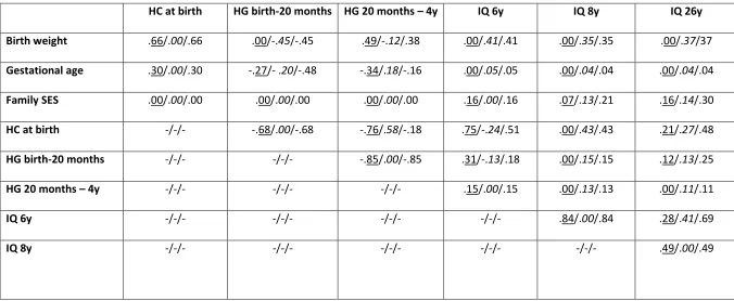
Table 2. Standardized direct, indirect, and total effects (i.e., path coefficients) of the predictors in the final overall Model 2 (N=401)
HC at birth HG birth-20 months HG 20 months – 4y IQ 6y IQ 8y IQ 26y Birth weight .66/.00/.66 .00/-.45/-.45 .49/-.12/.38 .00/.41/.41 .00/.35/.35 .00/.37/37 Gestational age .30/.00/.30 -.27/- .20/-.48 -.34/.18/-.16 .00/.05/.05 .00/.04/.04 .00/.04/.04
Family SES .00/.00/.00 .00/.00/.00 .00/.00/.00 .16/.00/.16 .07/.13/.21 .16/.14/.30
HC at birth -/-/- -.68/.00/-.68 -.76/.58/-.18 .75/-.24/.51 .00/.43/.43 .21/.27/.48
HG birth-20 months -/-/- -/-/- -.85/.00/-.85 .31/-.13/.18 .00/.15/.15 .12/.13/.25
HG 20 months – 4y -/-/- -/-/- -/-/- .15/.00/.15 .00/.13/.13 .00/.11/.11
IQ 6y -/-/- -/-/- -/-/- -/-/- .84/.00/.84 .28/.41/.69
IQ 8y -/-/- -/-/- -/-/- -/-/- -/-/- .49/.00/.49
Figure 1. Final Structural Equation Model 2. Neurocognitive cascades of head growth and
Figure 2. Multigroup Structural Equation Model 3. Neurocognitive cascades of head growth and
intelligence from birth to adulthood in VP/VLBW individuals only (n=203)
Please note: Solid lines represent significant paths, dotted lines represent non-significant paths
Figure 3. Multigroup Structural Equation Model 4. Neurocognitive cascades of head growth and
intelligence from birth to adulthood in term born individuals only (n=198)
Please note: Solid lines represent significant paths, dotted lines represent non-significant paths