Original Article
Expression and correlation of
matrix metalloproteinase-7 and
interleukin-15 in human osteoarthritis
Yulei Tao1, Xianxing Qiu2, Changbo Xu3, Bo Sun4, Changxiu Shi5
1Department of Orthopaedics, The First Clinical College of Shanxi Medical University, Taiyuan, Shanxi Province,
China; 2Guiyang Medical College, Guiyang, Guizhou Province, China; 3Department of Surgery, Public Health
Treatment Center of Guiyang, Guiyang, Guizhou Provence, China; 4Department of Orthopaedics, Guizhou
Libera-tion Army Hospital of The Second Group of The Third Camp, Reserve Infantry Division, Guizhou Provence, China;
5Luzhou Medical College, Luzhou, Sichuan Province, China
Received May 3, 2015; Accepted June 23, 2015; Epub August 1, 2015; Published August 15, 2015
Abstract: Objectives: To investigate the expression and correlation of matrix metalloproteinase (MMP)-7 and inter-leukin (IL)-15 in human osteoarthritis (OA). Methods: From October 2013 to December 2014, 30 patients with OA were enrolled. In addition, anther 30 patients with simple meniscus injury were collected as a control group. There were no significant differences in age and gender between the two groups. Articular cartilage tissue was obtained from both OA patients and control group patients. Protein, mRNA, and serum expression levels of MMP-7 and IL-15 in the both two groups were determined by immunohistochemical (IHC), in situ hybridization, and enzyme-linked im-munosorbent assay (ELISA) assay, respectively. Additionally, correlation between MMP-7 and IL-15 expression level in cartilage tissue and serum was assessed using Pearson correlation analysis. Results: Protein, mRNA, and serum expression levels of MMP-7 and IL-15 in patients with OA were all significantly increased in OA patients compared with the control group. Besides, there were strong positive relationships between articular MMP-7 level and serum MMP-7 level (R2 = 0.573, P = 0.018), between articular IL-15 level and serum IL-15 level (R2 = 0.861, P = 0.023),
and between serum IL-15 level and serum MMP-7 level (R2 = 0.602, P = 0.012). Conclusion: These results suggest
that MMP-7 and IL-15 might play important roles in the pathogenesis of OA, and IL-15 and MMP-7 has positive cor-relation in OA.
Keywords: Matrix metalloproteinase-7, interleukin-15, osteoarthritis
Introduction
Osteoarthritis (OA) is the most common joint disorder, which particularly affects the weight-bearing joints, predominantly in the knee [1]. It is characterized by progressive loss of cartila- ge matrix, subchondral bone remolding, and osteophyte formation, etc. [2, 3]. It has been reported that approximately 10% people over age 60 years in the world suffer from OA [2], and the prevalence is estimated to continue to grow [4]. Besides, OA not only impacts on an
individual’s health, but also carries out a signifi
-cantly financial and societal burden for families
and the society [3, 5]. However, effective man-agement of OA has not been acquired due to the unclear pathophysiology mechanism.
OA cartilage and MMP-7 may play a significant
role in the ECM degradation in OA [13]. Cytokines, such as interleukin (IL)-1, IL-6, IL-15
and tumor necrosis factor (TNF)-α, have been
involved in the pathogenic mechanism of OA [14-17]. Among the cytokines, IL-15 has been regarded as a candidate therapeutic target for OA [17]. However, little information is available regarding the associations of MMP-7 expres-sion and IL-15 expresexpres-sion in articular and serum of OA patients.
Therefore, we aimed to investigate the MMP-7 expression and IL-15 expression in both articu-lar and serum of OA patients, as well as the associations between articular and serum of MMP-7, the associations between articular and serum of IL-15 level, and the associations between serum of MMP-7 expression and IL-15 expression. Our study might contribute to ex-
Materials and methods
Participants and study design
This study was approved by the hospital medi-cal ethics committees, and informed consent was obtained from all participants. Between October 2013 and December 2014, thirty patients with knee OA (13 males and 17 females; mean age 65.16 ± 17.66, range 35-76) attending at our hospital were enrolled. Besides, thirty patients with simple meniscus injury (16 males and 14 females; mean age 63.60 ± 15.23, range 30-79) were collected as a control group. Diagnosis of OA was based on clinical and radiological evaluations revealed by the 1986 American College of Rheumatology (ACR) criteria [18]. Patients with obvious joint
injury or with inflammatory arthritis, previous
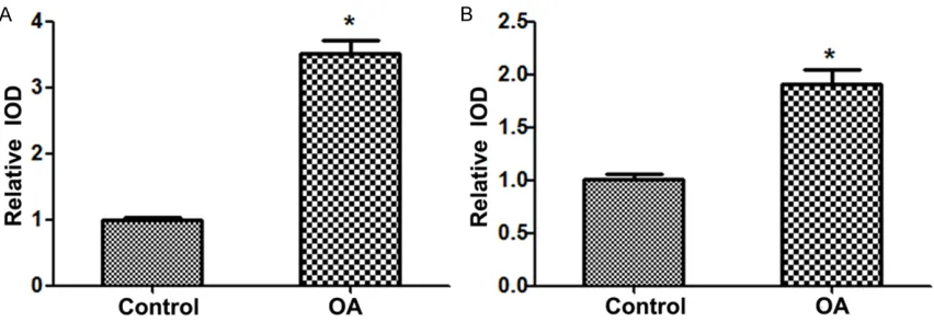
[image:2.612.90.522.75.218.2]knee surgery were excluded from the study. Figure 1. MMP-7 and IL-15 protein expression level in the two groups. A. MMP-7 protein expression level in the two groups; B. IL-15 protein expression level in the two groups. MMP, matrix metalloproteinase; interleukin, IL; OA, os-teoarthritis. *P < 0.05 vs. control group.
[image:2.612.93.519.280.427.2]costeroid or hyaluronan injections within 3 months of surgery were excluded.
Cartilage tissue samples
Articular cartilage was acquired from both OA patients and meniscus injury patients who were undergoing arthroscopic examination or arthroplastic surgery. The tissue sample was
fixed in 4% paraformaldehyde immediately for
24 h after biopsy. Then the tissue was
embed-ded in paraffin, sectioned, and dewaxed accord -ing to the standard methods [13]. Consecutive
4-μm- thickness sections were prepared.
Immunohistochemical (IHC) staining
IHC staining was performed using monoclonal anti-MMP-7 antibody (diluted 1:50; Chemicon, Temecula, CA) and anti-IL-15 antibody (diluted 1:200; Santa Cruz Biotechnologies). The slides were pretreated by microwave heating before the incubation. After incubation with primary
antibody, biotinylated secondary antibody was performed. Negative controls procedures were also performed (the primary antibody was replaced by phosphate-buffered saline (PBS) solution). Finally, chromogen 3,3-diaminobenzi-dine-hydrogen peroxide (DAB, Sigma- Alderich, S.R.L) was performed for visualized reaction.
In situ hybridization
In situ hybridization was performed on
4-μm-thickness sections according to a stan -dard method [19] with a MMP-7 kit (HPV-0610, Fuzhou Maixin Biotech. Co. Ltd) and a IL-15 kit
(MK1165, Boster, Wuhan, China). Briefly, the sections were deparaffinized, treated with
[image:3.612.97.520.73.214.2]0.01% Triton X-100 for 2 min, deproteinated with proteinase K (0.25 g/L) for 5 min and washed in 2 × saline sodium citrate (SSC) at 42°C. Then the slides were prehybridized at 42°C for 3-4 h in prehybridization solution. The probe of MMP-7 and IL-15, and 20 pg tRNA were mixed with the prehybridization solution Figure 3. MMP-7 and IL-15 serum expression level in the two groups. A. MMP-7 serum expression level in the two groups; B. IL-15 serum expression level in the two groups. MMP, matrix metalloproteinase; interleukin, IL; OA, osteo-arthritis. *P < 0.05 vs. control group.
[image:3.612.95.521.276.377.2]and incubated at 42°C overnight, and washed with 2 × SSC. Thereafter, 0.1% Triton X-100 was performed for 2 min and washed with buffer solution 1 (1 mol/L NACL, 0.1 mol/L Tris, PH7.5). The slides were then incubated with buffer solution 2 (buffer solution 1 and 3 g bovine serum albumin (BSA)) for 1 h. After the buffer solution 2 was discarded, avidin and alkaline phosphatase was added to the mixture for 20 min at 42°C. Finally, autoradiography was carried out by nitroblue tetrazolium (NBT).
Computer-assisted image analysis
Five representative areas of each sample (assessed by both IHC and in situ hybridiza- tion) were randomly chosen under a light micro-scope (Olympus, Tokyo, Japan). Quantitative analysis was performed with Image-Pro Plus 6.0 (Media Cybernetics, GE, USA). The positive
areas were regarded as specific brown yellow
color (for IHC) or blueviolet (for in situ hybridiza-tion). The integrated optical density (IOD; pixel area × the optical density) was measured after standard optical density (OD) calibration.
Enzyme-linked immunosorbent assay (ELISA) assay
Fasting blood samples (5 mL) were obtained from an antecubital vein. After clotting, the blood samples were separated by centrifuga-tion at 1,000 g for 15 min at 4°C. Serum sam-ples were subsequently stored at -80°C until use. The serum levels of MMP-7 and IL-15 were determined by an ELISA kit (Calbiochem, Cambridge, MA, USA) according to the
manu-facturer’s protocol. Briefly, 96-well plates were
coated with either anti-MMP-7 or anti-IL-15 antibody, and the appropriate diluent serum samples were added in duplicate, followed with incubation for 2 h at room temperature. After washing with wash buffer, the corresponding secondary antibody was applied. After incuba-tion for another 2 h, the plates were washed again. Spectrophotometric microplate reader was employed to determine the optical density at 490 nm. The concentration of MMP-7 and IL-15 was calculated from a standard curve.
Statistical analysis
Continuous variables were expressed as the mean ± standard deviation (SD). Statistical an- alysis was performed with statistical package
for the social sciences (SPSS) software (version 17.0; SPSS Inc., Chicago, IL). The 2-sample Student’s t test was used to compare continu-ous variables. Correlation analysis was
per-formed using Pearson test. A statistical signifi
-cance was defined when P < 0.05. Results
MMP-7and IL-15 protein and mRNA expression
IHC and in situ hybridization were performed to examine the protein and mRNA expression lev-els of MMP-7 and IL-15 in human cartilage tis-sue, respectively. Here we set the baseline of relative IOD in control group to be 1. The results showed that the expression protein levels of
MMP-7 and IL-15 were both significantly
in-creased in OA patients compared with control group (P < 0.05) (Figure 1A and 1B). Similarly, the results of in situ hybridization demonstrat-ed that the expression mRNA levels of MMP-7 and IL-15 were also both statistically increased in patients with OA compared with the control group (P < 0.05) (Figure 2A and 2B).
MMP-7and IL-15 serum expression
The serum expression levels of MMP-7 and IL-15 were showed in Figure 3A and 3B, respec-tively. As shown in Figure 3A, the average serum MMP-7 level was 5.617 ± 2.300 ng/ml (range 2.2-8.8 ng/ml) in the control group, whereas the average serum MMP-7 level was 20.117 ± 7.083 ng/ml (range 11.0-29.4 ng/
ml). There were significant differences in the
serum MMP-7 level between the two groups. Also, the serum IL-15 level (average 129.6 ± 39.922 pg/ml; range 85.0-176.0 pg/ml) in OA
patients was significantly higher than that in
the control group (average 576.8 ± 194.574 pg/ml; range 369.0-875.0 pg/ml) (Figure 3B).
Correlation analyses between MMP-7and IL-15
In order to confirm the clinical diagnostic value
MMP-7 level (R2 = 0.602, P = 0.012) (Figure 4A-C).
Discussion
In the present study, the expression and corre-lation of MMP-7 and IL-15 in human OA was investigated. The results showed that the pro-tein, mRNA, and serum expression levels of
MMP-7 and IL-15 in OA patients were all signifi -cantly higher than those in the control group. Besides, there were strong positive relation-ships between articular MMP-7 level and serum MMP-7 level, between articular IL-15 level and serum IL-15 level, and between serum IL-15 level and serum MMP-7 level. Our results indi-cated that MMP-7 and IL-15 might play impor-tant roles in the pathogenesis of OA, and IL-15 and MMP-7 has positive correlation in OA. MMPs are a group of proteolytic enzymes responsible for the degradation of ECM, which is involved in multiple physiological and patho-logical progressions of different tissues [20-22]. The degradation of ECM plays essential roles in the tissue resorption and remodeling [23]. It has been reported that approximately 95% of the dry weight of articular cartilage is ECM [24]. The imbalance of the cartilage ECM results in OA, therefore, a delicate balance between the breakdown and synthesis of ECM is of importance [25]. MMP-7, also known as matrilysin, is a unique member of the MMP family. Unlike other members, MMP-7 is the smallest member that only consists of the com-mon catalytic domain and zinc-binding region
[26]. It has a specific ability to degrade various
ECM components such as cartilage
proteogly-can [27]. Since Ohta has firstly reported that
MMP-7 is overexpressed in human OA cartilage [13], the functions and roles of MMP-7 in OA have been paid attention [28, 29]. In consistent with previous studies, our results also suggest-ed that the expression levels of MMP-7 were
significantly higher than those in the control
group, either in articular cartilage or in serum. Additionally, we found that there was a strong positive relationship between articular MMP-7 level and serum MMP-7 level.
The expression of MMPs is regulated by
cyto-kines [30]. Inflammatory reaction has been well
demonstrated in the development and progres-sion of OA, even in the early stage of the
dis-ease [31]. IL-15, a proinflammatory cytokine,
has been considered as a contributor to inflam -mation in OA [6]. Previous study has indicated that IL-15 has both potential diagnostic and prognostic biochemical marker for OA [28]. The
expression level of IL-15 was found significantly
increased in OA patient’s serum samples; besides, IL-15 level was positively correlated with a Western Ontario McMaster University Osteoarthritis Index (WOMAC) pain scores [32]. Moreover, IL-15 has been reported to induce MMP production, especially MMP-1 and MMP-9 [33]. Additionally, previous studies suggested that there were associations between IL-15 level with MMP-1 and MMP-3 [17]. Also, our
study confirmed the higher expression of IL-15
in OA patients than that in control patients, which was in line with previous studies. In addi-tion to the results, we found that serum IL-15 level was positively related with articular IL-15 level and serum MMP-7 level. One possible rea-son for that is MMP-7 is may be induced by IL-15, and both of them participate in the patho-genesis of OA.
In conclusion, our results suggest that both MMP-7 and IL-15 play important roles in the pathogenesis of OA, and IL-15 and MMP-7 has positive correlation in OA.
Disclosure of conflict of interest
None.
Address correspondence to: Dr. Yulei Tao, Depart-
ment of Orthopaedics, The First Clinical College of Shanxi Medical University, No. 85 Jiefang South Road, Taiyuan 030001, Shanxi Province, China. Tel: +86-13834569278; Fax: 0532-89751558; E-mail: taoyulei0988@126.com
References
[1] Fibel KH, Hillstrom HJ and Halpern BC. State-of-the-Art management of knee osteoarthritis. World J Clin Cases 2015; 3: 89-101.
[2] Pereira D, Peleteiro B, Araujo J, Branco J, San-tos RA and Ramos E. The effect of osteoarthri-tis definition on prevalence and incidence esti -mates: a systematic review. Osteoarthritis Cartilage 2011; 19: 1270-1285.
[3] Litwic A, Edwards MH, Dennison EM and Coo-per C. Epidemiology and burden of osteoarthri-tis. Br Med Bull 2013; 105: 185-199.
[5] Buckwalter JA, Saltzman C and Brown T. The impact of osteoarthritis: implications for re-search. Clin Orthop Relat Res 2004; S6-15. [6] Mabey T and Honsawek S. Cytokines as
bio-chemical markers for knee osteoarthritis. World J Orthop 2015; 6: 95-105.
[7] Goldring MB and Goldring SR. Osteoarthritis. J Cell Physiol 2007; 213: 626-634.
[8] Bluteau G, Conrozier T, Mathieu P, Vignon E, Herbage D and Mallein-Gerin F. Matrix metal-loproteinase-1, -3, -13 and aggrecanase-1 and -2 are differentially expressed in experimental osteoarthritis. Biochim Biophys Acta 2001; 1526: 147-158.
[9] Galasso O, Familiari F, De Gori M and Gaspari-ni G. Recent findings on the role of gelatinases (matrix metalloproteinase-2 and -9) in osteoar-thritis. Adv Orthop 2012; 2012: 834208. [10] Fosang AJ, Last K, Knauper V, Murphy G and
Neame PJ. Degradation of cartilage aggrecan by collagenase-3 (MMP-13). FEBS Lett 1996; 380: 17-20.
[11] Fosang AJ, Last K, Knauper V, Neame PJ, Mur-phy G, Hardingham TE, Tschesche H and Ham-ilton JA. Fibroblast and neutrophil collagenas-es cleave at two sitcollagenas-es in the cartilage aggrecan interglobular domain. Biochem J 1993; 295: 273-6.
[12] Wang M, Sampson ER, Jin H, Li J, Ke QH, Im HJ and Chen D. MMP13 is a critical target gene during the progression of osteoarthritis. Arthri-tis Res Ther 2013; 15: R5.
[13] Ohta S, Imai K, Yamashita K, Matsumoto T, Azumano I and Okada Y. Expression of matrix metalloproteinase 7 (matrilysin) in human os-teoarthritic cartilage. Lab Invest 1998; 78: 79-87.
[14] Benito MJ, Veale DJ, FitzGerald O, van den Berg WB and Bresnihan B. Synovial tissue in-flammation in early and late osteoarthritis. Ann Rheum Dis 2005; 64: 1263-1267.
[15] Youssef PP, Triantafillou S, Parker A, Coleman M, Roberts-Thomson PJ, Ahern MJ and Smith MD. Variability in cytokine and cell adhesion molecule staining in arthroscopic synovial bi-opsies: quantification using color video image analysis. J Rheumatol 1997; 24: 2291-2298. [16] Lee AS, Ellman MB, Yan D, Kroin JS, Cole BJ,
van Wijnen AJ and Im HJ. A current review of molecular mechanisms regarding osteoarthri-tis and pain. Gene 2013; 527: 440-447. [17] Scanzello CR, Umoh E, Pessler F, Diaz-Torne C,
Miles T, Dicarlo E, Potter HG, Mandl L, Marx R, Rodeo S, Goldring SR and Crow MK. Local cyto-kine profiles in knee osteoarthritis: elevated synovial fluid interleukin-15 differentiates ear -ly from end-stage disease. Osteoarthritis Carti-lage 2009; 17: 1040-1048.
[18] Altman R, Asch E, Bloch D, Bole G, Borenstein D, Brandt K, Christy W, Cooke TD, Greenwald
R, Hochberg M, et al. Development of criteria for the classification and reporting of os-teoarthritis. Classification of osteoarthritis of the knee. Diagnostic and Therapeutic Crite- ria Committee of the American Rheumatism Association. Arthritis Rheum 1986; 29: 1039-1049.
[19] Prosser IW, Stenmark KR, Suthar M, Crouch EC, Mecham RP and Parks WC. Regional het-erogeneity of elastin and collagen gene ex-pression in intralobar arteries in response to hypoxic pulmonary hypertension as demon-strated by in situ hybridization. Am J Pathol 1989; 135: 1073-1088.
[20] Peng WJ, Yan JW, Wan YN, Wang BX, Tao JH, Yang GJ, Pan HF and Wang J. Matrix metal- loproteinases: a review of their structure and role in systemic sclerosis. J Clin Immunol. 2012; 32: 1409-14.
[21] Jones CB, Sane DC and Herrington DM. Matrix metalloproteinases A review of their structure and role in acute coronary syndrome. Cardio-vascular research 2003; 59: 812-823. [22] Gialeli C, Theocharis AD and Karamanos NK.
Roles of matrix metalloproteinases in cancer progression and their pharmacological target-ing. FEBS J 2011; 278: 16-27.
[23] Gronski TJ Jr, Martin RL, Kobayashi DK, Walsh BC, Holman MC, Huber M, Van Wart HE and Shapiro SD. Hydrolysis of a broad spectrum of extracellular matrix proteins by human mac-rophage elastase. J Biol Chem 1997; 272: 12189-94.
[24] Alford JW and Cole BJ. Cartilage restoration, part 1: basic science, historical perspective, patient evaluation, and treatment options. Am J Sports Med 2005; 33: 295-306.
[25] Grimaud E, Heymann D and Redini F. Recent advances in TGF-beta effects on chondrocyte metabolism. Potential therapeutic roles of TGF-beta in cartilage disorders. Cytokine Gro- wth Factor Rev 2002; 13: 241-257.
[26] Yokoyama Y, Grunebach F, Schmidt SM, Heine A, Hantschel M, Stevanovic S, Rammensee HG and Brossart P. Matrilysin (MMP-7) is a novel broadly expressed tumor antigen recognized by antigen-specific T cells. Clin Cancer Res 2008; 14: 5503-5511.
[27] Wilson CL and Matrisian LM. Matrilysin: an epithelial matrix metalloproteinase with poten-tially novel functions. Int J Biochem Cell Biol 1996; 28: 123-136.
[28] Ling SM, Patel DD, Garnero P, Zhan M, Vaduga-nathan M, Muller D, Taub D, Bathon JM, Hoch-berg M, Abernethy DR, Metter EJ and Ferrucci L. Serum protein signatures detect early radio-graphic osteoarthritis. Osteoarthritis Cartilage 2009; 17: 43-48.
Me-talloproteinase and tissue inhibitor of metallo-proteinase expression in the murine STR/ort model of osteoarthritis. Osteoarthritis Carti-lage 2002; 10: 722-733.
[30] Matrisian LM and Hogan BL. Growth factor-regulated proteases and extracellular matrix remodeling during mammalian development. Curr Top Dev Biol 1990; 24: 219-259.
[31] Felson DT. Clinical practice. Osteoarthritis of the knee. N Engl J Med 2006; 354: 841-848.
[32] Sun JM, Sun LZ, Liu J, Su BH and Shi L. Serum interleukin-15 levels are associated with se-verity of pain in patients with knee osteoarthri-tis. Dis Markers 2013; 35: 203-206.