Interrelationships of body weight and bone weight
with densitometric properties of tibiotarsal bone
in geese during post-hatching development
A. Charuta
1, M.R. Tatara
2, M. Dzierzecka
3, E. Polawska
4,
I. Ptaszynska Sarosiek
51Faculty of Natural Science, Siedlce University of Natural Sciences and Humanities,
Siedlce, Poland
2Faculty of Veterinary Medicine, University of Life Sciences, Lublin, Poland
3Faculty of Veterinary Medicine, Warsaw University of Life Sciences, Warsaw, Poland 4Institute of Genetics and Animal Breeding PAS, Jastrzebiec, Poland
5Department of Forensic Medicine, Medical University of Bialystok, Bialystok, Poland
ABSTRACT: The aim of this study was to evaluate interrelationships of body weight and bone weight and densi-tometric properties of the tibiotarsus in White Koluda Geese (W31) in the post-hatching period. The study was performed using dual-energy X-ray absorptiometry (DEXA) and peripheral quantitative computed tomography (pQCT) at two different parts of tibia: proximal metaphysis and mid-diaphysis. The investigation was performed on 100 bones obtained from males and females at the age of 1, 14, 28, 42 and 56 days of life. All the calculations were performed using the Statistica 9.0 software (StatSoft, Inc. Tulsa, USA). Pearson’s correlation coefficient of body weight and bone weight with all the investigated variables of bone was determined. Depending on the method used for densitometric measurements – DEXA or pQCT, the current study has revealed significant differences in the number of correlations of bone weight and body weight with the evaluated densitometric parameters. Sex-related differences in the investigated interrelationships were also found. In the case of proximal epiphysis, negative cor-relations of vBMD, tBMC, CTR_DEN and CRT_CNT with body weight and bone weight dominated in one-day-old males. Based on the current observations and the negative correlations of body weight and vBMD, CRT_DEN and TRAB_DEN obtained in the mid-diaphysis of tibiotarsus at the age of 14 days of life, it was concluded that this bone is much more prone to deformations and fractures in males than in females.
Keywords: densitometry parameters; tibiotarsal bone; mineralisation; geese
List of abbreviation
BMC = bone mineral content; BMD = bone mineral density; CRT_CNT = cortical bone mineral content; CRT_DEN
= cortical bone mineral density; tBMC = total bone mineral content; TRAB_CNT = trabecular bone mineral content; TRAB_DEN = trabecular bone mineral density; vBMD = volumetric bone mineral density
Post-hatching development of meat-type poultry species is characterised by very rapid muscle tis-sue development and accumulation mainly due to skeletal muscles such as breast and leg muscles. Developmental skeletal muscle gain rate is very high both in relation to the internal organs of the body
and inert constituents of the locomotory system. Such intensive skeletal muscle gain is not propor-tional to the developmental formation of the other systems and tissues, including skeletal system and bones. Such developmental disproportion between skeletal muscle tissue mass and bone tissue mass of
the skeleton is strictly connected to breeding selec-tion toward higher slaughter efficacy. As the result of such breeding selection in meat-type poultry spe-cies, the relative percentage of slaughter waste is reduced in relation to the final body weight (Gille et al. 1999). The consequence of the imbalance between the development of skeletal muscles and bone mass is a higher incidence of pelvic limb bones deformities and fractures, due to an insufficient adaptation of the peripheral skeleton to heavy loading by excessive body weight (Crespo et al. 1999; Crespo et al. 2000; Horbanczuk et al. 2004; Cooper et al. 2008; Charuta et al. 2011; Tatara et al. 2011a; Charuta et al. 2012c; Charuta et al. 2013a; Charuta et al. 2014).
Studies on systemic development of internal or-gans and bones in broiler chickens, ducks and geese have revealed age-related increases in the percent-age content of skeletal muscles and simultaneous decreases in the percentage content of internal or-gans and bones in whole body weight (Murawska 2013). It was shown in experimental studies on geese that developmental body weight gain is as-sociated with significant decreases in the weight of pelvic limb bones; however, bone weight of the cervical vertebrae, wings and chest increase con-currently in relation to whole body weight (Bochno et al. 2006; Murawska 2013). Studies on peripheral skeletal system properties in growing meat-type poultry species have shown that body weight gain is associated with decreases in the amount of bone tissue as well as changes in densitometric prop-erties (Charuta and Cooper 2012; Charuta et al. 2012a; Charuta et al. 2012b; Charuta et al. 2012c; Charuta 2013; Charuta et al. 2013a; Charuta et al. 2013b). The observed developmental changes in long bones were found to be correlated both with the age and sex of birds (Beck and Hansen 2004).
Previous studies in meat-type turkeys at slaugh-ter age have shown that volumetric bone mineral density of the trabecular and cortical bone in distal epiphysis and mid-diaphysis of long bones of the pelvic limb is related to its mechanical endurance. The midshaft of long bones consists of cortical bone with the highest mineral density and may serve as the standard reference point for the evalu-ation of geometrical and densitometric parameters. Moreover, this reference point is the position for bone sample loading in the three-point bending test and for the determination of its mechanical properties (Tatara et al. 2004a; Tatara et al. 2004b; Tatara et al. 2006).
Considering that body weight and bone weight are the main factors contributing to the incidence of pelvic limb bone weakness and functional quality of the peripheral skeletal system in poultry spe-cies, the aim of this study was to evaluate inter-relationships of body weight and bone weight with densitometric parameters of tibia in growing geese.
MATERIAL AND METHODS
The slaughter and collection of material for re-search was conducted with the approval of the 3rd Ethics Committee concerning experiments on
animals of Warsaw University of Life Sciences, Warsaw, Poland (Resolution No. 23/2009).
The study was performed on one hundred (fif-ty females and fif(fif-ty males) White Koluda Geese (W31). All geese were divided into five age-dif-ferentiated groups (n = 10 in each group of males and females) and were slaughtered on Days 1, 14, 28, 42 and 56 of life. The left tibiotarsal bone was isolated from each bird, cleaned from soft tissues and kept frozen in plastic bags at a temperature of between –25 °C and –30 °C.
During growth, the geese were fed a standard concentrate that contained 19.5% protein at the beginning of fattening (11.72 MJ/kg) and 15.5% protein at the end of fattening (12.14 MJ/kg). The weight of the bones was determined using a precise laboratory balance AXIS AD 300 (AXIS, Poland) with a maximum range of 300 g and precision of 0.001 g. Bone mineral density (BMD) and bone min-eral content (BMC) of the bones from geese were measured using the dual-energy X-ray absorptiom-etry (DEXA) method and Norland Excell Plus den-sitometer (Fort Atkinson, WI, USA). A collimated X-ray beam and software developed for the exami-nation of the bones of small animals (Small Subject Scan, version 3.9.6) were used. Bones of one-day-old geese were scanned at a scanning resolution of 0.5 × 0.5 mm and scanning speed of 10 mm/s, while the bones obtained from the remaining groups of geese (14-, 28-, 42- and 56-day-old) were scanned using a scanning resolution of 1.5 × 1.5 mm and scanning speed of 30 mm/s. Bone mineral density was expressed in g/cm2 while bone mineral
Using peripheral quantitative computed tomog-raphy (pQCT) and a XCT Research SA Plus scan-ner (Stratec Medizintechnik GmbH, Pforzheim, Germany), densitometric parameters of the tibi-otarsal bones were analysed. The measurements were performed at two sites of the bone; at 18% of bone length at proximal metaphysis and at 50% of bone length at mid-diaphysis. Total bone mineral content (tBMC – mg/mm per 1 mm slice), volu-metric bone mineral density (vBMD – total bone mineral density, mg/cm3), cortical bone mineral
density (CRT_DEN – volumetric mineral density of the cortical bone, mg/cm3) and cortical bone
mineral content (CRT_CNT – the mineral con-tent within 1 mm slice of the cortical bone, mg/ mm) were determined. Furthermore, in the proxi-mal metaphysis, trabecular bone mineral density (TRAB_DEN – volumetric mineral density of the trabecular bone, mg/cm3) and trabecular bone
min-eral content (TRAB_CNT – the minmin-eral content within a 1 mm slice of the trabecular bone, mg/ mm) were measured.
Statistical analysis. All the calculations were performed using Statistica 9.0 software (StatSoft, Inc. Tulsa, USA). Pearson’s correlation coefficient of body weight and bone weight with all the in-vestigated variables of bone was determined and
P-values < 0.05 were considered as statistically significant.
RESULTS
Body weight values for males and females at ferent ages are presented in Table 1. Significant dif-ferences in body weight between males and females were found only in 42-day-old birds and the body
weight of males was higher by 450 g compared to that of females (P < 0.05). Gradual but significant increases body weight values were observed at each developmental stage both in males and females (P < 0.05). Significant increases in bone weight values were observed at each developmental stage both in males and females up to the age of 42 days of life (P < 0.05). Significantly higher bone weights were found in 28- and 42-day-old males compared to age-matched females (P < 0.05; Table 2).
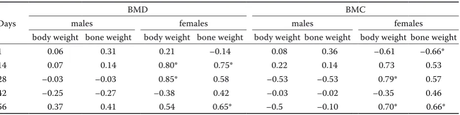
BMD in 14- and 28-day-old females was posi-tively correlated with body weight. A positive cor-relation of BMD with bone weight was found in females at the age of 14 and 56 days of life (P < 0.05). BMC in 28- and 56-day-old females was positively correlated with body weight. BMC was negatively correlated with bone weight in one-day-old females (P < 0.05). However, positive correlation between BMC and body weight was stated in 56-day-old females (P < 0.05; Table 3).
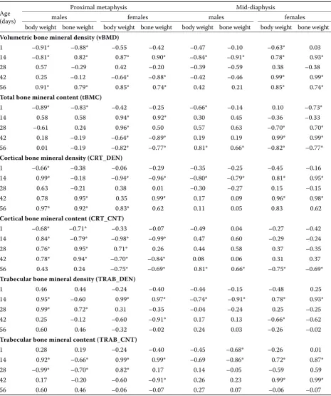
[image:3.595.63.537.609.722.2]vBMD of the proximal metaphysis of one-day-old males was negatively correlated with body weight and bone weight, while in two-week-old males this parameter correlated negatively with body weight and positively with bone weight (P < 0.05; Table 4). In 56-day-old males a positive correlation of with body weight and bone weight was found (P < 0.05). In 42-day-old females, the vBMD of the proximal metaphysis was negatively correlated with body weight and bone weight (P < 0.05). However, positive correlations of the vBMD of the proximal metaphysis with body weight and bone weight were observed in 14- and 56-day-old females (P < 0.05). A negative correlation between the vBMD of the mid-diaphysis and body weight and bone weight in 14-day-old males was revealed, while in females these correlations were positive (P < 0.05). vBMD
Table 1. Body weight (g) values in male and female geese at the age of 1, 14, 28, 42 and 56 days of life
Age (days)
Body weight
males females
min max –x ± SD min max –x ± SD
1 70 75 72A ± 5.83 61 68 65A ± 5.91
14 690 780 731B ± 58.18 680 740 708B ± 56.69
28 2140 2300 2220C ± 199.80 1940 2580 2130C ± 191.91
42 3300 4220 3760*,D ± 300.55 3100 3700 3310*,D ± 264.23
56 4140 4740 4450E ± 400.52 3760 4680 4165E ± 333.20
A–Emeans in each column designated with different superscript letters differ significantly at P < 0.05
of the mid-diaphysis in one-day-old females was negatively correlated with body weight (P < 0.05). Positive correlations of the vBMD of the midshaft with body weight and bone weight were observed in 42- and 56-day-old females (P < 0.05).
tBMC of the proximal metaphysis of one-day-old males was negatively correlated with body weight and bone weight (P < 0.05). In 14-day-old females, the tBMC of the proximal metaphysis of the tibio-tarsus was positively correlated with body weight and bone weight, while negative correlations of these parameters were observed in 42- and 56-day-old females (P < 0.05). Moreover, the tBMC of the proximal metaphysis of the tibiotarsus was posi-tively correlated with the body weight of 28-day-old females (P < 0.05).
The tBMC values of the midshaft were positively correlated with body weight and bone weight in 56-day-old males, whilst in one-day-old males a negative correlation with body weight was found (P < 0.05). Highly positive correlations of the tBMC of the midshaft with body weight and bone weight were ob-tained in 42-day-old females, while two weeks later
a negative correlation between these variables was found (P < 0.05). The tBMC values of the midshaft were negatively correlated with the body weight of 28-day-old females and the bone weight of one-day old females (P < 0.05). However, a positive correla-tion between the tBMC and bone weight of 28-day-old females was seen (P < 0.05).
[image:4.595.70.535.113.228.2]Positive correlations of CRT_DEN of the proxi-mal metaphysis of tibiotarsus and body weight in 14- and 56-day-old males were observed, while in one-day-old males a negative correlation was ob-served (P < 0.05). Positive correlations of CRT_DEN of the proximal metaphysis and bone weight were re-vealed in 42- and 56-day-old male geese (P < 0.05). In 14-day-old females, a negative correlation between CRT_DEN of the proximal metaphysis and body weight and bone weight was observed (P < 0.05). Moreover, positive correlations of CRT_DEN of the proximal metaphysis with body weight in 56-day-old females, and with bone weight of 42-day-old females were revealed (P < 0.05). In 14-day-old males, nega-tive correlations of CRT_DEN of the mid-diaphysis with body weight and bone weight were obtained,
Table 2. Bone weight (g) values in male and female geese at the age of 1, 14, 28, 42 and 56 days of life
Age (days)
Bone weight
males females
min max –x ± SD min max –x ± SD
1 0.73 0.85 0.83A ± 0.05 0.57 0.83 0.74A ± 0.07
14 7.05 8.13 7.78B ± 0.71 6.02 0.55 7.38B ± 0.66
28 23.98 24.34 24.16*,C ± 2.17 18.21 23.70 22.00*,C ± 1.54 42 32.87 35.15 34.06*,D ± 2.38 30.21 34.45 32.35*,D ± 2.26 56 33.54 35.08 34.85D ± 2.41 32.51 34.87 33.58D ± 2.35
A-Dmeans in each column designated with different superscript letters differ significantly at P < 0.05 *means in rows (males vs. females) differ significantly at P < 0.05
Table 3. Pearson’s correlation coefficient of bone mineral density (BMD) and bone mineral content (BMC) of the tibiotarsus with body weight and bone weight of males and females at the age of 1, 14, 28, 42 and 56 days of life
Days
BMD BMC
males females males females
body weight bone weight body weight bone weight body weight bone weight body weight bone weight
1 0.06 0.31 0.21 –0.14 0.08 0.36 –0.61 –0.66*
14 0.07 0.14 0.80* 0.75* 0.22 0.14 0.73 0.53
28 –0.03 –0.03 0.85* 0.58 –0.53 –0.53 0.79* 0.57
42 –0.25 –0.27 –0.38 0.42 –0.03 –0.02 –0.35 0.46
56 0.37 0.41 0.54 0.65* –0.5 –0.10 0.70* 0.66*
[image:4.595.66.533.618.735.2]while in females these correlations were positive (P < 0.05). Moreover, in 42-day-old females, positive cor-relations of CRT_DEN of the midshaft with body weight and bone weight were obtained (P < 0.05).
[image:5.595.64.530.124.680.2]CRT_CNT of the proximal metaphysis of one-day-old males was negatively correlated with body weight and bone weight, while in two-week-old males this parameter correlated negatively with
Table 4. Pearson’s correlation coefficient of the investigated densitometric parameters of the tibiotarsus with body weight and bone weight of males and females at the age of 1, 14, 28, 42 and 56 days of life
Age (days)
Proximal metaphysis Mid-diaphysis
males females males females
body weight bone weight body weight bone weight body weight bone weight body weight bone weight
Volumetric bone mineral density (vBMD)
1 –0.91* –0.88* –0.55 –0.42 –0.47 –0.10 –0.63* 0.03
14 –0.81* 0.82* 0.87* 0.90* –0.84* –0.91* 0.78* 0.93*
28 0.57 –0.29 0.42 –0.20 –0.39 –0.59 0.38 –0.38
42 0.25 –0.12 –0.64* –0.88* –0.42 –0.46 0.99* 0.99*
56 0.91* 0.79* 0.85* 0.74* 0.42 0.21 0.85* 0.74*
Total bone mineral content (tBMC)
1 –0.89* –0.83* –0.42 –0.25 –0.66* –0.14 0.10 –0.73*
14 0.58 0.58 0.94* 0.92* 0.30 0.45 –0.36 –0.33
28 –0.61 0.24 0.96* 0.50 0.57 0.63 –0.70* 0.70* 42 0.18 –0.19 –0.64* –0.89* 0.19 0.19 0.99* 0.99* 56 0.01 –0.19 –0.82* –0.77* 0.81* 0.66* –0.82* –0.77*
Cortical bone mineral density (CRT_DEN)
1 –0.66* –0.38 –0.06 –0.29 –0.35 –0.25 –0.45 –0.16
14 0.99* –0.18 –0.94* –0.96* –0.80* –0.79* 0.81* 0.95*
28 0.63 –0.21 0.38 0.01 –0.30 –0.27 0.15 –0.15
42 0.78 0.95* 0.35 0.99* 0.17 0.09 0.96* 0.98* 56 0.97* 0.92* 0.83* 0.62 0.11 0.05 0.83 0.62
Cortical bone mineral content (CRT_CNT)
1 –0.68* –0.71* –0.33 –0.07 –0.49 0.04 –0.27 –0.42
14 0.84* –0.79* –0.98* –0.99* 0.47 0.60 –0.29 –0.24
28 0.76* 0.95* 0.71* 0.26 0.44 0.58 0.37 –0.35
42 0.78* 0.94* –0.70* –0.84* 0.08 0.06 0.31 0.37
56 0.43 0.24 –0.75* –0.69* 0.81* 0.66* –0.75* –0.69*
Trabecular bone mineral density (TRAB_DEN)
1 0.46 0.44 –0.24 –0.40 –0.44 –0.15 –0.48 0.25
14 0.95* –0.60 0.99* 0.97* –0.74* –0.91* 0.78* 0.93*
28 0.99* 0.72* 0.31 –0.35 –0.04 –0.24 0.25 –0.25
42 0.25 –0.12 –0.60 –0.91* 0.17 0.13 –0.66* –0.62
56 0.60 0.46 –0.32 –0.02 0.24 0.03 –0.26 –0.02
Trabecular bone mineral content (TRAB_CNT)
1 0.28 0.19 –0.24 –0.40 –0.45 –0.68* –0.26 0.01
14 0.92* –0.66* 0.99* 0.99* –0.69 –0.86* 0.72* 0.87*
28 –0.99* –0.70* 0.82* 0.17 0.14 –0.05 –0.59 0.59
42 0.17 –0.20 –0.60 –0.91* 0.26 0.23 0.99* 0.99*
56 0.60 0.46 –0.06 –0.07 0.27 0.07 –0.06 –0.07
bone weight (P < 0.05). In 14-day old males, a posi-tive correlation between CRT_CNT of the proximal metaphysis and body weight was also observed. In 28- and 42-day-old males, CTR_CNT values of the proximal metaphysis were positively cor-related with both body weight and bone weight (P < 0.05). In 28-day-old females, CRT_CNT of the proximal metaphysis was positively correlated with body weight (P < 0.05). However, CRT_CNT of the proximal metaphysis was negatively correlated with body weight and bone weight of 14-, 42- and 56-day-old females (P < 0.05). In 56-day-old males, positive correlations of CRT_CNT of the midshaft with body weight and bone weight were obtained, while in fe-males the correlations were negative (P < 0.05).
In 14- and 28-day-old males, TRAB_DEN of proximal metaphysis was found to be positively correlated with body weight (P < 0.05). In 28-day-old males, TRAB_DEN was found to be also posi-tively correlated with bone weight (P < 0.05). The TRAB_DEN value of the proximal metaphysis was found to be positively correlated with body weight and bone weight in 14-day-old females (P < 0.05). However, in 42-day-old females TRAB_DEN was negatively correlated with bone weight (P < 0.05). In 14-day-old males, TRAB_DEN of the midshaft was negatively correlated with body weight and bone weight, while in females these correlations were positive (P < 0.05). Furthermore, in 42-day-old females TRAB_DEN of the midshaft was negatively correlated with bone weight (P < 0.05; Table 4).
TRAB_CNT in the proximal metaphysis was posi-tively correlated with the body weight of 14-day-old males but negatively correlated with body weight in 28-day-old males (P <0.05). A negative corre-lation of TRAB_CNT of the proximal metaphy-sis with bone weight in 14- and 28-day-old males was found (P < 0.05). TRAB_CNT in the proximal metaphysis was positively correlated with the body weight of 14- and 28-day-old females but nega-tively correlated with bone weight in 42-day-old females (P < 0.05). Moreover, a positive correlation between TRAB_CNT of the proximal metaphysis and bone weight in 14-day old females was obtained (P < 0.05). TRAB_CNT of the mid-diaphysis was negatively correlated with bone weight in one- and 14-day-old males (P < 0.05). Positive correlations of TRAB_CNT in the midshaft of the tibiotarsus and body weight in 14- and 42-day-old females were obtained (P < 0.05). At the age of 14 and 42 days of life, TRAB_CNT of the midshaft and bone
weight were also positively correlated in females (P < 0.05; Table 4).
DISCUSSION
Skeletal problems observed in farm birds often centre on pelvic limbs, which bear the entire body weight. A number of research tests have made it possible to determine that both the tibiotarsal and the tarsometarsal bone may serve as model bones in studies on the quality of the skeleton of farm birds (Tatara et al. 2005; Charuta et al. 2013c). As deformations and fractures of the tibiotarsal bones occurr frequently in geese bred for meat, the cur-rent study was focused on determination of inter-relationships of body weight and bone weight with densitometric parameters of tibia. In this study, the interrelationships were determined for both sexes of geese at five different developmental stag-es, between the 1st and 56th day of life. It should
densitometric parameters of males and females in both these parts of tibia. This study has also shown sex-related differences of the investigated interre-lationships. In the proximal metaphysis of females tBMC and CRT_CNT were negatively correlated with body and bone weights of 42- and 56-day old geese while neither positive nor negative correla-tions were found in age-matched males. Analogical results of correlations of tBMC and CRT_CNT with body and bone weights were observed in the mid-diaphysis. In 56-day-old females, negative correla-tions were found in this part of bone, while in males all these correlations were positive.
During the postnatal development, bones undergo a structural remodelling process, the aim of which is to adapt the bone structure in such way that it can resist gravitational and mechanical forces and play specific mechanical functions (Komosa et al. 2013). Adequate BMC, BMD and vBMD values, in particular at the metaphyses of tibiotarsal bones, are responsible
for mechanical resistance in the period of rapid gain of body weight. Beside BMD and BMC, long bone strength is determined by geometrical properties such as cross-sectional area, second moment of in-ertia, mean relative wall thickness and cortical index. However, the attainment of optimal densitometric and geometric properties of long bones which ensure maximum bone strength depend mainly on genetic, hormonal, nutritional, physiological and environmen-tal factors acting during the entire period of growth and development, as well as later in life in mature birds (Rath et al. 2000; Krupski and Tatara 2007). It is worth stressing that metabolic processes are the most intensive in the trabecular bone of proximal metaphyses (Czerwinski 1994).
The investigated geese were subjected to intensive breeding, as a result of which they were ready for slaughter at the eighth week of life, twice as quickly as in the case of traditional, semi-intensive breeding, where such maturity is attained between weeks 17 and 24 of life. Thus, due to their rapid growth and large increase in body weight, the skeletons of the geese investigated here were exposed to a signifi-cantly high risk of deformations and fractures.
In male quails, vBMD and BMC values at proxi-mal metaphyses decreased in the sixth week of life, and the BMD values of diaphyses decreased in the fourth week (Charuta et al 2013a). In male broiler chickens, in turn, vBMD values decreased in the fourth week of life at the two analysed sections of the bone (Charuta et al. 2013b).
Previously, the osseous structure of the tibia in geese reared in a semi-intensive system (up to the 16th week) was analysed using computed tomogra-phy and Trabelula® software (Charuta et al. 2012c). It was found that similarly to geese bred until the 8th week of life, in six-week-old males, a decrease in the number, volume and density of radiologically identified trabeculae was observed at the proximal metaphyses of the tibia. It should be underlined that these parameters make an important contribution to bone strength. The use of two different methods for tibia examination has proven that the critical moment in which the strength of the tibia in males decreased considerably was in the sixth week of life (Charuta et al. 2012c).
The Trabecula programme was also used to ana-lyse the tibia obtained from Peking domestic ducks. It was observed that in six-week-old female ducks the number and density of radiologically identi-fied trabeculae decreased significantly at proximal metaphyses. In male domestic ducks, in turn, no statistically significant decrease of these parameters characterising the trabecular bone compartment was found, unlike in geese (Charuta et al. 2011).
In the current study, numerous positive and neg-ative correlations of densitometric parameters of the tibiotarsus were found. In the case of proximal epiphysis, negative correlations of vBMD, tBMC, CTR_DEN and CRT_CNT with body weight and bone weight dominated in one-day old males, reveal-ing significant and characteristic interrelationships. Investigations of the densitometric parameters in the proximal metaphysis of the tibiotarsus in females and mid-diaphysis in males and females failed to re-veal such regular characteristic interrelationships. In other breeds of domestic birds, negative correlations were observed between vBMD and body weight and bone weight, for example in the proximal metaphy-ses of four- and six-week-old male quails (Charuta et al. 2013a). In the analysed one-day-old geese tBMC at the proximal metaphyses correlated negatively with body weight. In quails BMC correlated nega-tively with body weight, but only in six-week-old birds (Charuta et al. 2013a). In turkeys, in turn, no negative correlations were found between BMC at metaphyses and diaphyses of tibiotarsal bones and body weight (Charuta et al. 2012a). However, in an-other study on growing turkeys between the 4th and
20th week of life, tibia weight was positively
mineral density as well as maximum elastic strength and ultimate strength (Krupski and Tatara 2007). Densitometric parameters and their correlations with body weight and bone mass were also analysed in other domestic birds and other species of farm animals bred for meat, including pigs (Tatara et al. 2007, Tatara et al. 2012, Tymczyna et al. 2012), and Polish Merino Sheep (Tatara et al. 2011b). However, in the current study, the interrelationships between bone densitometric parameters and body weight and bone weight were varied more than in the earlier stud-ies on turkeys, pigs and sheep. It may be postulated that the differences observed between the results of the current study and previous studies may partially result from the different diagnostic techniques used (e.g. pQCT and QCT). Even though the principles are similar in both these methods, the QCT technique provides data collected from thicker measuring scans of bone tissues than pQCT, usually reaching a range of between 0.3–10 mm. In the case of pQCT, standard single measuring scan thickness varies between 0.07 and 0.5 μm; however, thicker measuring scans require significantly longer scanning procedures (Tatara et al. 2005).
In conclusion, the results obtained in this study have shown interrelationships of body weight and bone weight with densitometric properties of tibia in growing geese. The interrelationships were deter-mined for both sexes at five different developmental stages, between the 1st and 56th days of life. In the case
of one-day old geese, only negative correlations of the investigated parameters were found. Depending on the method used for densitometric measurements, DEXA or pQCT, the current study has revealed sig-nificant differences in the number of correlations of bone weight and body weight with the evaluated densitometric parameters. Sex-related differences of the investigated interrelationships were also found. Moreover, a higher number of negative correlations of the densitometric parameters at the proximal metaphysis than in the mid-diaphysis with the body weight of males and females confirmed that this part of the tibia is much more prone to the occurrence of metabolic disorders and mineralisation disturbances than the midshaft.
REFERENCES
Beck MM, Hansen KK (2004): Role of estrogen in avian osteoporosis. Poultry Science 83, 200–206.
Bochno R, Murawska D, Brzostowska U (2006): Age-related changes in the distribution of lean fat with skin and bones in goose carcasses. Poultry Science 85, 1987–1991. Charuta A, Dzierzecka M, Majchrzak T, Czerwinski E,
Cooper RG (2011): Computer-generated radiological im-agery of the structure of the spongious substance in the postnatal development of the tibiotarsal bones of the Peking domestic duck (Anas platyrhynchos var. domes-tica). Poultry Science 90, 830–835.
Charuta A, Cooper RG (2012): Computed tomographic and densitometric analysis of tibiotarsal bone mineral density and content in postnatal Peking ducks (Anas platyrhyn-chos var. domestica) as influenced by age and sex. Polish Journal of Veterinary Sciences 15, 537–545.
Charuta A, Cooper RG, Pierzchala M, Horbanczuk JO (2012a): Computed tomographic analysis of tibio-tarsal bone mineral density and content in postnatal turkeys (Meleagris gallopavo) as influenced by age and sex. Czech Journal of Animal Science 57, 572–578.
Charuta A, Dzierzecka M, Biesiada-Drzazga B (2012b): Evaluation of densitometric and geometric parameters of tibiotarsal bones in turkeys. Bulletin of the Veterinary Institute in Pulawy 56, 379–384.
Charuta A, Dzierzecka M, Czerwinski E, Cooper RG, Hor-banczuk J O (2012c): Sex-and age-related changes of tra-becular bone of tibia in growing domestic geese (Anser domesticus). Folia Biologica (Krakow) 60, 205–212. Charuta A (2013): Evaluation of densitometric and
geomet-ric parameters of the femur in 14-month-old ostgeomet-riches depending on sex with the use of computed tomography. Bulletin of the Veterinary Institute in Pulawy 57, 287–291. Charuta A, Dzierzecka M, Komosa M, Biesiada-Drzazga B,
Działa-Szczepanczyk E, Cooper RG (2013a): Age- and sex-related changes in mineral density and mineral content of the tibiotarsal bone in quails during post-hatching devel-opment. Kafkas Universitesi Veteriner Fakultesi Dergisi 19, 31–36.
Charuta A, Dzierzecka M, Komosa M, Kalinowski L, Pier-zchala M (2013b): Age- and sex - related differences of morphometric, densitometric and geometric parameters of tibiotarsal bone in Ross broiler chickens. Folia Bio-logica (Krakow) 61, 211–220.
Charuta A, Dzierzecka M, Pierzchala M, Cooper RG, Polaw-ska E, Horbanczuk JO (2013c): Sex-related differences of morphometric, densitometric and geometric parameters of tibia and tarsometatarsal bone in 14-month-old os-triches (Struthio camelus). Poultry Science 92, 2965– 2976.
tibial bone in the postnatal development in domestic geese. Animal Science Papers and Reports 32, 251–260. Cooper RG, Naranowicz H, Maliszewska E, Tennett A, Hor-banczuk JO (2008): Sex-based comparison of limb seg-mentation in healthy and tibiotarsal rotation ostriches aged 14 months. Journal of the South African Veterinary Association 79, 142–144.
Crespo R, Stover SM, Droual R, Chin RP, Shivaprasad HL (1999): Femoral fractures in young male turkeys breeder flock. Avian Diseases 43, 150–154.
Crespo R, Stover SM, Taylor KT, Chin RP, Shivaprasad HL (2000): Morphometric and mechanical properties of femora in young adult male turkey with and without femoral fractures. Poultry Science 79, 602–608.
Czerwinski E (1994): Quantitative evaluation of changes occurring in the spongious cortical bone influenced by
fluorine and their diagnostics meaning. [Habilitation
The-sis.] Collegium Medicum UJ, Krakow, Poland.
Gille U, Salomon FV, Ronnert J(1999): Growth of the
diges-tive organs in ducks with considerations on their growth
in birds in general. British Poultry Science 40, 194–202.
Horbanczuk JO, Huchzermeyer F, Parada R, Plaza K (2004): Four-legged ostrich (Struthio camelus) chick. Veterinary Record 154, 736.
Komosa M, Purzyc H, Wojnar M, Frackowiak H, Kobrync-zuk F (2013): Navicular syndrome in sport horses as a result of the disorder of biological tissue turnover rhythm: a review. Biological Rhythm Research 44, 339–351. Krupski W, Tatara MR (2007): Interrelationships between
densitometric, morphometric, and mechanical properties of the tibia in turkeys. Bulletin of the Veterinary Institute in Pulawy 51, 621–626.
Murawska D (2013): The effect of age on the growth rate of tissues and organs and the percentage content of ed-ible and ineded-ible components in Polish Koluda White Geese. Poultry Science 92, 1400–1407.
Rath NC, Huff G R, Huff WE, Balog J M (2000): Factors regulating bone maturity and strength in poultry. Poultry Science 79, 1024–1032.
Tatara MR, Majcher P, Krupski W, Studzinski T (2004a): Volumetric bone density, morphological and mechanical properties of femur and tibia in farm turkeys with leg deformities. Bulletin of the Veterinary Institute in Pulawy 48, 169–172.
Tatara MR, Pierzynowski SG, Majcher P, Krupski W, Brodzki A, Studzinski T (2004b): Effect of alpha-ketoglu-tarate (AKG) on mineralisation, morphology and me-chanical endurance of femur and tibia in turkey. Bulletin of the Veterinary Institute in Pulawy 48, 305–309. Tatara MR, Sierant-Rozmiej N, Krupski W, Majcher P, Sliwa
E, Kowalik S, Studzinski T (2005): Quantitative computer tomography for the assessment of mineralization of the femur and tibia in turkeys (in Polish). Medycyna Wetery-naryjna 61, 225–228.
Tatara MR, Sliwa E, Krupski W, Brodzki A, Pasternak K (2006): Ornithine alpha-ketoglutarate increases miner-alization and mechanical properties of tibia in turkeys. Bone 39, 100–105.
Tatara MR, Sliwa E, Krupski W, Worzakowska M, Studzin-ski T (2007): Interrelationships between morphometric, densitometric and mechanical properties of lumbar ver-tebrae in pig. Journal of Pre-Clinical and Clinical Re-search 1, 165–170.
Tatara MR, Krupski W, Jankowski M, Zdunczyk Z, Jankowski J, Studzinski T (2011a): Effects of dietary calcium content and vitamin D source on skeletal properties in growing turkeys. British Poultry Science 52, 718–729.
Tatara MR, Krupski W, Tymczyna B, Majcher P, Rosenbeiger P, Niedziela D (2011b): Interrelationships between den-sitometric, morphometric, and mechanical properties of the femur in male Polish Merino sheep. Bulletin of the Veterinary Institute in Pulawy 55, 795–799.
Tatara MR, Krupski W, Tymczyna B, Luszczewska-Siera-kowska I (2012): Biochemical bone metabolizm markers and morphometric, densitometric and biomechanical properties of femur and tibia in female and gonadecto-mised male polish landrace pigs. Journal of Pre-Clinical and Clinical Research 6, 14–19.
Tymczyna B, Bachanek T, Tatara MR, Krupski W, Tymc-zyna-Sobotka M, Luszczewska-Sierakowska I (2012): Interrelationships between morphometric, densitomet-ric, and mechanical properties of mandible in 6-month-old male pigs. Bulletin of the Veterinary Institute in Pulawy 56, 177–181.
Received: 2014–06–19 Accepted after corrections: 2015–10–16
Corresponding Author:
Anna Charuta, Siedlce University of Natural Sciences and Humanities, Faculty of Natural Sciences, Department of Zoology, Institute of Biology, Siedlce, Poland