Original Article
Effect of mesenchymal stem cell derived
exosomes carrying PDGFD on lung cancer
Feng Huang1, Yongliang Yao1, Jianhong Wu1, Liya Yu1, Shiyue Wu1, Xiongyong Pu1, Liang Xu1, Mei Wang2,
Longfei Xia2
1Department of Clinical Laboratory, The First People’s Hospital of Kunshan Affiliated with Jiangsu University, Su -zhou, China; 2School of Medicine, Jiangsu University, Zhenjiang, China
Received October 9, 2016; Accepted October 25, 2016; Epub January 1, 2017; Published January 15, 2017
Abstract: Mesenchymal stem cells (MSCs) are capable of promoting lung tumor progression, but the underlying mechanism remains unclear. In this study, we investigated the effect of bone marrow MSC-derived exosomes (MSC-exosomes) carrying PDGFD on in vitro and in vivo lung tumor growth and its underlying mechanism. MSC-exosomes carrying PDGFD promoted lung cancer cell migration, EMT, proliferation and PI3K signaling pathway. Recombinant PDGFD could mimic the effect of MSC-exosomes carrying PDGFD on lung cancer cells. These effects were abol-ished by a neutralizing antibody against PDGFD in vitro and in vivo. We found that MSC-exosomes were capable of regulating lung cancer cell progression through PDGFD MSC-exosomes carry. This study may pave the way for novel therapeutic strategy targeting the PDGFD pathway regulating MSC-exosome-mediated cell-cell interactions.
Keywords: MSC, exosome, PDGFD, lung cancer cells
Introduction
Non-small cell lung cancer (NSCLC) is the lead-ing cause of cancer-related mortality world-wide. Due to rapid disease development, pa- tients with NSCLC have poor prognoses [1]. However, the molecular indicators for NSCLC and its underlying mechanisms are still un- clear. Recent studies have revealed that can- cer associated fibroblasts (CAF) play important roles in cancer microenvironment and malig-nant transformation, and are closely related to prognosis [2-4].
Bone marrow derived mesenchymal stem cells (BM-MSCs) are multipotent progenitor cells characterized by their capacity to self-renew and differentiate into various cell types such as chondrocytes, osteocytes, adipocytes or fibro -blasts [5, 6]. MSCs are important sources of carcinoma-associated fibroblasts, and can be recruited to tumor microenvironment, thereby differentiate into tumor-associated fibroblasts and constitute tumor stroma [7-9]. It has been established that tumor progression partly de- pends on the interaction between cancer cells
and MSCs [10, 11]. Studies have shown that paracrine signals from MSCs significantly in-fluence tumor cell behavior [12, 13]; however, the mechanism underlying tumor progression mediated by MSCs is still not fully understood. Platelet-derived growth factors (PDGFs) are activated by two structurally related cell sur-face receptors tyrosine kinases α-PDGF recep -tor (PDGFR) and β-PDGFR, which regulate a variety of cellular processes, including cell inva-sion, proliferation, migration and transforma-tion during pathogenesis [14]. PDGF-D is a newly identified isoform of PDGF and primarily interacts with β-PDGFR. It is up-regulated in various cancers, and plays an important role in tumor metastasis, angiogenesis and growth [15, 16].
bio-active substances such as proteins, lipids, mRNAs, and microRNAs (miRNA). It has been found that they are major players in intercellu-lar communication. However, the mechanisms involved in the promotional roles of exosomes in tumor have not yet been fully elucidated. Therefore, the purpose of this study was to determine whether exosomes carrying PDGFD mediated the malignant transformation of BM- MSC to lung cancer.
Materials and methods
Isolation and culture of hBM-MSC
Human MSCs were isolated from the bone mar-row of normal donors with informed consent in accordance with the institutional guidelines under the approved protocol. hBM-MSCs were obtained and the characteristics of the isolat-ed hBM-MSCs were investigatisolat-ed as previously described [18, 19]. Osteogenic and adipogenic differentiation assays were also performed on the MSCs, respectively. hBM-MSCs at passage 3 were used for the experiments.
Lung cancer cell lines culture
Lung cancer cells A549 were obtained from cell banks of Shanghai Institutes of Biological Sciences, and maintained in Dulbecco’s modi-fied Eagle’s medium with low glucose (LG-DMEM, Invitrogen, Carlsbad, CA) supplement-ed with 10% fetal bovine serum (FBS, Invitro- gen, USA), at 37°C in a humidified atmosphere of 5% CO2. The cells were passaged every 2-3 days to maintain exponential growth.
Osteogenic and adipogenic differentiation of hBM-MSCs in vitro
hBM-MSCs in passage 3 were cultured in a medium that contained osteogenic (0.1 mM dexamethasone, 10 mM b-glycerophosphate, and 50 mM ascorbate-phosphate) or adipo- genic (0.5 mM isobutylmethylxanthine, 1 mM dexamethasone, 10 mM insulin, and 200 mM indomethacin) reagents, respectively. The rea- gents were from Sigma-Aldrich. Two weeks la- ter, osteogenic differentiation was assessed by the examination of neutrophil alkaline phos-phatase (NAP) with the NAP staining kit (Sun Bio, China) and adipogenic differentiation was examined via intracellular lipid accumulation, which was visualized using Oil-Red-O staining by an inverted microscope (Olympus, Japan).
Isolation and identification of hBMMSC-Exo -somes
When the MSCs reached 80%-90% confluence, they were cultured in conditioned medium for 48 h, and then the supernatants containing exosomes were harvested. The exosomes were isolated following the procedure described by Qu et al. with added modifications [20]. Briefly, the conditioned medium was collected and centrifuged at 1,000 g for 20 min to remove cell debris, followed by centrifugation at 2,000 g for 20 min and 10,000 g for 20 min. The supernatant was collected and concentrated using 100 KDa molecular weight cut off (MWCO) (Millipore) at 1,000 g for 30 min. The concen-trated supernatant was loaded upon 5 mL of 30% sucrose/D2O cushions, and then ultracen-trifuged at 100,000 g for 60 min (optimal-90K; Beckman Coulter). The exosome enriched frac-tion was diluted with PBS, and then centrifuged thrice at 1,000 g for 30 min using 100 KDa MWCO. Finally, the purified exosomes were sub -jected to filtration on a 0.22 μm pore filter (Millipore) and stored at -80°C. The protein con-tent of the concentrated exosomes was deter-mined using a BCA protein assay kit (Pierce). hBMMSCs-Ex was identified by transmission electron microscopy (FEI Tecnai 12; Philips), and CD9 (Bioworld Technology, Inc. Louis Park, MN) and CD81 (Epitomics, Burlingame, CA) were measured by western blotting.
Western blot analysis
anti-vimentin, anti-α-SMA (Abcam, USA); anti-N-cad -herin, anti-E-cad-herin, anti-PDGFD (Santa Cruz Biotechnology, USA); PI3K, p-PI3K (phosphory-lation of PI3K), proliferating cell nuclear antigen
and were then incubated for two weeks. At the end of the growth period, cells were fixed with methanol for 30 min and stained with crystal violet for 15 min. The cell colonies were
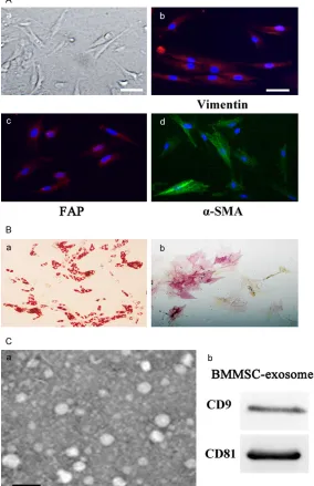
photo-Figure 1.Isolation and identification of MSCs and MSC-exosomes. A. Human BM-MSCs (a) were spindle-shaped and plastic adherent when maintained in Dulbecco’s modified Eagle’s medium with low glucose supplemented with 15% fetal bovine serum (magnification 200 ×; scale bar = 50 μm); hBM-MSCs (b, c, d) express high levels of fibroblast proteins such as vimentin (b), FAP (c) and α-SMA (d) (Magnification 200 ×; scale bar = 50 μm); B. Adipo -genic and osteo-genic differentiation of hBM-MSCs. Oil-Red-O staining (a) of lipids in hucMSCs; Neutrophil alkaline phosphatase (NAP) staining (b) of huc-MSCs. Cells were cultured for 14 days before staining (magnification 200 ×); C. Identification of hBMMSCs-exosome. Transmission electronic micrograph (a) of hBM-MSC-Ex, scale bar = 100 nm; Detection of hBMMSC-exosome CD9 and CD81 expression by western blot analysis (b).
(PCNA) (Signalway Antibody Co., Ltd.).
Enzyme-linked immunosor -bent assay
PDGF-D in the Dulbecco’s modified Eagle’s medium with low glucose (LG-DMEM) gro- up, MSC-Ex group were mea-sured using ELISA kit (R& D Systems, USA) in accor-dance with the manufactur-er’s instruction. Assays were performed in duplicate, and readings were compared with standard curves obtained with standard proteins pro-vided with the kits.
Transwell migration assay Following A549 cells treated with MSC-exosome for 72 h, A549 (1 × 10⁵/well) were
put into the upper chamber (8 mm) (Corning, NY, USA) in serum-free medium. The complete medium was pla- ced into the lower chamber. Following incubation at 37°C in 5% CO2 for 8 h, cells re- maining at the bottom of the polycarbonate membrane were wiped off with cotton swabs. The cells migrating to the lower surface of the mem-brane were then fixed with methanol for 30 min. The migrated cells were stained with crystal violet and count-ed in six random fields un-der the microscope (Olympus, Japan) for each assay.
[image:3.612.89.373.82.521.2]graphed and the number of colonies was counted.
Immunofluorescence staining
Following hBM-MSCs at 3 passage cultured for 48 h, the hBM-MSCs were washed 3 times with cold PBS, fixed with 4% paraformaldehyde for 20 min, permeabilized with 0.1% Triton X-100 for 5 min, blocked with 5% BSA (Boster Bioengineering, China) and incubated with anti-vimentin, anti-FAP, anti-α-SMA primary anti -body (Abcam, USA) at 4°C overnight and fol-lowed by Cy3-conjugated anti-rabbit second- ary antibody (Sigma, USA). The cells were then stained with DAPI for nuclear staining, and images were acquired using a Nikon Eclipse Ti-S microscope.
Immunohistochemistry
Formalin-fixed paraffin-embedded mouse tu-mor tissue sections were first deparaffinized in xylene, rehydrated through graded ethanol. The sections were boiled for 10 min in citrate buffer (pH 6.0, 10 mM) for antigen retrieval. The endogenous peroxidase activity was inhib-ited with exposure to 3% hydrogen peroxide for 10 min. Then the sections were blocked with 5% BSA and incubated with proper diluted PCNA primary antibody (1:200) at 37°C for 1 h.
4 groups. The animals were injected subcuta-neously with untreated A549 cells alone, A549 cells pretreated with MSC-exosome, A549 cells pretreated with MSC-exosome/NA-PDGFD and A549 cells pretreated with MSC-exosome/ CtrlA-PDGFD into the backside of mice respec-tively. Tumor growth was evaluated by mea- suring the length and width of the tumor mass with calipers every 4 days. Tumor volumes were calculated by the modified ellipsoidal formu-la: (length × width2)/2. Tumors were surgical-
ly removed, and photographed 40 days after injection.
Statistical analyses
All experiments were conducted at least in trip-licate. All data are expressed as the means ± SD. SPSS 11.0 software was used to analyze the data. The means of different treatment groups were compared by two-way ANOVA or the Student’s t-test. A P-value < 0.05 was considered to indicate a statistically signifi-cant difference.
Results
Isolation and identification of MSCs and MSC-exosomes
[image:4.612.89.376.76.282.2]Human bone marrow MSCs were isolated and confirmed positive for CD29, CD44, CD105,
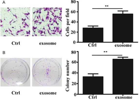
Figure 2. MSC-exosome promoted lung tumor migration and growth. A. Rep-resentative images showing that MSC-exosomes improved A549 cell pen-etration through membrane (magnification 200 ×; scale bar = 50 μm); B. Cell colony formation assay showing that MSC-exosome improved the ability of A549 cells to form colonies (**P < 0.01).
After washed with PBS, the sections were then incubated with diluted secondary anti-body for 20 min. Finally, sec-tions were visualized with 3, 3’-diaminobenzidine (DAB) and then counterstained with hematoxylin for examination under a light microscope (Oly- mpus, Japan).
Animal model
and HLA-I surface antigen expressions, and negative for CD34, CD38 and HLADR surface antigen expressions. BMMSCs presented a homogeneous population of spindle fibroblast-like cells. They possessed spindle shape and plastic adherent characteristics (Figure 1A). They also expressed high levels of fibroblast proteins, such as vimentin, fibroblast activation protein, FAP and α-SMA (Figure 1B-D). Von Kossa staining and Oil Red O staining were used to evaluate their osteogenic differentia-tion and adipogenic differentiadifferentia-tion potential, respectively. They showed spontaneous adi- pocyte or osteoblast formation (Figure 2B). MSC-exomes were isolated and purified using differential centrifugation on a sucrose cush-ion. By transmission electronic microscopy, it was found that MSC-exosomes were 30-100-nm microvesicles (MVs) (Figure 3A). MSC-exo- somes showed high levels of CD9 and CD81 expressions (Figure 3B).
To examine the molecular mechanism underly-ing MSC-exosome mediated promotion of tumor migration and proliferation, we investi-gated the expression of PDGFD in three differ-ent batches of MSC-exosome sources. Western blot and ELISA analyses revealed that PDGFD was highly expressed in exosome (Figure 3A). Because of the high expression of PDGFD in MSC-exosome, we investigated whether PDGFD was important for exosome in promoting the malignant transformation of lung cancer. To evaluate the effect of PDGFD on lung tumor cells, we performed migration and proliferation experiments after A549 cells were treated with recombinant PDGFD for one week. The results showed that the migration of cells was signifi -cantly increased compared with the control group (Figure 3B). We also found that the cell cloning ability was significantly enhanced. The untreated A549 cells were served as control in these experiments.
Figure 3. MSC-exosomes carrying PDGFD and the increased growth and mi-gration of PDGFD treated lung tumor cells. A. Western blot analysis (a) show-ing the expression of PDGFD in MSC-exosome (a). ELISA analysis showshow-ing (b) PDGF-DD level was much higher in MSC-exosomes than in the control cul-ture medium. B. Representative images showing that recombinant PDGF-DD (100 ng/ml) improved SGC-7901 cell penetration through membrane (mag-nification, 200 ×; scale bar = 50 μm). C. Cell colony formation assay showing that PDGF-DD could improve the cell colony forming ability of SGC-7901 (**P < 0.01, ***P < 0.001).
Influence of MSC-exosome on lung tumor migration and growth
The function of paracrine in- teractions between BM-MSCs and A549 was evaluated in
vitro by treating A549 cells with MSC-exosomes for 14 days. The A549 cell migration ability was evaluated by the transwell migration array. Af- ter A549 cells were cultured in the presence of MSC-exo- somes for 2 weeks, their mi- gration ability was significant -ly enhanced compared with the control group (Figure 2A). Moreover, the proliferation capacity of A549 cells was evaluated by the cell colony formation assay. Similarly, the results showed that cell pro- liferation was also significant -ly strengthened compared with the control group after A549 cells were treated with MSC-exosome for 2 weeks (Figure 2B).
Neutralizing antibody against PDGFD greatly alleviated MSC-exosome-induced lung tumor progression in vitro
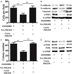
In order to study whether PDGFD was a key molecule in exosome mediated lung cancer development, we added either a neutralizing antibody against PDGFD (NA-PDGFD) or an iso-type-matched normal antibody (CtrlA-PDGFD) in the exosome group. Notably, this migration effect was abrogated by a PDGFD-specific blo-cking antibody, but not by a control antibody (Figure 4A). The results of western blot analy- sis also showed that the expression of E-cad- herin, an important feature of epithelial cell, was increased after A549 cells was treated with exosome in the presence of neutralizing antibody against PDGFD. The increased expre- ssion of E-cadherin was accompanied by de- creased expressions of the important
mesen-later, the xenograft tumors were removed, pho-tographed and weighed. Our data revealed that the size of the tumor from the neutralizing anti-body group was significantly smaller than those of the exosome group and the control antibody group; however, it was approximately equal to that in the control group (Figure 5A and 5B). In addition, we performed PCNA immunohis- tochemical staining on tissue sections from tumor tissue in each group. As shown by the increased number and intensity of PCNA-po- sitive cells, the results revealed that the per-centage of PCNA-positive cells in the neutraliz-ing antibody group was markedly lower than those in the exosome group and the control antibody group (Figure 5C).
Discussion
[image:6.612.93.376.74.363.2]Although MSCs is believed to be critically in- volved in tumor development and progression
Figure 4. Neutralizing antibody against PDGFD greatly alleviated MSC-ex-some-induced lung tumor progression in vitro. A. Transwell migration assay histogram of migration cell number in the exosome group, the NA-PDGFD (neutralizing antibody against PDGFD)/exosome group, and the CtrlA-PDGFD (isotype-matched normal antidody) group. B. Western blot analysis showing the expressions of E-cadherin, N-cadherin, α-SMA, and Vimentin in A549 cells in different groups. C. Cell colony formation assay histogram of A549 cells in the exosome group, the NA-PDGFD/exosome group, and the CtrlA-PDGFD group. D. Western blot analysis showing the expressions of PI3k, p-PI3k and PCNA in A549 cells in different groups (**P < 0.01).
chymal markers N-cadherin, Vimentin and α-SMA (Figure 4B). Similarly, compared with the control group, the pro- liferation ability of cells was significantly decreased in the PDGFD blocking antibody group (Figure 4C). In order to study the signal proteins associated with cell prolifera-tion, we tested p-PI3K, PI3K and PCNA. Western blot anal-ysis showed that the expre- ssions of p-PI3K and PCNA were weakened in the PDG- FD blocking antibody group (Figure 4D).
Neutralizing antibody against PDGFD abolished MSC-exosome growth-promoting
effect on lung cancer cells
tumor xenografts in vivo
[21, 22], the exact mechanism is still not very clear and poorly documented. The paracrine interactions that involve tumor growth between MSCs and cancer cells may be related to those responses. In addition to soluble factors, exo-somes have also been deemed as an important mechanism in the paracrine action of MSCs. Recent studies have found that exosomes con-tain functional nucleic acids and proteins when transferred into recipient cells, which indicated signal transmission for tumor progression [23, 24]. Here, we report an important finding that MSC-exosomes carrying PDGFD play a signifi -cant role in lung tumor progression.
In this study, we cultured BM-MSCs from hu- man bone specimens. The successful isolation and purification of exosomes from bone mar
-binant PDGFD for one week. The results showed that the effect of recombinant PDGFD on lung tumor progression was similar to that of MSC-exosome. Afterwards, we treated A549 cells with an antibody against PDGFD in the MSC-exsome group. The results demonstrated that the effect of MSC-exosome on migration and proliferation of tumor was greatly weakened compared to that that in the non-treated/con-trol-antibody group in vitro. It has been sug-gested that epithelial-mesenchymal transition (EMT) and migration are closely related [31]. Besides, PI3k signaling pathway is important for cell proliferation [32]. Western blot analysis demonstrated that EMT and PI3K signaling is significantly attenuated in the antibody against PDGFD group. Consistent with the in vitro
[image:7.612.93.378.70.395.2]results, our in vivo data showed that tumors
Figure 5. Neutralizing antibody against PDGFD abolished the growth pro-moting effect of MSC-exosome growth-propro-moting on lung cancer cell tumor xenografts in vivo. A. Representative photographs of tumor tissues in dif-ferent groups. B. The tumor volume of mice in difdif-ferent groups. C. Immu-nohistochemical analysis of PCNA expression in tumor tissues in different groups. (a) Representative photomicrograph; (b) percentage of proliferating cell nuclear antigen (PCNA)-positive cells (magnification, 200 ×; scale bar = 200 μm) (**P < 0.01).
row MSCs was by confirming their diameters (50-100 nm) and the protein expressions of CD9 and CD81. Carrying proteins, DNAs, mRNAs, and non-coding RNAs, exosomes play critical roles in tumori-genesis [25], growth [26], angiogenesis [27], metasta-sis [28] and drug remetasta-sistance [29]. At present, there is few research on the effect of MSC-exsomes on lung can- cer. Therefore, we investigat-ed whether MSC-exosomes promoted lung tumor migra-tion and growth in vitro. In order to study the mecha-nism, we evaluated the ex- pression of PDGFD in MSC-exsome. PDGFD is an isoform of the PDGF family, and may play a key role in the deve- lopment and progression of human cancers [30]. The re- sults of western blot and EL- ISA analyses showed high ex- pression of PDGFD in MSC- exosome.
recom-grew slower in the control and the antibody against PDGFD group than in the other two groups. Moreover, immunohistochemical analy-sis of PCNA expression in tumor tissues indi-cated that percentage of PCNA-positive cells was lower in the control and the antibody against PDGFD group than in the other two groups. These results suggest that PDGFD might play a key role in MSC-exosome medi- ated lung tumor progression. Further studies are needed to investigate whether there are other mechanisms involved.
In summary, it was revealed in this study that MSC-exosomes promote lung tumor progre- ssion through exosome carrying PDGFD. No- vel features of the complex dialogue between MSC-exosome and tumor cells were discover- ed in this study. These finding establishes an unprecedented link between both. This could be a lung tumor diagnostic biomarker and a novel selective therapeutic strategy targeting PDGFD exosomes carrying.
Acknowledgements
This work was supported by the Clinical Me- dicine Science and Technology Development Fund of Jiangsu University (grant no. JLY- 20140055) and the Develop the Health th- rough Science and Education of Suzhou You- th Science and Technology Project (grant no. kjxw2015052).
Disclosure of conflict of interest
None.
Address correspondence to: Longfei Xia, School of Medicine, Jiangsu University, 301 Xuefu Road, Zhenjiang 212013, China. Tel: 86-15850327201; E-mail: [email protected]
References
[1] Guo S, Reddy CA, Chao ST, Suh JH, Barnett GH, Vogelbaum MA and Videtic GM. Impact of non small cell lung cancer histology on sur-vival predicted from the graded prognostic as-sessment for patients with brain metastases. Lung Cancer 2012; 77: 389-393.
[2] Quail DF and Joyce JA. Microenvironmental regulation of tumor progression and metasta-sis. Nat Med 2013; 19: 1423-1437.
[3] Carnet O, Lecomte J, Masset A, Primac I, Maer- tens L, Detry B, Blacher S, Gilles C, Paupert J,
Foidart JM, Jerusalem G, Cataldo D and Noel A. Mesenchymal stem cells shed amphiregulin at the surface of lung carcinoma cells in a juxtacrine manners. Neoplasia 2015; 17: 552-563.
[4] Bremnes RM, Donnem T, Al-Saad S, Al-Shibli K, Andersen S, Sirera R, Camps C, Marinez I and Busund LT. The role of tumor stroma in cancer progression and prognosis: emphasis on carci-noma-associated fibroblasts and non-small cell lung cancer. J Thorac Oncol 2011; 6: 209-217.
[5] Huang J, Sha H, Wang G, Bao G, Lu S, Luo Q and Tan Q. Isolation and characterization of ex vivo expanded mesenchymal stem cells ob-tained from a surgical patient. Mol Med Rep 2015; 11: 1777-1783.
[6] Zhao K, Hao H, Liu J, Tong C, Cheng Y, Xie Z, Zang L, Mu Y and Han W. Bone marrow-derived mesenchymal stem cells ameliorate chronic high glucose-induced β-cell injury through modulation of autophagy. Cell Death Dis 2015; 6: e1885.
[7] Quante M, Tu SP, Tomita H, Gonda T, Wang SS, Takashi S, Baik GH, Shibata W, DiPrete B, Betz KS, Friedman R, Varro A, Tycko B and Wang TC. Bone marrow-derived myofibroblasts contrib -ute to the mesenchymal stem cell niche and promote tumor growth. Cancer Cell 2011; 19: 257-272.
[8] Kidd S, Spaeth E, Dembinski JL, Dietrich M, Watson K, Kloop A, Battula VL, Weil M, Andreeff M and Marini FC. Direct evidence of mesenchy-mal stem cell tropism for tumor and wounding microenvironments using in vivo biolumines-cent imaging. Stem Cells 2009; 27: 2614-2623.
[9] Kidd S, Spaeth E, Watson K, Burks J, Lu H, Klopp A, Andreeff M and Marini FC. Origins of the tumor microenvironment: quantita- tive assessment of adipose-derived and bone marrow-derived stroma. PLoS One 2012; 7: e30563.
[10] McAndrews KM, McGrail DJ, Ravikumar N and Dawson MR. Mesenchymal stem cells induce directional migration of invasive breast can- cer cells through TGF-β. Sci Rep 2015; 20: 16941-16943.
[11] Wang J, Wang Y, Wang S, Cai J, Shi J, Sui X, Cao Y, Huang W, Chen X, Cai Z, Li H, Bardeesi AS, Zhang B, Liu M, Song W, Wang M and Xiang AP. Bone marrow-derived mesenchymal stem cell-secreted IL-8 promotes the angiogenesis and growth of colorectal cancer. Oncotargel 2015; 6: 42825-42837.
mes-enchymal stem cells promote colorectal can-cer progression through paracrine neuregulin 1/HER3 signalling. Gut 2013; 62: 550-560. [13] Hogan NM, Dwyer RM, Joyce MR and Kerin MJ.
Mesenchymal stem cells in the colorectal tu-mor microenvironment: recent progress and implications. Int J Cancer 2012; 131: 1-7. [14] Jemal A, Siegel R, Xu J and Ward E. Cancer
sta-tistics, 2010. CA Cancer J Clin 2010; 60: 277-300.
[15] Wang Z, Kong D, Li Y and Sarkar FH. PDGF-D signaling: a novel target in cancer therapy. Curr Drug Targets 2009; 10: 38-41.
[16] Huang F, Wang M and Yang T. Gastric cancer-derived MSC-secreted PDGF-DD promotes gas-tric cancer progression. J Cancer Res Clin Oncol 2014; 140: 1835-1848.
[17] Roccaro AM, Sacco A, Maiso P, Azab AK, Tai YT and Reagan M. BM mesenchymal stromal cell-derived exosomes facilitate multiple myeloma progression. J Clin Invest 2013; 123: 1542-1455.
[18] Zhu W, Huang L, Li Y, Zhang X, Gu J, Yan Y, Xu X, Wang M, Qian H and Xu W. Exosomes de-rived from human bone marrow mesenchymal stem cells promote tumor growth in vivo. Cancer Lett 2012; 315: 28-37.
[19] Xu W, Zhang X, Qian H, Zhu W, Sun X, Hu J, Zhou H and Chen Y. Mesenchymal stem cells from adult human bone marrow differentiate into a cardiomyocyte phenotype in vitro. Exp Biol Med (Maywood) 2004; 229: 623-631. [20] Qu JL, Qu XJ, Zhao MF, Teng YE, Zhang Y, Hou
KZ, Jiang YH, Yang XH and Liu YP. Gastric can-cer exosomes promote tumour cell prolifera-tion through PI3K/Akt and MAPK/ERK activa-tion. Dig Liver Dis 2009; 41: 875-880.
[21] Xu WT, Bian ZY, Fan QM, Li G and Tang TT. Human mesenchymal stem cells (HMSCs) tar-get osteosarcoma and promote its growth and pulmonary metastasis. Cancer Lett 2009; 281: 32-41.
[22] Spaeth EL, Dembinski JL, Sasser AK, Watson K, Klopp A, Hall B, Andreeff M and Marini F. Mesenchymal stem cell transition to tumor-as-sociated fibroblasts contributes to fibrovascu -lar network expansion and tumor progression. PLoS One 2009; 4: e4992.
[23] Luga V, Zhang L, Viloria-Petit AM, Ogunjimi AA, Inanlou MR, Chiu E, Buchanan M, Hosein AN, Basik M and Wrana JL. Exosomes mediate stromal mobilization of autocrine Wnt-PCP sig-naling in breast cancer cell migration. Cell 2012; 151: 1542-1546.
[24] Lim PK, Bliss SA, Patel SA, Taborga M, Dave MA, Gregory LA, Greco SJ, Bryan M, Patel PS and Rameshwar P. Gap junction-mediated im-port of microRNA from bone marrow stromal cells can elicit cell cycle quiescence in breast cancer cells. Cancer Res 2011; 71: 1550-1560.
[25] Abd Elmageed ZY, Yang Y, Thomas R, Ranjan M, Mondal D, Moroz K, Feng Z, Reck BM, Moparty K, Sikka SC, Sartor O and Abdel-Mageed AB. Neoplastic reprogramming of pa-tient-derived adipose stem cells by prostate cancer cell-associated exosomes. Stem Cells 2014; 32: 983-997.
[26] Soldevilla B, Rodriguez M, San Millan C, Garcia V, Fernandez-Perianez R, Gil-Calderon B, Martin P, Garcia-Grande A, Silva J, Bonilla F and Dominguez G. Tumor-derived exosomes are enriched in Delta Np73, which promotes oncogenic potential in acceptor cells and cor-relates with patient survival. Hum Mol Genet 2014; 23: 467-478.
[27] Zhou W, Fong MY, Min Y, Somlo G, Liu L, Palomares MR, Yu Y, Chow A, O’Connor ST, Chin AR, Yen Y, Wang Y, Marcusson EG, Chu P, Wu J, Wu X, Li AX, Li Z, Gao H, Ren X, Boldin MP, Lin PC and Wang SE. Cancer-secreted miR-105 destroys vascular endothelial barriers to pro-mote metastasis. Cancer Cell 2014; 25: 501-515.
[28] Zomer A, Maynard C, Verweij FJ, Kamermans A, Schafer R, Beerling E, Schiffelers RM, Wit E, Berenguer J, Ellenbroek SI, Wurdinger T, Pegtel DM and van Rheenen J. In vivo imaging reveals extracellular vesicle-mediated phenocopying of metastatic behavior. Cell 2015; 161: 1046-1057.
[29] Wei Y, Lai X, Yu S, Chen S, Ma Y, Zhang Y, Li H, Zhu X, Yao L and Zhang J. Exosomal miR-221/222 enhances tamoxifen resistance in recipient ER-positive breast cancer cells. Breast Cancer Res Treat 2014; 147: 423-431. [30] Wang Z, Kong D, Banerjee S, Li Y, Adsay V and
Abbruzzese JL. Down-regulation of platelet de-rived growth factor-D inhibits cell growth and angiogenesis through inactivation of notch-1 and nuclear factor-κB signaling. Cancer Res 2007; 67: 11377-11385.
[31] Liu Z, Long J, Du R, Ge C, Guo K and Xu Y. miR-204 regulates the EMT by targeting snai1 to suppress the invasion and migration of gastric cancer. Tumour Biol 2016; 37: 8327-8335. [32] Gao J, Zhou XL, Kong RN, Li LM, He LL and