Original Article
The value of performing dual-source dual-energy
computed tomography in analysis strategy for
urinary mixed stone
Xiao-Gang Wang1*, Bo Fan1*, Wei Wang1, Yi-Ying Jin1, Yan Jin2, Zhi-Qiang Yang3, Xian-Cheng Li1,4
1Department of Urology, The Second Affiliated Hospital of Dalian Medical University, Dalian, Liaoning, China;
Departments of 2Laboratory Medicine, 3Cardiac Imaging, 4Urology, The First Affiliated Hospital of Dalian Medical
University, Dalian, Liaoning, China. *Equal contributors.
Received November 6, 2016; Accepted November 21, 2016; Epub February 1, 2017; Published February 15, 2017
Abstract: The aim of this study was to characterize and explore the mixed urinary stones by using Dual-Source Dual-Energy Computed Tomography (DSDECT) in vitro. Study scanned 134 human pure and mixed stones that were embedded into fresh porcine kidneys. Image analysis of these stones gave us the mean attenuation values
in Hounsfield units (HU) and energy index (DEI) at 80 kVp and 140 kVp. The mean of attenuation values, dual-energy and DEI were statistically different between uric acid (UA) containing and other stones, including mixed stones. For mixed stone, COX/UA and COX/PH had significantly difference in DEI (P<0.05), and the HU of 80 kVp, 140 kVp, and dual-energy were significantly different among COX/PH and PH/UA groups. The 80 kVp HU, 140 kVp HU and dual energy HU were significantly higher in COX/PH group compared with PH/UA group (1316.31 vs 813.23, 717.60 vs 440.03, 951.49 vs 557.38, respectively).The value of 80 kVp HU, 140 kVp HU and dual energy HU in COX/ UA was 1052.54, 704.12, 802.94 respectively. DSDECT was precise in the differentiation of UA-containing stones
and gave promising results for mixed stones of different types.
Keywords: Dual-source dual-energy computed tomography, urinary stone, stone composition
Introduction
Urinary calculi is a worldwide disease with an increasingly morbidity and a high recurrence rate. Incidence of urinary calculi is up to 1~5% in China; it could be even higher in some spe-cial area [1]. The following factors contribute to the etiopathogenesis and development of uri-nary calculi: (a) congenital factors, such as sex and age [2]; (b) dietary factors [3]; (c) abnormal factors, such as metabolic disorder (hypercal-cemia or hyperoxaluria), urinary tract obstruc-tion and urinary tract infecobstruc-tion [4]; and (iv) genetic and geographic factors [2-5].
Although patient’s symptoms and stone size are two important factors for selection of appro-priate treatment options, knowledge of chemi-cal composition of the stone would be crucial to determine the optimal management, assess the effectiveness of therapy, and formulate strategies to prevent recurrence [6-8]. In the case of calcareous stones (calcium oxalate
cal-culus or calcium phosphate calcal-culus), the treat-ment may require surgical intervention with shock wave lithotripsy (SWL), ureteroscopy or percutaneous nephrolithotomy (PCNL), and not adjunct medical therapy [9, 10]. In the case of non-calcareous stones, the treatment typically includes adjunct medical therapy that is also different according to the characteristics of each stone type. For example, UA-containing calculus may be treated with urinary alkaliniza-tion and low purine diet; and patients with stru-vite stones will often be treated with antibiotics before intervention [11-13]. The recent advanc-es in computed tomography (CT) attracted attention of the urologists and radiologists [14] that led to the progress in the management of 10% to 25% patients after reliable identification of stone composition [15].
col-lection. When image is collected at low and high-energy x-ray spectra, it has different atten-uation values that allow to detect different materials [16, 17]. Several studies on the pre-dictive power of dual-energy CT (DECT) for anal-ysis of pure stones have been recently pub-lished [16, 18, 19]. However, there is a lack of information about the predictive power of DSDECT for mixed stones. In this work, we used DSDECT to explore the characteristics of mixed urinary stones in vitro and to develop criteria for distinguishing of different types of stones.
Materials and methods
Study design and patients
We performed a study with 212 urinary calculi obtained from the sample bank of The First Affiliated Hospital of Dalian Medical University
Medical University. These samples have been collected during surgical and endoscopic inter-ventions between December 2007 and June 2015. The stone’s diameter ranged from 0.1 to 6.3 cm (the mean size was 1.4 cm). After exclu-sion of the stones with a diameter less than 4 mm (stones of this size usually is ejected spon-taneously and without treatment) [20], we obtained a dataset of 134 stones.
[image:2.612.95.522.69.436.2]Figure 2. The image of DSDECT. Radiologists measured attenuation value in image at 80 kVp (A) and 140 kVp (B).
Three images of stones in sagittal (C), coronal (D), and axial planes (E) and settings dialog (F). In diagram, four stone types (calcium oxalate, hydroxyapatite, cystine, and uric acid) are described by four small circles (CT density at 140
kVp on x-axis and CT density at 80 kVp on y-axis). Blue line splits plane in two parts: uric acid (red) and nouric acid
[image:4.612.92.311.156.249.2](blue).
Table 1. Characteristics of patients and stones Mean stone size ± SD cm 1.4±1.1 (0.1-6.3)
Stone location Bladder stone 49 19%
Kidney stone 133 65%
Ureteral stone 30 16%
Sex Male 124 58%
Female 88 42%
Age 5-87
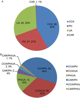
carbonate, phosphate and uric acid (CAR/PH/ UA), calcium oxalate, phosphate and uric acid (COX/PH/UA), calcium oxalate and phosphate (COX/PH), carbonate and phosphate (CAR/PH) containing stones.
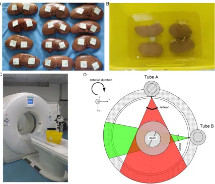
All stones were embedded into fresh porcine kidneys (obtained from a slaughterhouse) after hydrating in distilled water for 24 hours. The incision of the stones was coronal and implan-tation was performed in a water bath to keep air bubbles from entering into the collecting system. The location of each stone embedded in the kidneys was marked. The kidneys were placed in a 20-cm-deep water phantom and then scanned (Figure 1).
Dual-Source Dual-Energy Computed Tomography (DSDECT)
All examinations were performed with a DSDECT device (Somatom Definition, Siemens Healthcare). Technical parameters for the dual-energy scan were as follows: tube voltage, 80 kVp and 140 kVp; reference tube current, 96 mA and 400 mA with automatic exposure con-trol; acquisition slice thickness, 5 mm; recon-struction slice thickness, 1.5 mm; reconstruc-tion increment, 1.5 mm; gantry rotareconstruc-tion time, 0.5 second; filter kernel, B30f (medium smooth); and detector configuration, 32 × 0.6 mm. Image analysis was performed by two independent radiologists (ZQY, WW) who had years of experience in abdominal imaging and were blinded to the stone composition (Figure 2). The readout was carried out on a dedicated remote workstation (Leonardo, Siemens Heal- thcare) with a commercial software set (Syngo Dual Energy Viewer, Siemens Healthcare).
Regions of interest (ROI) of measured attenu-ation value for each urinary stone were detected with the software mentioned above. The dual-energy index (DEI) [22] was calcu-lated from DECT data (80/140 kVp), accord -ing to the follow-ing formula:
DEI
CT number (80 kVp) CT number (140 kVp) 2000 CT number (80 kVp) CT number (140 kVp) =
+ +
-Statistical analysis
Statistical analysis was performed using com-mercially available statistical software (SPSS, version 21.0, Chicago, IL). One-way ANOVA was conducted to compare the attenuation values, DEI values and overlap values among the differ-ent compositions of urinary stones. To adjust for multiplicity, Fisher’s least significant differ -ence t test correction was used when multiple comparisons were performed. Pearson correla-tion was used to analyze attenuacorrela-tion values and mixed stone sizes. Samples were consid-ered statistically different, if P value was <0.05.
Results
Urinary stones were obtained from the 212 patients during surgical or endoscopic inter-ventions. Characteristics of patients and stones are shown in Table 1.
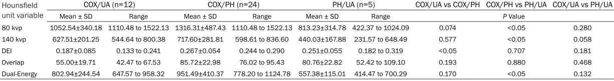
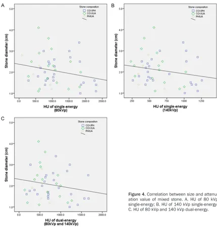
among COX/PH and PH/UA groups. The 80 kVp HU, 140 kVp HU and dual-energy HU were sig -nificantly higher in COX/PH group compared with PH/UA group (1316.31 vs 813.23, 717.60 vs 440.03, 951.49 vs 557.38, respectively). The value of 80 kVp HU, 140 kVp HU and dual-energy HU in COX/UA was 1052.54, 704.12, 802.94 respectively. The date are summarized in the Table 3. Mixed stones did not show a cor -relation between size and attenuation value overall, but it may have such tending (Figure 4).
Discussion
Nowadays, urinary stone disease has been an increasing problem. The choice of method for the clinical treatment of urinary tract stones depends not only on stone size, location, and brittleness, but also on stone composition [23]. Therefore, the ability to predict stone composi-tion before the treatment is a crucial feature for the selection of an optimal treatment. During
their method in an in vivo cohort is still not clear [28].
The application of energy information in DECT imaging has become a popular area of research with the development of radiological technolo-gy. There are two main DECT types, such as a dual-source DECT and single-source DECT. The single-source DECT relies on fast kilovoltage switching and a single-source dual-energy scanner with dual detector layers. Most of the studies are based on the application of dual-source DECT. Each material has a specific change in attenuation between images with a high-energy spectrum and with a low-energy spectrum.
[image:5.612.90.368.72.412.2]This attenuation differentiates one type stone from the others with a nuanced characteriza-tion. DSDECT has two separate x-ray tubes that can be operated at two different tube poten-tials. DSDECT’s two separate detectors can Figure 3. Distribution of mineral composition of pure stones (A) and mixed
stones (B).
Table 2. 80 kVp HU, 140 kVp HU, DEI, overlap value and dual-energy HU from pure stones
Hounsfield unit variable COX (n=34) UA (n=33) PH (n=26) UA vs COX UA vs PH PH vs COX
Mean ± SD Range Mean ± SD Range Mean ± SD Range P Value
80 kvp 1236.26±496.62 1062.99 to 1409.54 442.75±188.27 376.01 to 509.49 1233.47±470.31 1033.50 to 1413.44 <0.05 <0.05 0.906 140 kvp 704.12±259.86 550.94 to 748.78 423.28±132.71 550.94 to 748.78 649.86±244.91 550.94 to 748.78 <0.05 <0.05 0.363
DEI 0.238±0.118 0.196 to 0.297 0.009±0.092 -0.023 to 0.042 0.272±0.067 0.245 to 0.299 <0.05 <0.05 0.132
Overlap 100.02±126.01 56.05 to 143.99 -14.18±16.64 -21.08 to -8.27 76.37±23.44 66.90 to 85.83 <0.05 <0.05 0.174
Dual-Energy 889.32±352.71 766.25 to 1012.39 404.23±137.12 355.61 to 452.85 819.57±321.01 689.91 to 949.23 <0.05 <0.05 0.381
Table 3. 80 kVp HU, 140 kVp HU, DEI, overlap value and dual-energy HU from mixed stones
Hounsfield
unit variable
COX/UA (n=12) COX/PH (n=24) PH/UA (n=5) COX/UA vs COX/PH COX/PH vs PH/UA COX/UA vs PH/UA
Mean ± SD Range Mean ± SD Range Mean ± SD Range P Value
80 kvp 1052.54±340.18 1110.48 to 1522.13 1316.31±487.43 1110.48 to 1522.13 813.23±314.78 422.37 to 1024.09 0.074 <0.05 0.280 140 kvp 627.51±201.25 544.64 to 800.38 717.60±281.81 598.61 to 836.60 440.03±167.88 231.57 to 648.49 0.577 <0.05 0.058
DEI 0.187±0.085 0.133 to 0.241 0.267±0.054 0.244 to 0.290 0.251±0.055 0.182 to 0.319 <0.05 0.707 0.181
Overlap 55.00±19.71 42.47 to 67.53 85.72±22.98 76.02 to 95.43 80.76±22.82 52.42 to 109.10 0.193 0.880 0.468
[image:6.792.91.704.205.286.2]al for prediction of three types of stone, such as calcium oxalate-, cysteine- and uric acid-con-taining stones [20]. In addition, Li et al [16] found that the differences in mean of calcium density, calcium-water ratio, and radiodensity were statistically significant among five groups (uric acid, struvite, cystine, calcium phosphate and calcium oxalate) and demonstrated dual energy spectral CT provides a novel method for better characterization of pure urinary stones. Although the studies which applied DECT to predict stone composition were gradually increasing, most of them were focused on the predictive power of DECT for analysis of pure stones [16, 30-32]. The increased appearance acquire two different image datasets.
[image:7.612.90.520.67.535.2]Low-energy scans can be obtained at 80 or 100 kVp simultaneously, and high-energy scans can be obtained at 120 or 140 kVp. DSDECT can opti -mize image quality because the capacity of its two separate x-ray sources allows beam filtra -tion and adjustment of the current in each tube. In recent years, DECT with either a single or dual x-ray tube had advances in determination of the stone composition. Qu et al improved separation in CT number ratio between the five stone groups (uric acid, cystine, struvite, calci-um oxalate, carbonate apatite and hydroxyapa-tite) using 128-slice DSDECT scanner [29]. DSDECT was successfully used by Manglaviti et
Figure 4. Correlation between size and
of mixed stones in clinic requires the develop-ment of the reliable methods for the detection of these stones in vivo. In our study, we ana-lyzed the composition of mixed stones using DSDECT and distinguished COX/UA and COX/ PH groups, as well as COX/PH and PH/UA groups. We speculated that uric acid might play an important role in this differentiation, because uric acid stones are made of low molecular weight compound and they have a higher attenuation value at higher voltages. However, other stones containing calcium oxa-late, phosphate or cystine are made of molecular-weight compounds and have a high-er HU value at lowhigh-er voltages. In a result, uric acid mixed stones containing uric acid can be easily differentiated. However, we found that we cannot distinguish PH/UA and COX/UA stones by our method. The patients with both these stone types, PH/UA and COX/UA, can have with urinary alkalization or allopurinol therapy before subsequent PCNL or extracor-poreal SWL.
The relation between attenuation value of stones and effectiveness of chosen treatment (PCNL/SWL) were analyzed in several studies. Michio Tanaka’s study indicated that stone’s attenuation value less than 780 HU might have a successful result on SWL treatment. The combination of stone cross-sectional area and stone attenuation value was useful in deter-mining the SWL treatment for patients with uri-nary calculi [33]. Kawahara et al showed that stones with higher CT densities are more resis-tant to ESWL than those with lower CT densi-ties [34]. There was no high HU threshold above which clinicians would not consider SWL as a treatment option for urinary stones [33, 35, 36]. Largo et al reported that stones’ HU/DEI ratio was a significant and independent predic -tor for extracorporeal SWL in the number of required shock waves [35]. At the same time, Gucuk et al indicated that the attenuation value was one of independent predictors of the failure of the PCNL. The cut-off value was 677.5, and having a HU value under the cut-off value increased the likelihood of procedure fail-ure by 2.65 times [37]. According to above studies, we could consider that stones with low HU value were advantageous to extracorporeal SWL compared with PCNL. In our study, PH/UA stones have lowest HU value, suggesting that patients with PH/UA will have a better result
when treated with extracorporeal SWL rather than PCNL.
Several limitations of the present study should be considered. Although COX and PH had been identified by DSDECT, COX contained calcium oxalate monohydrate and calcium oxalate dehy-drate. Meanwhile, subtypes of PH were made of struvite, brushite and calcium phosphate. It suggested that further investigations will be required to differentiate subtypes of calcium stones. Moreover, a receiver operating charac -teristic study were not performed to establish a threshold for sensitivity and specificity, as the sample size of each mixed stone group (such as PH/UA and COX/UA ) was small. Large-scale studies in the future are required to establish the threshold values for quantitative evaluation of the stone types. In addition, the measure-ments of stones performed in vitro could give inaccurate results in comparison with mea-surements performed in vivo. Phantoms may not accurately reflect the anatomic surround -ings of renal stones. Future studies with a larg-er sample size should include in vivo study to determine clinical usefulness.
Conclusion
DSDECT differentiates uric and non-uric acid-containing stones, and also allows analysis of characteristics among mixed stones. It offers us a chance to make a better treatment plan for the patients with urinary stone disease.
Acknowledgements
This work was partly supported by the Natural Science Foundation of China (code 81572505).
Disclosure of conflict of interest
None.
Address correspondence to: Dr. Xian-Cheng Li,
Department of Urology, The Second Affiliated Hospital of Dalian Medical University, Shahekou
District Zhongshan Road no. 467 of Dalian, Liaoning, China. Tel: 0411-84671291; Fax: 0086-0411- 84671291; E-mail: [email protected]
References
[1] Zeng G, Mai Z, Zhao Z, Li X, Zhong W, Yuan J,
experi-ence with 12,482 consecutive patients over
20 years. Urolithiasis 2013; 41: 225-229.
[2] Moe OW. Kidney stones: pathophysiology and
medical management. Lancet (London, Eng-land) 2006; 367: 333-344.
[3] Ferraro PM, Curhan GC, Gambaro G, Taylor EN. Total, Dietary, and Supplemental Vitamin C In -take and Risk of Incident Kidney Stones. Am J Kidney Dis 2016; 67: 400-7.
[4] Astroza GM, Neisius A, Tsivian M, Preminger GM, Lipkin ME. Treatment Response in Stone Patients with Low Urinary pH and Hypocitratu
-ria Stratified by Body Mass Index. J Urol 2016;
195: 653-7.
[5] Skolarikos A, Straub M, Knoll T, Sarica K, Seitz C, Petřík A, Türk C. Metabolic evaluation and
recurrence prevention for urinary stone
pa-tients: EAU guidelines. Eur Urol 2015; 67:
750-763.
[6] Wiener SV, Deters LA, Pais VM Jr. Effect of
stone composition on operative time during ureteroscopic holmium: yttrium-aluminum-gar-net laser lithotripsy with active fragment
re-trieval. Urology 2012; 80: 790-794.
[7] Ramaswamy K, Killilea DW, Kapahi P, Kahn AJ,
Chi T, Stoller ML. The elementome of
calcium-based urinary stones and its role in
urolithia-sis. Nat Rev Urol 2015; 12: 543-557.
[8] Scherer K, Braig E, Willer K, Willner M, Fingerle AA, Chabior M, Herzen J, Eiber M, Haller B, Straub M, Schneider H, Rummeny EJ, Noël PB,
Pfeiffer F. Non-invasive differentiation of
kid-ney stone types using X-ray dark-field radiogra -phy. Sci Rep 2015; 5: 9527.
[9] Holmes RP, Knight J, Assimos DG. Lowering uri-nary oxalate excretion to decrease calcium
ox-alate stone disease. Urolithiasis 2016; 44:
27-32.
[10] Orhan N, Onaran M, Şen İ, Işık Gönül İ, Aslan M. Preventive treatment of calcium oxalate crystal deposition with immortal flowers. J Eth -nopharmacol 2015; 163: 60-67.
[11] Joshi HB, Kumar PV, Timoney AG. Citric acid
(solution R) irrigation in the treatment of re-fractory infection (struvite) stone disease: is it
useful? Eur Urol 2001; 39: 586-590.
[12] Friedlander JI, Moreira DM, Hartman C, Elsam -ra SE, Smith AD, Okeke Z. Comparison of the
metabolic profile of mixed calcium oxalate/uric
acid stone formers to that of pure calcium
oxa-late and pure uric acid stone formers. Urology
2014; 84: 289-294.
[13] Heilberg IP. Treatment of patients with uric
acid stones. Urolithiasis 2016; 44: 57-63.
[14] Ngo TC, Assimos DG. Uric Acid nephrolithiasis: recent progress and future directions. Rev Urol
2007; 9: 17-27.
[15] Kulkarni NM, Eisner BH, Pinho DF, Joshi MC, Kambadakone AR, Sahani DV. Determination
of renal stone composition in phantom and
pa-tients using single-source dual-energy comput-ed tomography. J Comput Assist Tomogr 2013; 37: 37-45.
[16] Li X, Zhao R, Liu B, Yu Y. Gemstone Spectral Imaging Dual-energy Computed Tomography: A
Novel Technique to Determine Urinary Stone Composition. Urology 2013; 81: 727-730.
[17] Duan X, Li Z, Yu L, Leng S, Halaweish AF,
Fletcher JG, McCollough CH. Characterization of Urinary Stone Composition by Use of
Third-Generation Dual-Source Dual-Energy CT With Increased Spectral Separation. AJR Am J Roentgenol 2015; 205: 1203-1207.
[18] Wisenbaugh ES, Paden RG, Silva AC,
Hum-phreys MR. Dual-energy vs Conventional Com -puted Tomography in Determining Stone
Com-position. Urology 2014; 83: 1243-1247.
[19] Selby MG, Vrtiska TJ, Krambeck AE, McCol -lough CH, Elsherbiny HE, Bergstralh EJ, Lieske
JC, Rule AD. Quantification of asymptomatic
kidney stone burden by computed tomography for predicting future symptomatic stone
events. Urology 2015; 85: 45-50.
[20] Manglaviti G, Tresoldi S, Guerrer CS, Di Leo G, Montanari E, Sardanelli F, Cornalba G. In Vivo Evaluation of the Chemical Composition of Uri
-nary Stones Using Dual-Energy CT. AJR Am J
Roentgenol 2011; 197: W76-W83.
[21] Ye YW. Clincial Laboratory Diagnostics.
Peo-ple’s Medical Publishing House; 1989.
[22] Graser A, Johnson TR, Bader M, Staehler M, Haseke N, Nikolaou K, Reiser MF, Stief CG,
Becker CR. Dual energy CT characterization of urinary calculi: initial in vitro and clinical expe-rience. Invest Radiol 2008; 43: 112-119. [23] Li XH, Zhao R, Liu B, Yu YQ. Determination of
urinary stone composition using dual-energy spectral CT: Initial in vitro analysis. Clin Radiol 2013; 68: e370-e377.
[24] Smith RC, Rosenfield AT, Choe KA, Essenmach
-er KR, V-erga M, Glickman MG, Lange RC. Acute flank pain: comparison of non-contrast-en -hanced CT and intravenous urography. Radiol-ogy 1995; 194: 789-794.
[25] Fielding JR, Silverman SG, Samuel S, Zou KH,
Loughlin KR. Unenhanced helical CT of ureter -al stones: a replacement for excretory urogra-phy in planning treatment. AJR Am J Roentgen-ol 1998; 171: 1051-1053.
[26] Hamm M, Wawroschek F, Weckermann D, Knöpfle E, Häckel T, Häuser H, Krawczak G, Harzmann R. Unenhanced helical computed tomography in the evaluation of acute flank pain. Eur Urol 2001; 39: 460-465.
[27] Motley G, Dalrymple N, Keesling C, Fischer J, Harmon W. Hounsfield unit density in the de
[28] Deveci S, Coşkun M, Tekin MI, Peşkircioglu L, Tarhan NC, Ozkardeş H. Spiral computed to -mography: role in determination of chemical compositions of pure and mixed urinary
stones--an in vitro study. Urology 2004; 64:
237-240.
[29] Qu M, Ramirez-Giraldo JC, Leng S, Williams JC, Vrtiska TJ, Lieske JC, McCollough CH. Dual-En -ergy Dual-Source CT With Additional Spectral Filtration Can Improve the Differentiation of
Non–Uric Acid Renal Stones: An Ex Vivo Phan -tom Study. Am J Roentgenol 2011; 196: 1279-1287.
[30] Stewart G, Johnson L, Ganesh H, Davenport D,
Smelser W, Crispen P, Venkatesh R. Stone Size Limits the Use of Hounsfield Units for Predic -tion of Calcium Oxalate Stone Composi-tion.
Urology 2015; 85: 292-295.
[31] Hidas G, Eliahou R, Duvdevani M, Coulon P, Le -maitre L, Gofrit ON, Pode D, Sosna J. Determi-nation of renal stone composition with dual-energy CT: in vivo analysis and comparison with x-ray diffraction. Radiology. 2010; 257: 394-401.
[32] Torricelli FC, Marchini GS, De S, Yamaçake KG, Mazzucchi E, Monga M. Predicting urinary
stone composition based on single-energy noncontrast computed tomography: the
chal-lenge of cystine. Urology 2014; 83:
1258-1263.
[33] Tanaka M, Yokota E, Toyonaga Y, Shimizu F, Ishii Y, Fujime M, Horie S. Stone attenuation
value and cross-sectional area on computed tomography predict the success of shock wave
lithotripsy. Korean J Urol 2013; 54: 454-459.
[34] Kawahara T, Miyamoto H, Ito H, Terao H, Kak
-izoe M, Kato Y, Ishiguro H, Uemura H, Yao M, Matsuzaki J. Predicting the mineral composi -tion of ureteral stone using non-contrast
com-puted tomography. Urolithiasis 2016; 44:
231-9.
[35] Largo R, Stolzmann P, Fankhauser CD, Poyet C, Wolfsgruber P, Sulser T, Alkadhi H, Winklhofer S. Predictive value of low tube voltage and du-al-energy CT for successful shock wave
litho-tripsy: an in vitro study. Urolithiasis 2016; 44:
271-6.
[36] Wang LJ, Wong YC, Chuang CK, Chu SH, Chen CS, See LC, Chiang YJ. Predictions of outcomes of renal stones after extracorporeal shock wave lithotripsy from stone characteristics de-termined by unenhanced helical computed to-mography: a multivariate analysis. Eur Radiol 2005; 15: 2238-2243.