Original Article
Downregulation of miR-181a alleviates renal
fibrosis in diabetic nephropathy mice
Jia Zhang1, Cong Wu2*, Jie Dong2, Jian Liu3*, Xiaowen Wei2
1School of Medicine, Shandong University, Jinan 250012, China; 2Department of Geriatric Endocrinology, The First Affiliated Hospital of Zhengzhou University, Zhengzhou 450003, China; 3Zhengzhou University, Zhengzhou 450001, China. *Equal contributors.
Received January 22, 2018; Accepted March 8, 2018; Epub August 1, 2018; Published August 15, 2018
Abstract: Accumulating evidence suggests that microRNAs are important regulators in the pathology of diabetes and its relevant renal injures. Little is known about the role of miR-181a in development of diabetic nephropathy. The aim of our present study was to investigate levels of miR-181a in diabetic nephropathy and explore its underlying mechanism. In the present study, Db/db and db/m mice were randomized into groups with 12 mice in each: db/m group, db/db group, and antagomiR-181a-treated db/db group. Changes in renal cortical sections were studied by histopathology. Mouse mesangial cells transfected with miR-mimic or miR-inhibitor and cell growth was measured using MTT assay. Levels of miR-181a expression were detected using qRT-PCR under different conditions. Indexes were measured using qRT-PCR and Western blot. Our results show that downregulation of miR-181a could alleviate pathological changes of diabetic nephropathy in mice. miR-181a expression was significantly upregulated in mouse mesaginal cells in vitro (P<0.05). Overexpression of miR-181a promoted extracellular matrix under high glucose by measuring related indexes such as collagen I, collagen IV, and fibronectin, which could be reversed by miR-181a in -hibitors (P<0.05). Upregulation of miR-181a suppressed expression of TβRIII by binding with 3’-UTR. These findings suggest miR-181a plays as an important role in renal fibrosis of diabetic nephropathy in an animal model.
Keywords: Diabetic nephropathy, miR-181a, mouse mesangial cells
Introduction
Diabetes mellitus, commonly referred to as dia-betes, is a metabolic disease characterized by hyperglycemia due to insulin-secreting or insu-lin-action disorders [1]. Persistent hyperglyce-mia and chronic metabolic disorders can lead to systemic organ dysfunction and failure, es- pecially in the eyes [2], kidneys [3], cardiovas-cular [4] and nervous [5] systems.
Diabetic nephropathy (DN) is one of the most serious and dangerous chronic complications caused by diabetes. Pathophysiologic abnor-malities in diabetic nephropathy begin with long-standing poorly controlled blood glucose levels. This is followed by multiple changes within the glomerulus which include a thicken-ing of the basement membrane, a widenthicken-ing of slit membranes of the podocytes, an increase in the number of mesangial cells, and an accu-mulation of extracellular matrix proteins [6, 7].
microRNAs (miRNAs) are a class of endogeno- us non-coding RNAs (about 21-24 nucleotides) that exhibit biological functions by binding to
the 3’ untranslated region (3’-UTR) of target
genes [8]. miRNAs could participate in a series of physiological and pathological processes by inhibiting target gene expressions including ce- ll differentiation, metabolism, proliferation, and apoptosis [9].
[15]. Recombinant FGF1 could normalize blood glucose levels and restore insulin sensitivity in diabetic rodents [16]. Guang Liang et al. have demonstrated that FGF1 could attenuate devel-opment of diabetic nephropathy by preventing
inflammatory responses [17].
The aim of our present study was to investigate levels of miR-181a in development of DN and explore its underlying mechanism.
Materials and methods
Animals
C57BL/6 db/db and control db/m mice (8 we- eks old, nine each group) were acquired fr- om the laboratory animal center of Zhengzhou University. The animal room was controlled at 20-22°C and 40-60% humidity with a 12 hour light/12 hour dark cycle. Mice were given standard chow and autoclaved water ad libi-tum. Mouse models of diabetes were con-structed by intraperitoneal STZ injections (100 mg/kg) for three consecutive days [18]. Db/db and db/m mice were randomized into groups with 12 mice in each: (1) db/m group, (2) db/db group, and (3) antagomiR-181a-treated db/ db group. Tail vein blood glucose levels >300 mg/dl was considered diabetic, measured by Glucose LiquiColor Test (Stanbio Laboratory, Boerne, TX, USA) every 4 weeks. Urinary sam-ples (24 hours) were collected by metabolic cage every 4 weeks and urine albumin concen-tration was determined by competitive ELISA, according to manufacturer instructions (Exo- cell, PA). Renal cortical tissues were dissected from kidneys, as previously described [19]. One
piece of fresh kidney tissue sample was fixed
by neutral formalin and the remaining tissues were stored at -80°C for future analysis. All ani-mal experiments were performed according to institutional, local, and national guidelines on animal research and ethics.
Histological studies
Renal cortical tissues were fixed for 48 hours,
dehydrated through a graded series of ethanol,
embedded in paraffin wax, and cut into 4 μm
sections. The samples were stained with hema-toxylin-eosin (HE) and periodic acid Schiff (PAS) to observe glomerular morphological changes.
Four random fields were chosen by light
mi-croscope.
Cell culture and transfection
293T cells were cultured in DMEM with 5 mmol/l glucose and 10% fetal bovine serum
at 37°C in a humidified 5% CO2 atmosphere. Mouse mesangial cells (MMCs) cultured in DMEM were treated with 25 and 5 mmol/l glu-cose respectively to mimic diabetic pathologi-cal and normal physiologipathologi-cal environments, as previously described [20]. For transfection, ce- lls were seeded into plates and transfected with miRNAs or siRNAs (Gene Pharm, Shang- hai, China) mixed with Lipofectamine 2000 re- agent (Invitrogen, Carlsbad, MA, USA), accord-ing to manufacturer protocol.
Cell proliferation assay
Cells (3×10³) were cultured in 96-well plates, incubated for 24 hours, and stained with 0.5 mg/ml MTT for 4 hours. Supernatant was
dis-carded and 200 μl of dimethylsulfoxide
(DM-SO) was added to dissolve precipitates. Abs- orption values were measured at 490 nm.
Real time quantitative PCR analysis
Total RNA was isolated from tissues and cells using TRIzol Reagent (Invitrogen, Carlsbad, CA, USA), according to manufacturer protocol. Complementary DNA (cDNA) was synthesized from 50 ng of total RNA using a miRCURY LNA Universal cDNA synthesis kit (Exiqon, Vedbaek,
Denmark). β-actin was used as an internal con -trol gene to normalize target genes. All primers were designed and synthesized by GenePharma (Shanghai, China). Relative levels of gene ex-
pression were expressed relative to β-actin and calculated using 2-ΔΔCt method.
Luciferase activity assay
The 3’ untranslated region (UTR) of FGF1 con
-taining miR-181a binding sites was amplified
and cloned into the psiCHECK-2 luciferase
vec-tor (Promega, USA). Similarly, mutant 3’-UTR of
FGF1 was cloned into the same vector. Cells maintained in 96-well plates were co-transfect-ed with miR-141a mimic or miR-NC. Transfectco-transfect-ed cells were detected using Dual-Luciferase Re- porter Assay System (Promega) 48 hours later.
Western blotting analysis
protease inhibitor mixture. After
[image:3.612.91.375.69.571.2]electrophore-sis, the protein samples were incubated wi- in db/db mice, significantly improved by an-tagomiR-181a (P<0.05) (Figure 1C). Overall,
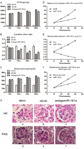
Figure 1. Role of miR-181a in renal function and pathological changes of DN in db/db mice. A, B: The ACR increased and the Ccr decreased signif-icantly with the progression of DN. When db/db mice were injected with antagomiR-181a, there was a significant decrease of ACR and an increase in Ccr (P<0.05). C: The level of blood glucose was upregulated remarkably in db/db mice, which was significantly improved by antagomiR-181a (P<0.05). D-F: miR-181a expression was positively correlated with ACR and blood glucose (r=0.689, r=0.759; P<0.05) and negatively correlated with Ccr (r=-0.685, P<0.05). G: Specimens showed increased mesangial matrix in db/ db mice and the degree of pathological changes was lightened when db/db mice received antagomiR-181a treatment.
th primary antibody (anti-FGF1, anti-Collagen I,
anti-Col-lagen IV, and anti-fibronectin,
1:1000 dilution). Samples we- re incubated with secondary antibodies conjugated by HRP.
Bands were quantified using
ImageJ software. Statistical analysis
Results are expressed as me-
an ± standard error. Student’s
t-test and ANOVA were per-formed among the different groups. All calculations were performed using SPSS 17.0 software (IBM Software, Chi- cago, IL, USA) and GraphPad (vision 6.0, USA). Corr-elation analysis of continuous vari-ables was based on Spear-
man’s test correlation meth -od. A value of P<0.05
indicat-ed a statistically significant
diff-erence. Results
Role of miR-181a in renal
function and pathological changes of DN in db/db mice
ACR and Ccr were considered clinical indexes of renal func-tion in DN [11, 21]. In order to examine miR-181a function in progression of DN, expression of these indexes was analyzed using qPCR. Results showed that ACR increased and Ccr
decreased significantly with
progression of DN (P<0.05) (Figure 1A, 1B). When db/db mice were injected with anta- gomiR-181a, there was a
sig-nificant decrease of ACR and
miR-181a expression was positively correlated with ACR and blood glucose (r=0.689, r= 0.759; P<0.05) (Figure 1D, 1E) and negatively correlated with Ccr (r=-0.685, P<0.05) (Figure 1F). To characterize the role of miR-181a in
renal fibrosis in DN, specimens obtained from
renal cortex at 20 weeks showed increased mesangial matrix in db/db mice. PAS and HE staining also showed the degree of pathologi-cal changes as they were lightened when db/db mice received antagomiR-181a treat-ment (Figure 1G).
Expression of miR-181a in MMCs under high glucose condition
To further investigate effects of miR-181a on development of DN, expression of miR-181a in MMCs was analyzed by RT-qPCR. MMCs were induced by high glucose (25 mM) for 12, 24, 48, and 72 hours. Figure 2A shows that
miR-181a mRNA expression was significantly
upre-gulated in MMCs under high glucose
condi-As major components of ECM in MMCs,
expres-sions of collagen I, collagen IV, and fibronectin
were detected to predict the accumulation of ECM under high glucose conditions by RT-PCR and Western blot. The results indicated that
expression of these indexes mRNA was signifi -cantly increased in MMCs (P<0.05) (Figure 3A). Additionally, overexpression of miR-181a ele-vated levels of these indexes (P<0.05) (Figure 3D). This means that overexpression of
miR-181a aggravated fibrosis of MMCs. Also, a
small interfering RNA (si-RNA) against miR-181a was successfully designed and assessed by qRT-PCR (P<0.05) (Figure 3B). Results of
qRT-PCR confirmed that expression of these indexes was significantly reduced in MMCs
(P<0.05) (Figure 3C, 3D). Taken together, this
suggests that miR-181a regulates fibrosis of
MMCs in vitro.
miR-181a targets with 3’-UTR of FGF1
Bioinformatics analysis was used to predict candidate targets of miR-141. Results revealed
Figure 2.Expression of miR-181a in MMCs under high glucose conditions.
MMCs were induced by high glucose (25 mM) for 12, 24, 48, and 72 hours. A: Showed that miR-181a mRNA expression was significantly upregulated in MMCs under high glucose conditions. B: miR-181a level was signifi-cantly elevated in MMCs transduced with miR-181a mimic compared with miR-NC (P<0.001). C: The growth rate of MMCs in high glucose conditions was increased, which could be further elevated by BANCR over-expression (P<0.05).
tions, implying the elevated level of miR-181a mRNA indu- ced by high glucose was time-dependent. Moreover, miR-18- 1a-overexpression in MMCs
was firstly established by ret -rovirus infection. Results of qRT-PCR manifested that
miR-181a levels were significantly
elevated in MMCs transduced with miR-181a mimic com-pared with miR-NC (P<0.001) (Figure 2B). To explore the role of miR-181a in cell prolifera-tion, MMT assay was per-formed to evaluate viability of MMCs under high and low glu-cose conditions, which mimic diabetic pathological and nor-mal physiological environme- nts, respectively. The results indicated that the growth rate of MMCs in high glucose con-ditions was increased, which could be further elevated by BANCR overexpression (P< 0.05) (Figure 2C).
miR-181a promotes
extracel-lular matrix accumulation in
3’UTR of FGF1 was highly conserved to bind with miR-181a. The 3’-UTR binding sites can be
seen in Figure 4A. Luciferase reporter assay
showed transfection of miR-181a could signifi -cantly restrict relative luciferase activity in MMCs (P<0.05) (Figure 4B), suggesting that miR-181a has inhibitory effects on FGF1 ex-
pression via interaction with 3’-UTR of FGF1.
After cells were transfected with miR-181a
mimic, FGF1 expression levels were
significa-ntly downregulated (P<0.05) (Figure 4C). Ov- erall, our study discovered that miR-181a sup-pressed expression of FGF1 by binding with
3’-UTR.
Discussion
[image:5.612.93.521.74.457.2]microRNAs (miRNAs) are small non-coding RNA molecules that function in RNA silencing and post-transcriptional regulation of gene expres- sion [22]. miR-181a is derived from an overlap-ping gene locus that is highly conservative among mammalian species [23]. miR-181a participates in various gene regulatory process-es such as development, differentiation, and immune modulation [24]. Zhou, B et al. report-ed that miR-181a could improve hepatic insulin sensitivity and glucose homeostasis, perhaps providing a potential new therapeutic strategy
for treating insulin resistance and type 2 diabe-tes [14]. However, to date, few studies have evaluated the role of miR-181a in DN deve- lopment.
Our present study focused on the role of
miR-181a in renal fibrosis of DN in db/db mice.
Results showed that miR-181a expression was positively correlated with ACR and negatively correlated with Ccr, indicating the value of miR-181a as a predictor for progression of DN. When db/db mice received antagomiR-181a
injections, renal fibrosis histopathological
ch-anges were slightly alleviated. miR-181a mRN-
A expression was significantly upregulated in
MMCs under high glucose conditions. MMT as- say showed that overexpression of miR-181a accelerated proliferation of MMCs. Furthermo- re, upregulated miR-181a promoted expression
pathogenesis of DN and provides a novel thera-peutic target for treatment of DN.
Acknowledgements
This work was supported by The First Affiliated
Hospital of Zhengzhou University. Disclosure of conflict of interest
None.
[image:6.612.89.371.71.392.2]Address correspondence to: Dr. Cong Wu, De- partment of Geriatric Endocrinology, The First Affi-liated Hospital of Zhengzhou University, No. 1 Jian- she East Road, Zhengzhou 450003, China. E-mail: cong0805@qq.com; Dr. Jian Liu, Zhengzhou Uni- versity, No. 100 Science Road, Zhengzhou 450001, China. E-mail: liujian_MD@163.com
Figure 4. miR-181a targets with 3’-UTR of FGF1. Bioinformatics analysis was used to predict the candidate targets of miR-141. The 3’-UTR binding sites can be seen in A. B: Luciferase reporter assay showed transfection of miR-181a could significantly restrict relative luciferase activity in MMCs (P<0.05), suggesting miR-181a has inhibitory effects on FGF1 expression via interaction with the 3’-UTR of FGF1. C: After cells were transfected with miR-181a mimic, FGF1 expression levels were significantly downregulated (P<0.05).
of fibrotic indexes mRNA and
protein levels. This was rever- sed, however, by using knock-down of miR-181a. These da- ta suggest that miR-181a
pro-motes renal fibrosis in devel -opment of DN.
FGF-1 belongs to FGF family and was discovered as a
mito-gen for cultured fibroblasts
[25]. FGF-1 has traditionally been believed to be a
pro-fibrotic factor promoting fibro -blasts proliferation [25, 26]. Nevertheless, strong evidence has demonstrated that FGF-1
has an anti-fibrotic role. FGF-1 could induce fibroblast apop -tosis and inhibit type I
colla-gen expression in lung fibrosis
[27, 28]. As for diabetic ne- phropathy, bioinformatics an- alysis showed miR-181a
tar-gets 3’-UTR of FGF1 closely.
These results demonstrate that overexpression of miR-181a could lower FGF1 mRNA and related protein produc-tion. These data suggest that miR-181a may induce renal
fibrosis in DN through modula -tion of FGF1.
References
[1] Rother KI. Diabetes treatment--bridging the di-vide. N Engl J Med 2007; 356: 1499-1501. [2] Zhang X, Zhao L, Deng S, Sun X and Wang N.
Dry eye syndrome in patients with diabetes mellitus: prevalence, etiology, and clinical characteristics. J Ophthalmol 2016; 2016: 8201053.
[3] Alicic RZ and Tuttle KR. Novel therapies for dia-betic kidney disease. Adv Chronic Kidney Dis 2014; 21: 121-133.
[4] Nakanishi T and Kato S. Impact of diabetes mellitus on myocardial lipid deposition: an au-topsy study. Pathol Res Pract 2014; 210: 1018-1025.
[5] Dehghani C, Srinivasan S, Edwards K, Pritchard N, Russell AW, Malik RA and Efron N. Presence of peripheral neuropathy is associated with progressive thinning of retinal nerve fiber layer in type 1 diabetes. Invest Ophthalmol Vis Sci 2017; 58: BIO234-BIO239.
[6] Wang M, Wang S, Yao D, Yan Q and Lu W. A novel long non-coding RNA CYP4B1-PS1-001 regulates proliferation and fibrosis in diabetic nephropathy. Mol Cell Endocrinol 2016; 426: 136-145.
[7] Tervaert TW, Mooyaart AL, Amann K, Cohen AH, Cook HT, Drachenberg CB, Ferrario F, Fogo AB, Haas M, de Heer E, Joh K, Noel LH, Rad-hakrishnan J, Seshan SV, Bajema IM and Bruijn JA. Pathologic classification of diabetic nephropathy. J Am Soc Nephrol 2010; 21: 556-563.
[8] Valencia-Sanchez MA, Liu J, Hannon GJ and Parker R. Control of translation and mRNA deg-radation by miRNAs and siRNAs. Genes Dev 2006; 20: 515-524.
[9] Rigoutsos I and Furnari F. Gene-expression fo-rum: decoy for microRNAs. Nature 2010; 465: 1016-1017.
[10] Conserva F, Pontrelli P, Accetturo M and Gesu-aldo L. The pathogenesis of diabetic nephrop-athy: focus on microRNAs and proteomics. J Nephrol 2013; 26: 811-820.
[11] Wang J, Gao Y, Ma M, Li M, Zou D, Yang J, Zhu Z and Zhao X. Effect of miR-21 on renal fibrosis by regulating MMP-9 and TIMP1 in kk-ay dia-betic nephropathy mice. Cell Biochem Biophys 2013; 67: 537-546.
[12] Zhang L, He S, Guo S, Xie W, Xin R, Yu H, Yang F, Qiu J, Zhang D, Zhou S and Zhang K. Down-regulation of miR-34a alleviates mesangial proliferation in vitro and glomerular hypertro-phy in early diabetic nephropathy mice by tar-geting GAS1. J Diabetes Complications 2014; 28: 259-264.
[13] Collares CV, Evangelista AF, Xavier DJ, Rassi DM, Arns T, Foss-Freitas MC, Foss MC, Puthier
D, Sakamoto-Hojo ET, Passos GA and Donadi EA. Identifying common and specific microR -NAs expressed in peripheral blood mononucle-ar cell of type 1, type 2, and gestational diabe-tes mellitus patients. BMC Res Nodiabe-tes 2013; 6: 491.
[14] Zhou B, Li C, Qi W, Zhang Y, Zhang F, Wu JX, Hu YN, Wu DM, Liu Y, Yan TT, Jing Q, Liu MF and Zhai QW. Downregulation of miR-181a upregu-lates sirtuin-1 (SIRT1) and improves hepatic insulin sensitivity. Diabetologia 2012; 55: 2032-2043.
[15] Itoh N and Ornitz DM. Evolution of the Fgf and Fgfr gene families. Trends Genet 2004; 20: 563-569.
[16] Holmes D. Therapy: FGF1 restores blood glu-cose levels and insulin sensitivity in diabetic mice. Nat Rev Endocrinol 2014; 10: 576. [17] Liang G, Song L, Chen Z, Qian Y, Xie J, Zhao L,
Lin Q, Zhu G, Tan Y, Li X, Mohammadi M and Huang Z. Fibroblast growth factor 1 amelio-rates diabetic nephropathy by an anti-inflam -matory mechanism. Kidney Int 2018; 93: 95-109.
[18] Yokoi H, Mukoyama M, Mori K, Kasahara M, Suganami T, Sawai K, Yoshioka T, Saito Y, Oga-wa Y, KuOga-wabara T, SugaOga-wara A and Nakao K. Overexpression of connective tissue growth factor in podocytes worsens diabetic nephrop-athy in mice. Kidney Int 2008; 73: 446-455. [19] Putta S, Lanting L, Sun G, Lawson G, Kato M
and Natarajan R. Inhibiting microRNA-192 ameliorates renal fibrosis in diabetic nephrop -athy. J Am Soc Nephrol 2012; 23: 458-469. [20] Mahimainathan L, Das F, Venkatesan B and
Choudhury GG. Mesangial cell hypertrophy by high glucose is mediated by downregulation of the tumor suppressor PTEN. Diabetes 2006; 55: 2115-2125.
[21] Viberti G and Wheeldon NM. Microalbuminuria reduction with valsartan in patients with type 2 diabetes mellitus: a blood pressure-indepen-dent effect. Circulation 2002; 106: 672-678. [22] Bartel DP. MicroRNAs: genomics, biogenesis,
mechanism, and function. Cell 2004; 116: 281-297.
[23] Pichler M, Winter E, Ress AL, Bauernhofer T, Gerger A, Kiesslich T, Lax S, Samonigg H and Hoefler G. miR-181a is associated with poor clinical outcome in patients with colorectal cancer treated with EGFR inhibitor. J Clin Pathol 2014; 67: 198-203.
[24] Tekirdag KA, Korkmaz G, Ozturk DG, Agami R and Gozuacik D. MIR181A regulates starva-tion- and rapamycin-induced autophagy through targeting of ATG5. Autophagy 2013; 9: 374-385.
-[28] Ramos C, Montano M, Becerril C, Cisneros-Lira J, Barrera L, Ruiz V, Pardo A and Selman M. Acidic fibroblast growth factor decreases al -pha-smooth muscle actin expression and in-duces apoptosis in human normal lung fibro -blasts. Am J Physiol Lung Cell Mol Physiol 2006; 291: L871-879.
lations by a fibroblast growth factor, dexameth -asone, and insulin. Proc Natl Acad Sci U S A 1974; 71: 4584-4588.
[26] Armelin HA. Pituitary extracts and steroid hor-mones in the control of 3T3 cell growth. Proc Natl Acad Sci U S A 1973; 70: 2702-2706. [27] Becerril C, Pardo A, Montano M, Ramos C,