Original Article
Increased SPARC expression in skeletal muscle
and adipose tissue of db/db mice
Hai-Yan Song1, Xiao-Yao Yang1, Lei Ding2, Lu-Jie Yang1, Hua Xie1, Yan-He Wang1
1Department of Endocrinology and Metabolism, The Second Affiliated Hospital of Harbin Medical University, Harbin, China; 2Department of Basic Medical Science, Wuxi Medical School, Jiangnan University, Jiangsu, China Received May 8, 2016; Accepted June 22, 2016; Epub August 1, 2016; Published August 15, 2016
Abstract: Secreted protein acidic and rich in cysteine (SPARC), also known as osteonectin or basement mem-brane-40, is involved in osteogenesis, angiogenesis, wound healing, and tumorigenesis. SPARC is predominantly secreted by adipose tissue, which has been recently linked with insulin resistance and glucose metabolism. Skeletal muscle is responsible for more than 75% of glucose disposal in response to insulin. In the current study, we ex-amined SPARC expression in insulin-resistant skeletal muscle and adipose tissue. Specifically, SPARC mRNA and protein levels were measured in adipose tissue and skeletal muscle of db/db and control groups. Increased SPARC expression was observed in adipose tissue and skeletal muscle of db/db mice, suggesting that the protein partici-pated in insulin resistance and glucose uptake. Our findings collectively indicated that SPARC contributed to insulin resistance and regulation of glucose metabolism, supporting its utility in the prevention and treatment of type 2 diabetes mellitus (T2DM) and metabolic syndrome.
Keywords: Secreted protein acidic and rich in cysteine, AMP-activated protein kinase, insulin resistance, adipose tissue, skeletal muscle, glucose uptake
Introduction
Secreted protein acidic and rich in cysteine (SPARC) is the protein product of a single gene localized at the chromosomal region 5q31-33 which is closely proximal to the genes encoding macrophage colony-stimulating factor 1, inter-leukin 3, platelet-derived growth factor and
β2-adrenergic receptor [1]. SPARC, also known
as osteonectin or basement membrane-40, is a member of the matricellular protein family, which was initially detected in bone and
endo-thelial cells [2, 3]. The protein was subsequent
-ly identified in a variety of tissues during devel
-opment, tissue repair and remodeling [4],
including notochord, somites, embryonic skel-eton, differentiating chondrocytes, megakaryo-cytes, and macrophages, at the sites of tissue
injury [5]. Interestingly, SPARC is a multifunc -tional protein involved in osteogenesis, angio-genesis, wound healing, tumoriangio-genesis, and
pathogenesis of fibrosis of the kidney [6, 7] and liver [8]. The role of SPARC in modifying the
ECM, particularly its effect on collagen
produc-tion and assembly, is well established [9, 10]. SPARC additionally contributes to collagen fibril
formation in the dermis, and consistently, SPARC knockout mice display reduced collagen
content in the dermis [11]. Incidentally, there is
increasing evidence to support the involvement of SPARC in regulating the tumor microenviron-ment, tumor developmicroenviron-ment, invasion,
metasta-sis, angiogenemetasta-sis, and inflammation [12].
SPARC is secreted by adipose cells, and its cir-culatory level is positively correlated with BMI in
humans [13, 14]. Moreover, the protein is pro -posed to contribute to the pathogenesis of obe-sity-associated disorders, including T2DM and its complications, mainly through promoting insulin resistance. Although several activities are attributed to SPARC, little is known about its
specific role in glucose metabolism in skeletal
muscle. Intriguingly, SPARC overexpression in skeletal muscle has been reported, leading to the hypothesis that the protein plays a key role in the glucose transport system. However, this
Diabetes is associated with dysfunction of glu-cose metabolism and insulin resistance. The objective of this study was to assess the conse-quences of SPARC expression in skeletal mus-cle and adipose tissue, and ascertain whether SPARC contributed to insulin resistance and glucose metabolism.
Materials and methods
Materials
Ten-week-old male db/db mice and their het-erozygote littermates were donated by the Model Animal Research Center of Nanjing University. The mice were housed in a tempera-ture-controlled (21°C) room under a dark-light
cycle of 12 h each. Following sacrifice, rat tis -sues were removed and stored at -80°C until further processing. This study was approved by the Animal Care and Handling Committee of Harbin Medical University.
Double antibody sandwich enzyme linked im-munosorbent assay (ELISA)
The prepared serum was taken from the -80°C refrigerator, which underwent natural coagula-tion under the room temperature for 30 min, followed by centrifugation at 1000×g for 15 min under the room temperature. The sample serum was diluted by 40 times, and double antibody sandwich enzyme linked immunosor-bent assay was employed for the determina-tion. The absorbance value (OD value) of the standard and sample serum was read by the multi-functional micro-plate reader. The stan-dard curve was plotted by the concentration and OD value of the standard serum, and SPARC concentration in the sample serum was
calculated, which was finally multiplied by the
dilution times.
Semi-quantitative RT-PCR
Specific primers were synthesized by Invitrogen,
including SPARC forward,5’-GAA GGT ATG CAG CAA TGA CAA CAA-3’, and SPARC reverse, 5’-TTC
GGT CAG CTC GGA ATC CA-3’ (leading to amplifi -cation of a 160 bp SPARC DNA fragment), and ß-actin forward, 5’-CCC AGC ACA ATG AAG ATC AAG ATC AT-3’, and ß-actin reverse, 5’-ATC TGC TGG AAG GTG GAC AGC GA-3’. Total RNA was extracted from adipose and skeletal muscle of
each sample using TRIzol reagent (Invitrogen).
cDNA was generated from total RNA (1 μg)
using the Reverse Transcription system (Prome-
ga), and further amplified via 35 cycles of PCR using Tag PCR Master Mix (Takara) and specific
primers. The reverse transcription reaction involved incubation at 42°C for 15 min, at 99°C for 5 min, and at 5°C for 5 min. Subsequent PCR reactions were performed under the following conditions: initial incubation at 95°C for 2 min; denaturation at 94°C for 15 s, anneal-ing at 55°C for 15 s, and extension at 72°C for
15 s, 32 cycles; final extension at 72°C for 2
min. PCR products were analyzed by electro-phoresis on 2% agarose gels. Band intensities
were quantified using the gel documentation
system.
Western blot analysis
Protein extracts (30 μg) were separated by
electrophoresis on the 10% SDS gel run at 100 V for 80 min. Fractionated proteins from sam-ple homogenates were transferred to nitrocel-lulose membrane, as described previously. Membranes were blocked for 70 min in TBS-T (Tris-buffered saline with 0.1% Tween-20) con-taining 5% non-fat dry milk at the room temper-ature, and incubated overnight at 4°C with anti-SPARC and anti-ß-actin primary antibodies (1:1000 dilution). Subsequently, membranes were incubated with the appropriate anti-rabbit secondary antibody diluted in TBS-T (1:4000)
for 1 h. Proteins were visualized on Kodak film
using an enhanced chemiluminescence (ECL) Western Blot Detection Kit according to the manufacturer’s instructions.
Immunofluorescence analysis
Skeletal muscle and adipose tissues were fixed
onto glass slides, and examined using an
epi-fluorescence microscope with the appropriate filters and photographic attachment.
Results
Quantitative detection of serum SPARC con -centration
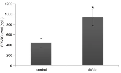
The serum SPARC level in db/db mice was
sig-nificantly higher than that in control mice (P <
0.01, Figure 1).
Expression levels of SPARC mRNA and protein
We analyzed both mRNA (Figure 2A and 2B) and protein (Figure 3A and 3B) expression lev-els of SPARC in skeletal muscle and adipose tissue in db/db mice, and compared them with the controls. Semi-quantitative PCR revealed elevation of SPARC mRNA in db/db mice com-pared to the controls. Western blot analysis was performed to ascertain whether there was increased expression of SPARC protein. SPARC protein expression data were consistent with those of semi-quantitative PCR
Immunofluorescence of SPARC
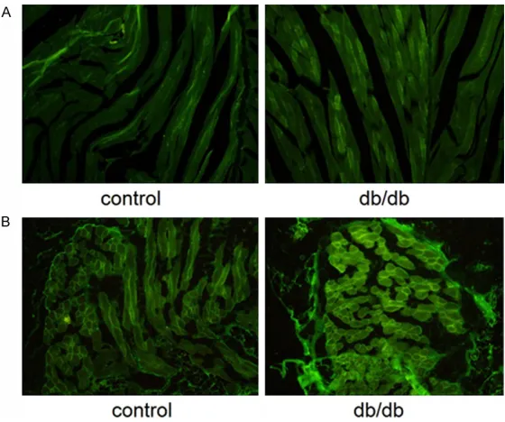
Immunofluorescence analysis for SPARC was
performed in skeletal muscle (Figure 4A) and adipose tissue (Figure 4B). The distribution and
intensity of fluorescence in these regions were
increased in db/db mice, compared with the controls, indicating upregulation of SPARC expression.
Discussion
SPARC is closely linked with the elevated risk of metabolic disorders, especially T2DM and its
complications, which are characterized by insu-lin resistance. Previous studies have suggested possible involvement of SPARC in insulin resis-tance through regulating lipometabolism. Overexpression of SPARC has been reported in
rodent models of diabetic nephropathy [15],
and SPARC-null mice are protected against
renal fibrosis [16]. Furthermore, SPARC expres -sion is reduced in early diabetes-related kidney
growth [17]. SPARC is expressed in retinal
endothelial cells and associated with diabetic
retinopathy [18]. In addition, diabetes-related
mesenteric vascular hypertrophy is associated with increased SPARC expression in the vessel
wall [19].
Insulin resistance in skeletal muscle and adi-pose tissue is linked to the development of metabolic diseases, such as obesity and hyper-glycemia. Accordingly, recent research has focused on identifying natural bioactive com-pounds, with a view to improve insulin resis-tance associated with T2DM.
In the current study, we aimed to determine whether elevated SPARC expression was asso-ciated with insulin resistance and T2DM. Analysis of SPARC mRNA and protein sion levels clearly disclosed increased expres-sion in adipose tissue and skeletal muscle of db/db mice compared with the controls. Earlier investigations have shown that insulin resistance is derived from adipose tissue, lead-ing to increased tissue decomposition and decreased synthesis. SPARC is an active com-ponent derived from adipocytes, which limits adipogenesis by inhibiting the differentiation of preadipocytes to mature adipocytes, suggest-ing its involvement in the regulation of adipo-cyte differentiation and insulin resistance. One possible mechanism is via stimulation of the
wnt/β-catenin signaling pathway, which trig -gers increased osteogenesis along with decreased adipogenesis. Adipogenic transcrip-tion factors, including CAAT/enhancer-binding
protein α, CAAT/enhancer-binding protein β,
and peroxisome proliferator-activated receptor
γ, are significantly decreased by SPARC, along
with adipocyte genes, such as leptin and
lipo-protein lipase [20].
[image:3.612.91.290.69.190.2]Skeletal muscle not only plays an important role in glucose homeostasis, providing an effec-tive therapeutic target for T2DM, but also acts
Figure 1. Plasma levels of SPARC. The graphs represent mean ± SEM. *P < 0.01, compared to con
Notably, skeletal muscle is the major site of insulin-stimulated glucose utilization. The ability of SPARC inducing glucose uptake possibly depends on both the genetic makeup and metabolic properties of mus-cle. Glycogen phosphorylase activity excee-ds glycogen syn-thesis activity in skeletal mus-cle, ensuring continued capac-ity for glycogenolysis. Data from the present study indicat-ed that increasindicat-ed SPARC expression in skeletal muscles was associated with insulin resistance and T2DM.
Two possible mechanisms underlie SPARC involvement in insulin resistance and
glu-cose metabolism, specifically,
AMP-activated protein kinase (AMPK) and insulin signal- ing pathways. Previous studi- es have demonstrated that SPARC and AMPK co-interact with each other and enhance each other. SPARC may regu-late glucose metabolism via
AMPK activation [21]. AMPK
plays a key role in sensing intracellular ATP levels, and acts as a crucial component in maintaining the energy bal-ance within cells. Stimulation of AMPK triggers an increase in the intracellular AMP: ATP
ratio [22]. Overexpression of
SPARC may increase the intra-cellular AMP: ATP ratio, thus enhancing glucose uptake via
AMPK activation. Recent find -ings have shown that expres-sion and secretion of SPARC
are influenced by glucose, insulin and leptin [13]. Skeletal
muscle and adipose tissue represent the major sites of insulin-stimulated glucose uti-lization. Both PI3-kinase and AKT are linked to the insulin signaling pathway. Notably, SPARC promotes AKT and
[image:4.612.91.374.72.156.2]PI3-Figure 2. SPARC mRNA expression in skeletal muscle (A) and adipose tissue (B). Total RNA was extracted, and mRNA expression was evaluated using RT-PCR, with β-actin as the control. RT-PCR was conducted using specific primers. The amplified product was run on the 2% agarose gel and visual -ized with the gel documentation system.
Figure 3. SPARC protein expression in skeletal muscle (A) and adipose tissue (B). Protein extracts were prepared for Western blot analysis, and SPARC expression was estimated by immunoblotting with the appropriate antibodies. The β-actin protein served as the control.
Figure 4. Immunofluorescence analysis of SPARC in skeletal muscle (A) and adipose tissue (B). Fixed tissues were permeabilized for 5 min in PBS con-taining 0.1% Triton X-100. Tissues were re-washed with PBS and incubated with mouse anti-human SPARC, followed by goat anti-mouse IgG-FITC. Dis-tribution and intensity of fluorescence were increased in db/db mice com -pared with the controls.
as a major target of insulin. Thus it is commonly
[image:4.612.91.374.238.317.2] [image:4.612.91.371.388.622.2]Since AMPK signaling is independent of insulin
signaling [23], we hypothesize that cross-talk
occurs between the two pathways.
In summary, our findings provide foundation for
clarifying the relationship between SPARC and insulin resistance in T2DM, and extend our understanding about its roles in fat and glu-cose metabolism. We propose that SPARC presents an attractive agent for the treatment of metabolic disorders and prevention of insu-lin resistance in T2DM.
Acknowledgements
This work was supported by the National Natural Science Foundation of China (No. 81370903 to Haiyan Song).
Disclosure of conflict of interest
None.
Address correspondence to: Hai-Yan Song, De- partment of Endocrinology and Metabolism, The Second Affiliated Hospital of Harbin Medical University, 246 Xuefu Road, Harbin 150080, China. Tel: +86-451-86297160; Fax: +86-451-86413857; E-mail: [email protected]
References
[1] Sage H, Johnson C and Bornstein P. Character-ization of a novel serum albumin-binding glyco-protein secreted by endothelial cells in culture. J Biol Chem 1984; 259: 3993-4007.
[2] Termine JD, Kleinman HK, Whitson SW, Conn KM, McGarvey ML and Martin GR. Osteonec-tin, a bone-specific protein linking mineral to collagen. Cell 1981; 26: 99-105.
[3] Saltman DL, Dolganov GM, Warrington JA, Wasmuth JJ and Lovett M. A physical map of 15 loci on human chromosome 5q23-q33 by two-color fluorescence in situ hybridization. Genomics 1993; 16: 726-732.
[4] Lane TF and Sage EH. The biology of SPARC, a protein that modulates cell-matrix interactions. FASEB J 1994; 8: 163-173.
[5] Tai IT and Tang MJ. SPARC in cancer biology: Its role in cancer progression and potential for therapy. Drug Resist Update 2008; 11: 231-246.
[6] Reed MJ and Sage EH. SPARC and the extra-cellular matrix: implications for cancer and wound repair. Curr Top Microbiol Immunol 1996; 213: 81-94.
[7] Bradshaw AD, Reed MJ and Sage EH. SPARC-null mice exhibit accelerated cutaneous wound closure. J Histochem Cytochem 2002; 50: 1-10.
[8] Camino AM, Atorrasagasti C, Maccio D, Prada F, Salvatierra E, Rizzo M, Alaniz L, Aquino JB, Podhajcer OL, Silva M and Mazzolini G. Adeno-virusmediated inhibition of SPARC attenuates liver fibrosis in rats. J Gene Med 2008; 10: 993-1004.
[9] Bradshaw AD. The role of SPARC in extracellu-lar matrix assembly. J Cell Commun Signal 2009; 3: 239-246.
[10] Seet LF, Su R, Barathi VA, Lee WS, Poh R, Heng YM, Manser E, Vithana EN, Aung T, Weaver M, Sage EH and Wong TT. SPARC deficiency re -sults in improved surgical survival in a novel mouse model of glaucoma filtration surgery. PLoS One 2010; 5: e9415.
[11] Bradshaw AD, Puolakkainen P, Dasgupta J, Da-vidson JM, Wight TN, Helene Sage E. SPARC-null mice display abnormalities in the dermis characterized by decreased collagen fibril di -ameter and reduced tensile strength. J Invest Dermatol 2003; 120: 949-955.
[12] Chlenski A and Cohn SL. Modulation of matrix remodeling by SPARC in neoplastic progres-sion. Semin Cell Dev Biol 2010; 21: 55-65. [13] Kos K, Wong S, Tan B, Gummesson A, Jernas
M, Franck N, Kerrigan D, Nystrom FH, Carlsson LM, Randeva HS, Pinkney JH and Wilding JP. Regulation of the fibrosis and angiogenesis promoter SPARC in human adipose tissue by weight change, leptin, insulin and glucose. Dia-betes 2009; 58: 1780-1788.
[14] Takahashi M, Nagaretani H, Funahashi T, Nishizawa H, Maeda N, Kishida K, Kuriyama H, Shimomura I, Maeda K, Hotta K, Ouchi N, Ki-hara S, Nakamura T, Yamashita S and Matsu-zawa Y. The expression of SPARC in adipose tissue and its increased plasma concentration in patients with coronary artery disease. Obes Res 2001; 9: 388-393.
[15] Taneda S, Pippin JW, Sage EH, Hudkins KL, Takeuchi Y, Couser WG and Alpers CE. Amelio-ration of diabetic nephropathy in SPARC-null mice. J Am Soc Nephrol 2003; 14: 968-980. [16] Socha MJ, Manhiani M, Said N, Imig JD and
Motamed K. Secreted protein acidic and rich in cysteine deficiency ameliorates renal inflam -mation and fibrosis in angiotensin hyperten -sion. Am J Pathol 2007; 171: 1104-1112. [17] Gilbert RE, McNally PG, Cox A, Dziadek M,
Rumble J, Cooper ME and Jerums G. SPARC gene expression is reduced in early diabetes-related kidney growth. Kidney Int 1995; 48: 1216-1225.
[18] Munjal ID, McLean NV, Grant MB and Blake DA. Differences in the synthesis of secreted proteins in human retinal endothelial cells of diabetic and nondiabetic origin. Curr Eye Res 1994; 13: 303-310.
gene expression is increased in diabetes- related mesenteric vascular hypertrophy. Mi-crovasc Res 2000; 59: 61-71.
[20] Nie J and Sage EH. SPARC inhibits adipogene-sis by its enhancement of betacatenin signal-ing. J Biol Chem 2009; 284: 1279-1290. [21] Song H, Guan Y, Zhang L, Li K and Dong C.
SPARC interacts with AMPK and regulates GLUT4 expression. Biochem Biophys Res Com-mun 2010; 396: 961-966.
[22] Carling D. The AMP-activated protein kinase cascade-a unifying system for energy control. Trends Biochem Sci 2004; 29: 18-24. [23] Kurth-Kraczek EJ, Hirshman MF, Goodyear LJ