Case Report
Mandibular central leiomyosarcoma
with high telomerase activity: a case report
Tomoki Sumida1, Akiko Ishikawa2, Hiroyuki Nakano1, Yosuke Kobayashi1, Tomohiro Yamada1, Yoshihide Mori1
1Department of Oral and Maxillofacial Surgery, Kyushu University, Fukuoka, Japan; 2Department of Oral and
Maxillofacial Surgery, Ehime University, Ehime, Japan
Received December 19, 2015; Accepted April 25, 2016; Epub February 1, 2018; Published February 15, 2018
Abstract: Leiomyosarcoma is a malignant lesion of smooth muscle origin, and rare in the oral region. This report presents an extremely rare case of intraosseous leiomyosarcoma of the mandible. After visiting other general hospi-tal, a 29-year-old man was referred to our hospital because of a pain in the left mandibular region with paresthesia of the left mental region. The left mandibular third molar had already been extracted in another hospital, and a brownish mass occupied the corresponding region. A panoramic radiograph showed osteolytic destruction around the left mandibular angle and ramus. A computed tomography scan and magnetic resonance image revealed per-foration of the lingual and buccal cortex of the mandible. A non-epithelial malignant tumor was diagnosed from a biopsy specimen. Immediately, we resected the tumor and reconstructed the titan plate under general anesthesia. A final diagnosis of leiomyosarcoma was made from a surgical specimen based on findings showing a proliferation of hyperchromatic spindle cells, which were positive for the markers α- smooth muscle actin, calponin, HHF35, and desmin. The S-100, epithelial membrane antigen, and cytokeratin markers were negative. The patient had 3 courses of adjuvant chemotherapy after the operation, and showed no evidence of recurrence during the follow-up at the outpatient clinic. However, 2 years after the first operation, lung metastases and local recurrence were de-tected. Additional chemotherapy was not effective. Finally, the patient died almost 3 years after the first operation.
Keywords: Leiomyosarcoma, intraosseous, immunohistochemistry, telomerase
Introduction
Leiomyosarcoma is a malignant neoplasm de- rived from smooth muscle, and accounts for 5-10% of non-epithelial malignant tumors [1]. It generally appears in the uterus, digestive tract, or skin [1], and occurrence in the oral region is believed to be rare [2], with involvement of the mandible being particularly rare.
A certain amount of the reports of leiomyosar-coma can be found in the oral region. The leio-myosarcoma of tongue [3], maxillary region [4, 5], nasal sinus [6] and other site has been reported. However, leiomyosarcoma of with mandibular centrality can be found with diffi-culty. Herein, we report a case of leiomyosar-coma with mandibular centrality.
Case report
A 29-year-old man with no significant medical
ment of a local hospital with the chief complaint of pain in the left mandibular third molar area. Panoramic radiography revealed an area with an indistinct boundary extending from the third molar region into the mandibular ramus, and mandibular osteomyelitis originating at the third molar was diagnosed. After administration of an anti-inflammatory agent, left mandibular third molar extraction and curettage under gen-eral anesthesia were planned. Because the mandibular tissue had a highly enhanced area, strongly suspected of being a neoplastic lesion after the extraction, only the biopsy was per-formed and the curettage was canceled. After the histopathological diagnosis of a non-epithe-lial malignant tumor, the patient was referred to our hospital for a complete evaluation.
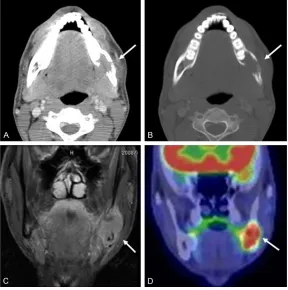
men-lymphadenopathy. A hyperplastic brownish solid tumor was observed in the left mandibu-lar third momandibu-lar extraction socket (Figure 1B). There was no trismus. Panoramic radiography showed bone resorption from the left mandibu-lar third momandibu-lar area to the mandibumandibu-lar angle and ramus (Figure 2). Contrast-enhanced com-puted tomography (CT) showed an osteoclastic lesion centering on the left mandibular retro-molar area, with destruction of cortical bone extending to the buccolingual side, and a soft tissue shadow invading the surrounding area (Figure 3A, 3B). Magnetic resonance imaging of the left mandibular body showed muscle tis-sue and isointense signals on a T1-enhanced image, and high signals on a T2-enhanced image; gadolinium contrast-enhanced imaging showed a solid tumor with an indistinct bound-ary and enhancement (Figure 3C). The tumor measured approximately 41 × 33 × 20 mm.
probability of leiomyosarcoma in the non-epi-thelial malignant tumor. In February, we per-formed a tumorectomy (mandibular region re- section) and reconstructive surgery using a titanium plate. The resection was perform- ed according to the imaging findings and a 3-dimensional stereoscopic model (Figure 4A,
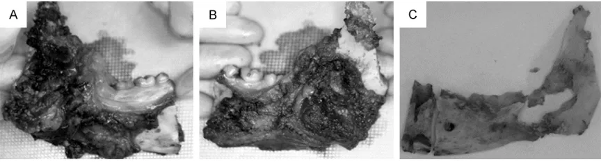
4B). Although the anterior resection stump was originally planned for the distal canine area, we progressed further into the bone while perform-ing rapid pathology tests, and the resection ultimately almost reached the midline. The resection extended to subcutaneous adipose tissue on the outer and rear sides, directly under the articular process on the upper side and the pterygoid muscle on the inner side (Figure 5A-C). The resection margin was 15 mm. While no tumor was apparent on the re- section stump, histopathological progression toward the outside of the mandible was particu-larly advanced on the inner side; therefore, the safety margin for a non-epithelial malignant tumor was inadequate. To avoid local reoccur-rence and remote metastasis, we proposed systemic chemotherapy for the patient. After obtaining consent, we performed postopera-tive adjuvant chemotherapy (cisplatin: 120 mg/ body, doxorubicin hydrochloride: 50 mg/body, and caffeine: 2400 mg/body), for a total of three courses from April to June. Adverse ev- ents included hair loss and vomiting, as well as plate exposure in the area corresponding to the mandibular angle. After the completion of che-motherapy, we performed plate removal under general anesthesia, following which chewing, swallowing, and ingestion were possible, even though the jawbone was transected.
Figure 1.Figure of first visit to our hospital. A. Patient’s frontal view. B. Oral cavity at initial diagnosis. A hyperplastic brownish tumor is visible in the left mandibular third molar extraction socket (arrow).
Figure 2. Panoramic radiograph at initial diagno-sis.Radiograph revealing an area with an indistinct boundary, extending from the left mandibular body to the mandibular ramus, mandibular angle, and coronoid process (arrow).
Positron emission tomogra-phy-CT showed fluorodeoxy-glucose-enhanced uptake wi- th a maximum standardized uptake value of 9.0 in the ret-romolar area, which is be- lieved to indicate centrality at the mandibular angle and ramus (Figure 3D). There was no evidence of cervical lymph node or systemic metasta- ses.
Histopathology involved hematoxylin-eosin st- aining and various immunohistological tests of the resected specimen. On hematoxylin-eosin staining, heterocysts arrayed in bundles with stained nuclei within spindle-shaped cells, and fibrous cytoplasm, were visible. High cell
den-rived from surrounding vascular smooth mus-cle in the mandibular canal. In this particular case, as decreased sensation was noted in the mentum in the initial evaluation, the tumor was thought to originate from blood vessels in the mandibular canal or from surrounding
branch-Figure 3.Imaging findings. A, B. Computed tomography scan showing an os-teoclastic lesion centering on the left mandibular retromolar area, with corti-cal bone destruction extending to the buccolingual side, and a soft tissue shadow extending into the surrounding area (arrow). C. Gadolinium contrast-enhanced magnetic resonance image showing a solid tumor with enhance-ment and an indistinct boundary (arrow). D. Positron emission tomography-computed tomography scan showing enhanced fluorodeoxyglucose uptake with a maximum standardized uptake value of 9.0 and centrality localized to the mandibular angle (arrow).
Figure 4.Three-dimensional reality model. A. Inner side surface view; B. Out-er side surface view and pre-vending titanium plate.
[image:3.612.90.378.72.362.2]sity, nuclear atypia, and nu- clear fission images were al- so seen (Figure 6). Immuno- histological staining results are shown in Figure 6 and
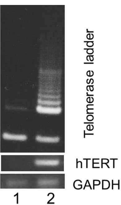
Table 1. From these findings and surgical specimens, we diagnosed mandibular-origin leiomyosarcoma. Generally, sarcomas have lower levels of telomerase activity, however, this leiomyosarcoma has an abundant telomerase activity, which is one of the marker of cell proliferation (Figure 7). Following satisfactory prog-ress, the patient was dis-charged from the hospital in June, and returned to our department for regular out- patient follow-up. CT scans taken two years after the sur-gery revealed multiple metas-tases in the lungs. There was no response to additional ch- emotherapy, and the patient died about three years after the initial surgery.
Discussion
[image:3.612.89.375.484.594.2]es; however, the exact origin could not be identified.
In general, a definitive pathological diagnosis is extremely important for selecting an appropri-ate treatment method and determining the prognosis for non-epithelial malignant tumors. This is also important for soft tissue sarcomas because effective tissue-based chemotherapy regimens are available. While our case was a leiomyosarcoma, fibrosarcoma or osteosarco-ma wasere also suggested. Therefore, immuno-histological tests were necessary for differen-tial diagnosis. While the cells were negative for epithelial markers such as cytokeratin or epi-thelial membrane antigen, they were positive for smooth muscle cell markers, such as α-smooth muscle actin, calponin, HHF35, des-min, and caldesmon; thus, a definitive diagno-sis of leiomyosarcoma could be made. More- over, the proliferative marker is also useful information at the treatment of this kind of tumor. Ki-67 index was high. Moreover, this tumor has a telomerase activity and expressed hTERT mRNA [5, 8]. This indicated that this tumor has a great potential in tumor cell pro- liferation.
For leiomyosarcoma, the first choice for treat-ment is complete resection [9, 10], and the prognosis is expected to be particularly favor-able when a wide initial resection is possible. Chemotherapy is adjunctive or palliative. In this particular case, while a wide mandibular resec-tion including soft tissue was performed, the tumor invasion was more extensive than ex- pected, and chemotherapy was performed to avoid postoperative recurrence or metastasis. We consulted an orthopedic surgeon for the chemotherapy regimen and administered drugs commonly used for leiomyosarcoma with the concomitant use of caffeine to augment cispla-tin activity. With no definite residual tumor or remote metastasis, efficacy could not be de- termined.
Ethunandan et al. reported that the five-year survival rate for leiomyosarcoma in the oral region is about 55% [11]. However, the survival rate decreases for cases with bone invasion
[image:5.612.91.290.136.283.2]Figure 6.Histopathological images. A. Hematoxylin and eosin staining (×100); B. Calponin immunostaining (×100); C. α-smooth muscle actin immunostaining (×200); D. Caldesmon immunostaining (×100); E. Ki-67 immunostaining (×200); F. Desmin immunostaining (×200).
Table 1. Immunohistochemica
Markers Reactivity
α-smooth muscle actin +
Calponin +
HHF35 +
Desmin +
Caldesmon +
Cytokeratin
-Epithelial membrane antigen
-S-100
-CD34
-CD246
[image:5.612.92.287.301.632.2][11]. As the number of reported cases with mandibular centrality is small, we were unable to discern a trend, but these included extreme-ly malignant cases in which metastasis to other organs had already occurred at the time of ini-tial diagnosis. Unfortunately, although follow-up was conducted every three weeks, and imaging including positron emission tomogra-phy-CT was performed as needed, the patient developed respiratory failure due to lung me- tastasis, and died about three years after the surgery. Thus, as with many other sarcomas, the choice of initial treatment is critical, but control is extremely difficult when a recurrence or remote metastasis develops.
Acknowledgements
This work was supported by JSPS KAKENHI Grant Number 16K20587. The authors grate-fully acknowledge this financial support. We would like to thank Editage (www.editage.jp) for English language editing.
Disclosure of conflict of interest
None.
Address correspondence to: Dr. Tomoki Sumida, Section of Oral and Maxillofacial Surgery, Division of Maxillofacial Diagnostic and Surgical Sciences, Faculty of Dental Science, Kyushu University, 3-1-1, Maidashi, Higashi-ku, Fukuoka, 812-8582, Japan. Tel: +81-92-642-6452; Fax: +81-92-642-6392; E-mail: [email protected]
References
[1] Enzinger FM and Weiss SW. Soft tissue tu-mors. 3rd Edition. In: Enzinger FM, editor. St Louis: Mosby Co; 1995. pp. 491-510.
[2] Farman AG and Kay S. Oral leiomyosarcoma. Report of a case and review of the literature pertaining to smooth-muscle tumors of the oral cavity. Oral Surg Oral Med Oral Pathol 1977; 43: 402-409.
[3] Ahn JH, Mirza T and Ameerally P. Leio- myosarcoma of the tongue with multiple me-tastases: a case report and review of litera-ture. J Oral Maxillofac Surg 2012; 70: 1745-1750.
[4] Shokouh TZ, Taghipour Z, and Naser SS. Leiomyosarcoma of the maxilla in a 24-year-old man who initially presented with odontal-gia, and suffered from tumour mismanage-ment. BMJ Case Rep 2013; 2013: bcr2013- 200933.
[5] Sumida T, Hamakawa H, Otsuka K and Tanioka H. Leiomyosarcoma of the maxillary sinus wi- th cervical lymph node metastasis. J Oral Maxillofac Surg 2001; 59: 568-571.
[6] Fitzpatrick SG, Woodworth BA, Monteiro C and Makary R. Nasal sinus leiomyosarcoma in a patient with history of non-hereditary unilater-al treated retinoblastoma. Head Neck Pathol 2011; 5: 57-62.
[7] Nikitakis NG, Lopes MA, Bailey JS, Blanchaert RH Jr, Ord RA and Sauk JJ. Oral leiomyosarco-ma: review of the literature and report of two cases with assessment of the prognostic and diagnostic significance of immunohistochemi-cal and molecular markers. Oral Oncol 2002; 38: 201-208.
[8] Sumida T, Hamakawa H, Sogawa K, Sugita A, Tanioka H and Ueda N. Telomerase compo-nents as a diagnostic tool in human oral le-sions. Int J Cancer 1999; 80: 1-4.
[9] Mindell RS, Calcaterra TC and Ward PH. Leiomyosarcoma of the head and neck: a re-view of the literature and report of two cases. Laryngoscope 1975; 85: 904-910.
[10] Brandjord RM, Reaume CE and Wesley RK. Leiomyosarcoma of the floor of the mouth: re-view of the literature and report of case. J Oral Surg 1977; 35: 590-594.