Original Article
Correlations between VEGF-A expression and prognosis
in patients with gastric adenocarcinoma
Bo Wei1, Yang Tai1, Huan Tong1, Shi-Lei Wen2,3, Shi-Hang Tang1, Hui Huan1, Zhi-Yin Huang1, Rui Liu2, Ying-Mei
Tang4, Jin-Hui Yang4, Cheng-Wei Tang1,2, Jin-Hang Gao1,2
1Department of Gastroenterology, 2Division of Peptides Related with Human Diseases, West China Hospital,
Sich-uan University, Chengdu, China; 3Department of Human Anatomy, Academy of Preclinical and Forensic Medicine,
West China Medicine College, Sichuan University, Chengdu, China; 4Department of Gastroenterology, The Second
Affiliated Hospital of Kunming Medical University, Kunming, Yunnan Province, China
Received May 21, 2017; Accepted July 14, 2017; Epub August 1, 2017; Published August 15, 2017
Abstract: Angiogenesis induced by vascular endothelial growth factor A (VEGF-A) plays a critical role in tumor growth and metastasis. The study aimed to evaluate the expression of VEGF-A in gastric adenocarcinoma and investi-gate its correlations with tumor clinicopathological features and prognostic significance. VEGF-A expression was detected by immunohistochemistry on a tissue microarray containing 90 pairs of human gastric adenocarcinoma and paracancerous tissues. Levels of VEGF-A in gastric adenocarcinoma were significantly higher than those in paracancerous tissues (P=0.018). Furthermore, the result was coincident with that of human gastric adenocarci-noma xenografts in nude-mice (P<0.01). In addition, the VEGF-A expression was positive correlation with TNM stage (P=0.047), tumor size (P=0.028), positive lymph nodes (P=0.002) and lymphovascular invasion (P=0.001). Finally, Kaplan-Meier survival analysis showed that VEGF-A up-regulation indicated a poor prognosis for overall survival (P=0.039). In conclusions, VEGF-A may be used as a biomarker for evaluating both the biological behavior of tumor and the prognosis in patients with gastric adenocarcinoma.
Keywords: Vascular endothelial growth factor, gastric adenocarcinoma, prognosis, biomarker, overall survial
Introduction
Gastric cancer is the fifth most common can- cer and the third leading cause of cancer-relat-ed deaths globally. An estimatcancer-relat-ed 951,600 new gastric cancer cases and 723,100 deaths oc- curred in 2012 worldwide [1]. The incidence of gastric cancer is generally about twice as high in men as in women and varies tremen-dously throughout the world. The highest inci-dence countries are in Eastern Asia (particu-larly in Korea, Japan, and China) [2, 3]. Gastric adenocarcinoma accounts for the vast ma- jority of gastric cancer cases. Gastric adeno- carcinoma clinically has no characteristic mani-festations in early stage and further progress to weight loss, epigastric pain or discomfort, anorexia, vomiting and gastrointestinal bleed-ing. When these symptoms emerge, the cancer has often reached an advanced stage and may also have regional invasion or metasta-
sis. Despite the development in diagnosis and treatment, advanced gastric adenocarcinoma carries a highly unfavorable prognosis, with a 5-year survival rate of about 20%-30% [4]. Therefore, identification of novel biomarkers to estimate prognosis early and correctly may provide useful information for clinical mana- gement.
Table 1. Clinicopathological characteristics of 90 gastric adenocarcinoma cases
Characteristics Number of cases Percentage (%)
Total 90 100
Average years 62.1±12.3
<65 52 57.8
≥65 38 42.2
Gender
Male 53 58.9
Female 37 41.1
Histologic grade
I~II 21 23.3
II~III 69 76.7
TNM stage
I 9 10
II 27 30
III 50 55.6
IV 4 4.4
Tumor size (cm) 5.7±2.7
<5 cm 35 38.9
≥5 cm 55 61.1
Lymphovascular Invasion 29 32.2 Positive lymph nodes 67 74.4
Distant metastasis 4 4.4
em, VEGF-A is a dimeric glycoprotein which plays a significant role in promoting blood ves-sel dilation and permeability. And VEGF-A is considered to be the main and dominant induc-er to the growth of new blood vessels. Recently, increasing evidences suggest up-regulation of VEGF-A in several malignant solid tumors tis-sues, as well as the elevation of circulating lev-els of VEGF-A in patients with cancer [10-15]. The impact of VEGF-A expression on prognosis has been demonstrated by meta-analysis in hepatocellular carcinoma [11] and pancreatic cancer [12]. Many studies have evaluated whether up-regulation of VEGF-A may be a prog-nostic factor for survival in patients with gastric cancer. However, the results of the studies are inconclusive and no consensus has been achieved [16]. In the present study, we detect-ed the expression of VEGF-A in both human gastric adenocarcinoma tissues and in situ xenografts tissues of human gastric carcinoma cells in nude mice, as well as their corres- ponding paracancerous tissues, respectively. Additionally, we analyzed whether the
expres-sion of VEGF-A is correlated with tumor clinico-pathological features and clinical outcome in patients with gastric adenocarcinoma.
Materials and methods
The animal experiment was approved by the Animal Use and Care Committee of Sichuan University and conducted according to the regulations.
Tissue microarray (TMA)
Gastric adenocarcinoma TMA implied in the present study was obtained from National Engineering Center for Biochip at Shanghai (Shanghai, China). TMA samples include a total of 90 gastric adenocarcinoma patients who had undergone a complete surgical resection of the gastric tumor between August 2008 and March 2009. Written informed consent forms for the tissue specimens were received from all participants, and the study was ap- proved by the ethical committee of biobank center related hospitals. Detailed clinical and pathologic information of these 90 patients was collected (Table 1). All patients were fol-lowed up from the date of surgery until April 2014.
Cell culture and treatments
Human gastric adenocarcinoma cell SGC7901 was obtained from the Type Culture Collection of the Chinese Academy of Sciences (Shanghai, China). Cells were cultured in RPMI-1640 (Hy- Clone, Logan, UT, USA) supplemented with 10% fetal bovine serum (Gibco, Logan, UT, USA) and 100 U/ml penicillin, 100 U/ml streptomy- cin (HyClone, Logan, UT, USA) in a humidified atmosphere at 37°C with 5% CO2 in air. SG- C7901 cells were trypsinized and resuspended in normal saline when cell density reached 80~90%.
Human gastric cancer xenografts
Ten healthy male BAL b/c nude mice, weighting 18-23 g, were obtained from the Experimental Animal Center of Sichuan University (Chengdu, China). The mice were kept under 12 hours’ light-dark cycles at a constant temperature and humidity with free access to chow and water. Briefly, 1 × 10⁷ of SGC7901 cells were
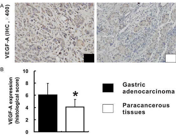
Figure 1. Expression of VEGF-A in human gastric adenocarcinoma tissues and paracancerous tissues. A. Shows the immunohistochemical staining of VEGF-A expression in human gastric adenocarcinoma tissues and para-cancerous tissues (all images were taken at 400 × magnification). B and C. Reveal the differences in expression levels of VEGF-A in tumor tissues and paracancerous tissues based on the histological score.
mice. Tumors were aseptically dissected and mechanically minced after 4 weeks. A piece of tumor (~2 mm3) was transplanted under gastric
serosa in each of the rest 8 nude mice and all mice were sacrificed under anesthesia after 4 weeks. Tumor tissues and corresponding para-cancerous tissues were fixed in 4% neutral buffered paraformaldehyde for histopathologic and immunohistochemical examinations.
Immunohistochemistry (IHC) and scoring
For IHC, sections of TMA and gastric cancer xenografts were deparaffinized in xylene, rehy-drated with graded ethanol dilutions. Antigen retrieval was performed at high temperature under high pressure in 10 mM sodium citrate buffer for 10 minutes. After blocking of endog-enous peroxidase activity by H2O2, the sec- tions were incubated with rabbit anti-VEGF-A (1:100, Santa Cruz Biotechnology, Santa Cruz, CA, USA) overnight at 4°C followed by incuba-tion with horseradish peroxidase (HRP)-con- jugated secondary antibody kits (ZSGB Bio, Beijing, China) for 30 minutes at room tempera-ture. Finally, sections were stained with a
solu-was considered to be low expression of VEGF-A, whereas a total score of 5-9 was defined as high expression.
Statistical analysis
All data were expressed as mean ± standard deviation and were analyzed by SPSS 13.0 soft-ware (SPSS, Chicago, IL, USA). Quantitative data were analyzed by using one-way ANOVA. The X2 test was applied to analyze the dif-
ference of clinicopathological parameters be- tween low and high VEGF expression groups. Survival curves were evaluated by using the Kaplan-Meier method and compared by the log-rank test. A value of P<0.05 was conside- red significant.
Results
Patient characteristics
The clinicopathological characteristics of the patients enrolled in this study were summa-rized in Table 1. The TMA consisted of 90 pa- tients with a diagnosis of gastric
adenocarci-tion of 3,3’-diaminobenzidine tetrahydrochloride and coun-terstained with hematoxylin. Ne- gative control slides in which the primary antibodies were omitted were included in all as- says.
Figure 2. Expression of VEGF-A in human gastric adenocarcinoma xe-nografts tissues and paracancerous tissues. A. Shows the immunohisto-chemical staining of VEGF-A expression in human gastric adenocarcinoma xenografts tissues and paracancerous tissues in nude mice (400 × magni-fication). B. Reveals the differences in expression levels of VEGF-A in xeno-grafts tissues and paracancerous tissues based on the histological score.
noma, among who included 53 males (58.9%) and 37 females (41.1%) with an average age of 62.1±12.3 years-old. In evaluating tumor his- tologic differentiation, 21 cases (23.3%) were in grade I~II, while the remaining 69 cases (76.7%) were in grade II~III. According to the TNM staging system, 9 (10%) cases were in stage I, 27 (30%) in stage II, 50 (55.6%) in stage III, and 4 (4.4%) in stage IV. The average tumor size was 5.7 cm, with 35 (38.9%) tumors <5 cm and 55 (61.1%) tumors ≥5 cm. Positive lymph nodes and distant metastasis were 67 (74.4%) and 4 (4.4%) of 90 patients, respectively. Lymphovascular invasion was observed in 29 cases (32.2%).
Expression of VEGF-A in gastric adenocarci-noma tissues and corresponding paracancer-ous tissues
In TMA slide, the positive staining of VEGF-A was mainly located to the cytoplasm in both tumor and paracancerous tissues (Figure 1A). Base on the staining index described above, the expression of VEGF-A in gastric adenocarci-noma tissues was significantly higher in 53 cases, equivalent in 8 cases, and lower in 29 cases, compared with that in corresponding paracancerous tissues (Figure 1B). There was
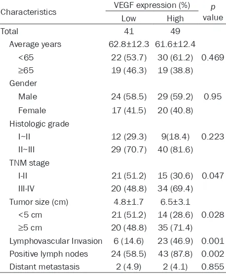
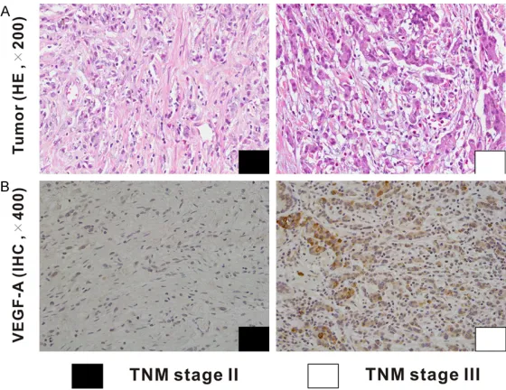
Among 90 gastric adenocarcinoma patients, 41 (45.6%) cases were identified as low VEGF-A expression group, and 49 (54.4%) cases as high VEGF-A expression group. The relation-ships between the expression of VEGF-A and clinicopathological features were analyzed (Table 2). A significantly positive correlation was observed between VEGF-A expression and TNM stage (P=0.047, Figure 3 and Table 2). The rate of patients with TNM stage I~II was higher in low VEGF-A expression group (51.2%) than that in high VEGF-A expression group (30.6%). Whereas, the rate of patients with TNM stage III~IV was lower in low VEGF-A expression group than that in high VEGF-A expression group (48.8% vs. 69.4%). Besides, the expression of VEGF-A was significantly associated with tumor size (P=0.028). The rate of patients with tumor size ≥5 cm was lower in low VEGF-A expression group compared with that in high VEGF-A expression group (48.8%
vs. 71.4%). Furthermore, high VEGF-A expres-sion increased the risk of positive lymph nodes (P=0.002) and lymphovascular invasion (P=0.001). However, the results showed that there was no significant correlation between the expression of VEGF-A and other clinicopath-ological factors, including age, gender, histo-a significhisto-ant difference in the expression of VEGF-A between human gastric adenocarcino-ma tissues and paracancerous tissues (5.0±2.1 vs. 4.3±1.9;
P=0.018, Figure 1C). Further- more, the result was coinci-dent with that of human gastric adenocarcinoma xenografts in node mice, which suggested that stronger expression of VE- GF-A was observed in xeno-grafts tissues, compared with that in paracancerous tissues (6.1±1.9 vs. 4.1±1.2; P<0.01,
Figure 2). Overall, the levels of VEGF-A were higher in gastric adenocarcinoma tissues than those in paracancerous tis- sues.
Table 2. Relationships between VEGF-A expression and clinicopathological characteristics in 90 gastric adenocarcinoma cases
Characteristics VEGF expression (%) valuep
Low High
Total 41 49
Average years 62.8±12.3 61.6±12.4 <65 22 (53.7) 30 (61.2) 0.469 ≥65 19 (46.3) 19 (38.8) Gender
Male 24 (58.5) 29 (59.2) 0.95 Female 17 (41.5) 20 (40.8) Histologic grade
I~II 12 (29.3) 9(18.4) 0.223 II~III 29 (70.7) 40 (81.6) TNM stage
I-II 21 (51.2) 15 (30.6) 0.047 III-IV 20 (48.8) 34 (69.4) Tumor size (cm) 4.8±1.7 6.5±3.1
<5 cm 21 (51.2) 14 (28.6) 0.028 ≥5 cm 20 (48.8) 35 (71.4) Lymphovascular Invasion 6 (14.6) 23 (46.9) 0.001 Positive lymph nodes 24 (58.5) 43 (87.8) 0.002 Distant metastasis 2 (4.9) 2 (4.1) 0.855
logic grade, and distant metastasis (P>0.05,
Table 2).
Survival analysis
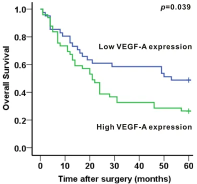
The survival analysis showed that the 5-years survival rate of patients was 36.7%. The medi-an overall survival was 24 months. The medimedi-an survival time was 53 months for low VEGF-A expression group and 21 months for high VEGF-A expression group. In addition, the 5- year survival rates were 54.1% and 24.5% in patients with low-expression and high-expres-sion of VEGF-A, respectively. A significant asso-ciation between VEGF-A expression and tumor prognosis in human gastric adenocarcinoma had been established. Patients with low expres-sion levels of VEGF-A achieved better survival than patients with high expression levels of VEGF-A (P=0.039, Figure 4).
Discussion
Angiogenesis, which induced by VEGF and its receptor (VEGFR), plays a critical role in tumor growth and metastasis. VEGF-A, also referred
to as VEGF, is the best characterized and the most frequently studied subtype of the VEGF family during the past decades [18]. Up-expression of VEGF-A has been ob- served in numerous solid tumor tissues [8], including gastric adenocarcinoma. Shi [19] conducted a retrospective study of 232 gastric adenocarcinoma tissues and 60 noncancerous tissues, which showed that compared with paired noncancerous tis-sues, expression of VEGF-A and microves-sel density in carcinoma tissues were obvi-ously increased. Yang [14] reported VEGF-A expression in gastric carcinoma tissues was significantly higher than that in normal gastric mucosal tissues with distance >5 cm from the tumor margin. Furthermore, recent researches [15, 20] have suggested the VEGF-A levels in serum were signifi-cantly increased preoperatively in gastric cancer patients, compared with apparently healthy volunteers. In this study, we detect-ed the expression of VEGF-A by IHC method in gastric adenocarcinoma tissues and paracancerous tissues in both TMA and nude-mice xenografts. According to the semi-quantitatively method, the levels of VEGF-A expression in adenocarcinoma tis-sues were higher than that in paracancerous tissues, indicating the correlation between VEGF-A expression and tumor biological behav-ior in gastric adenocarcinoma.
Figure 3. Correlation between the VEGF-A expression and the TNM stage in human gastric adenocarcinoma. Representing images for HE and IHC of VEGF-A in gastric adenocarcinoma patients with TNM stage II and III are shown.
The value of VEGF-A expression as an indepen-dent prognostic indicator has been evaluated by numerous studies [26-29]. However, these studies have shown conflicting and opposite results and no consensus has been reached. Recently, an extensive meta-analysis [16] has been published showing VEGF-A up-expression indicates a poor prognosis for overall survival and disease-free survival in patients with gas-tric carcinoma. In this study, we also confirmed a significant prognostic value of VEGF-A in gas-tric adenocarcinoma using a Kaplan-Meier sur-vival analysis. Patients with low expression lev-els of VEGF-A achieved better survival than patients with high expression levels of VEGF-A. The current TNM staging system has some restrictions for the evaluation of the prognosis in gastric carcinoma patients with identical clin-ical or pathologclin-ical stages may differ widely in their clinical evolution. Thus, detecting VE- GF-A expression in gastric carcinoma tissues might be helpful for predicting patient progno-sis. Inhibition of VEGF-A pathway can effectively suppress tumor angiogenesis in vitro and in vivo [30]. Monoclonal antibodies or receptor tyrosine kinase inhibitors with anti-VEGF-A activity are in clinical trials or are currently approved by the United States Food and Dr- ug Administration. The above medicines have been prove to improved progression-free or adenocarcinoma including TNM stage, tumor
size, positive lymph nodes and lymphovascular invasion. High expression of VEGF-A was ac- companied by elevated TNM stage and tumor size, as well as increased the risk of lympho- vascular invasion and positive lymph nodes. These results suggest that VEGF-A plays an essential role in tumor growth, regional inva-sion and lymphatic metastasis.
VEGF-A and its receptors regulate tumor bio-logical behavior through angiogenesis pathway [23]. VEGF-A exerts its effects primarily throu- gh transmembrane tyrosine kinase receptors, VEGFR-1 and VEGFR-2. When binding to the extracellular domain of the receptor, a cascade of downstream proteins is activated after phos-phorylation of the intracellular receptor tyro-sine kinases. This process stimulates neovas-cularisation by promoting endothelial cell pro- liferation and migration. The newborn vessels can supply nutrition oxygen for tumors and discharge the metabolic wastes, which stimu-late the growth of tumor and provide vascular pathway for tumor metastasis. The biological processes provide the reliable basis for the explanation of the relationship between VEGF-A expression and tumor size as well as vascular invasion and metastasis. The lack of correla-tion between VEGF-A expression and distant
mation assessed each slide independently by using a semi-quantitatively scoring system. In conclusion, the present study showed that VEGF-A expression was significantly up-regulat-ed in gastric adenocarcinoma tissues. Mean- while, the expression of VEGF-A was intimately relevant to clinicopathological features includ-ing TNM stage, tumor size, positive lymph nodes and lymphovascular invasion. This study also indicated that the VEGF-A up-expression was significantly correlated with poor survival. Therefore, VEGF-A may be of use as a biomark-er for evaluating both the biological behavior of tumor and the prognosis in patients with gas-tric adenocarcinoma.
Acknowledgements
This study is supported by the National Natural Science Foundation of China (81670551 and 81400637), the Chinesisch-Deutsches Zentr- um fṻr Wissenschaftsfὅrderung (GZ1065) and the Science and Technology Support Program of Sichuan province (2016SZ0041).
Disclosure of conflict of interest
None.
Address correspondence to: Dr. Jin-Hang Gao, Di- vision of Peptides Related with Human Diseases, West China Hospital, Sichuan University, 37 Guo Xue Xiang, Chengdu 610041, China. Tel: 86-28-85164011; Fax: 86-28-85582944; E-mail: gjh731@ foxmail.com
References
[1] Torre LA, Bray F, Siegel RL, Ferlay J, Lortet-Tieu-lent J, Jemal A. Global cancer statistics, 2012. CA Cancer J Clin 2015; 65: 87-108.
[2] Bertuccio P, Chatenoud L, Levi F, Praud D, Fer-lay J, Negri E, Malvezzi M, La Vecchia C. Recent patterns in gastric cancer: a global overview. Int J Cancer 2009; 125: 666-673.
[3] Rahman R, Asombang AW, Ibdah JA. Charac-teristics of gastric cancer in Asia. World J Gas-troenterol 2014; 20: 4483-4490.
[4] Fang WL, Huang KH, Chen JH, Lo SS, Hsieh MC, Shen KH, Li AF, Niu DM, Chiou SH, Wu CW. Comparison of the survival difference between AJCC 6th and 7th editions for gastric cancer patients. World J Surg 2011; 35: 2723-2729. [5] Carmeliet P, Jain RK. Molecular mechanisms
[image:7.612.95.292.72.256.2]and clinical applications of angiogenesis. Na-ture 2011; 473: 298-307.
Figure 4. Association between VEGF-A expression and 5-year overall survival in human gastric adeno-carcinoma. Patients with low expression levels of VEGF-A achieved better survival than patients with high expression levels of VEGF-A (P<0.05).
overall survival in patients with metastatic colorectal cancer, metastatic breast cancer, and metastatic non-squamous non-small-cell lung cancer combined with chemotherapeutic drugs [31]. Several phase II [32, 33] or III [34] clinical trials conducted to evaluate therapies targeting the VEGF-A pathway have shown ben-efits in certain patients with advanced gastric carcinoma from the addition of bevacizumab to chemotherapy. Nevertheless, further resear- ches will be needed to identify eligible patients to receive therapies targeting the VEGF-A path-way and the ideal time window to initiate this target therapy.
infor-[6] Ilson DH. Angiogenesis in gastric cancer: hit-ting the target? Lancet 2014; 383: 4-6. [7] Javle M, Smyth EC, Chau I. Ramucirumab:
suc-cessfully targeting angiogenesis in gastric can-cer. Clin Cancer Res 2014; 20: 5875-5881. [8] Ferrara N, Gerber HP, LeCouter J. The biology
of VEGF and its receptors. Nat Med 2003; 9: 669-676.
[9] Holmes DI, Zachary I. The vascular endothelial growth factor (VEGF) family: angiogenic factors in health and disease. Genome Biol 2005; 6: 209.
[10] Dvorak HF. Vascular permeability factor/vas-cular endothelial growth factor: a critical cyto-kine in tumor angiogenesis and a potential target for diagnosis and therapy. J Clin Oncol 2002; 20: 4368-4380.
[11] Schoenleber SJ, Kurtz DM, Talwalkar JA, Rob-erts LR, Gores GJ. Prognostic role of vascular endothelial growth factor in hepatocellular car-cinoma: systematic review and meta-analysis. Br J Cancer 2009; 100: 1385-1392.
[12] Smith RA, Tang J, Tudur-Smith C, Neoptolemos JP, Ghaneh P. Meta-analysis of immunohisto-chemical prognostic markers in resected pan-creatic cancer. Br J Cancer 2011; 104: 1440-1451.
[13] Wang X, Chen X, Fang J, Yang C. Overexpres-sion of both VEGF-A and VEGF-C in gastric can-cer correlates with prognosis, and silencing of both is effective to inhibit cancer growth. Int J Clin Exp Pathol 2013; 6: 586-597.
[14] Yang S, Zhao Z, Wu R, Lu H, Zhang X, Huan C, Wang C, Wu X, Guan G. Expression and bio-logical relationship of vascular endothelial growth factor-A and matrix metalloprotein-ase-9 in gastric carcinoma. J Int Med Res 2011; 39: 2076-2085.
[15] Halmaciu I, Gurzu S, Dobreanu M, Suciu BA, Brinzaniuc K. [Preliminary results regarding vascular endothelial growth factor (VEGF-A) levels in the serum of gastric cancer patients]. Rev Med Chir Soc Med Nat Iasi 2012; 116: 446-451.
[16] Ji YN, Wang Q, Li Y, Wang Z. Prognostic value of vascular endothelial growth factor a expres-sion in gastric cancer: a meta-analysis. Tu-mour Biol 2014; 35: 2787-2793.
[17] Ohuchida K, Mizumoto K, Ishikawa N, Fujii K, Konomi H, Nagai E, Yamaguchi K, Tsuneyoshi M, Tanaka M. The role of S100A6 in pancreatic cancer development and its clinical implica-tion as a diagnostic marker and therapeutic target. Clin Cancer Res 2005; 11: 7785-7793. [18] Otrock ZK, Makarem JA, Shamseddine AI. Vas-cular endothelial growth factor family of li-gands and receptors: review. Blood Cells Mol Dis 2007; 38: 258-268.
[19] Shi H, Xu JM, Hu NZ, Xie HJ. Prognostic signifi-cance of expression of cyclooxygenase-2 and vascular endothelial growth factor in human gastric carcinoma. World J Gastroenterol 2003; 9: 1421-1426.
[20] Al-Moundhri MS, Al-Shukaili A, Al-Nabhani M, Al-Bahrani B, Burney IA, Rizivi A, Ganguly SS. Measurement of circulating levels of VEGF-A, -C, and -D and their receptors, VEGFR-1 and -2 in gastric adenocarcinoma. World J Gastroen-terol 2008; 14: 3879-3883.
[21] Yang Q, Ye ZY, Zhang JX, Tao HQ, Li SG, Zhao ZS. Expression of matrix metalloproteinase-9 mRNA and vascular endothelial growth factor protein in gastric carcinoma and its relation-ship to its pathological features and prognosis. Anat Rec (Hoboken) 2010; 293: 2012-2019. [22] Liu YF, Guo S, Zhao R, Chen YG, Wang XQ, Xu
KS. Correlation of vascular endothelial growth factor expression with tumor recurrence and poor prognosis in patients with pN0 gastric cancer. World J Surg 2012; 36: 109-117. [23] Abdel-Rahman O. Targeting vascular
endothe-lial growth factor (VEGF) pathway in gastric cancer: preclinical and clinical aspects. Crit Rev Oncol Hematol 2015; 93: 18-27.
[24] Kondo K, Kaneko T, Baba M, Konno H. VEGF-C and VEGF-A synergistically enhance lymph node metastasis of gastric cancer. Biol Pharm Bull 2007; 30: 633-637.
[25] Ding S, Li C, Lin S, Han Y, Yang Y, Zhang Y, Li L, Zhou L, Kumar S. Distinct roles of VEGF-A and VEGF-C in tumour metastasis of gastric carci-noma. Oncol Rep 2007; 17: 369-375.
[26] Vidal O, Soriano-Izquierdo A, Pera M, Elizalde JI, Palacin A, Castells A, Pique JM, Volant A, Metges JP. Positive VEGF immunostaining in-dependently predicts poor prognosis in cura-tively resected gastric cancer patients: results of a study assessing a panel of angiogenic markers. J Gastrointest Surg 2008; 12: 1005-1014.
[27] Lee SJ, Kim JG, Sohn SK, Chae YS, Moon JH, Kim SN, Bae HI, Chung HY, Yu W. No associa-tion of vascular endothelial growth factor-A (VEGF-A) and VEGF-C expression with survival in patients with gastric cancer. Cancer Res Treat 2009; 41: 218-223.
[28] Suzuki S, Dobashi Y, Hatakeyama Y, Tajiri R, Fujimura T, Heldin CH, Ooi A. Clinicopathologi-cal significance of platelet-derived growth fac-tor (PDGF)-B and vascular endothelial growth factor-A expression, PDGF receptor-beta phos-phorylation, and microvessel density in gastric cancer. BMC Cancer 2010; 10: 659.
A, Morgagni P, Farsi M, Roviello F, De Manzoni G, Bechi P, Arcangeli A; Gruppo Italiano di Ri-cerca Cancro Gastrico (GIRCG). VEGF-A clinical significance in gastric cancers: immunohisto-chemical analysis of a wide Italian cohort. Eur J Surg Oncol 2014; 40: 1291-1298.
[30] Jain RK. Normalization of tumor vasculature: an emerging concept in antiangiogenic thera-py. Science 2005; 307: 58-62.
[31] Lyseng-Williamson KA, Robinson DM. Spotlight on bevacizumab in advanced colorectal cer, breast cancer, and non-small cell lung can-cer. Bio Drugs 2006; 20: 193-195.
[32] Shah MA, Jhawer M, Ilson DH, Lefkowitz RA, Robinson E, Capanu M, Kelsen DP. Phase II study of modified docetaxel, cisplatin, and fluo-rouracil with bevacizumab in patients with metastatic gastroesophageal adenocarcino-ma. J Clin Oncol 2011; 29: 868-874.
[33] El-Rayes BF, Zalupski M, Bekai-Saab T, Heilb-run LK, Hammad N, Patel B, Urba S, Shields AF, Vaishampayan U, Dawson S, Almhanna K, Smith D, Philip PA. A phase II study of bevaci-zumab, oxaliplatin, and docetaxel in locally ad-vanced and metastatic gastric and gastro-esophageal junction cancers. Ann Oncol 2010; 21: 1999-2004.