Original Article
CTHRC1 mediates multiple pathways regulating
cell invasion, migration and adhesion in glioma
Peng-Jin Mei1,2*, Jin Bai2*, Fa-An Miao1, Chen Chen1, Yi-Shuo Zhu1, Zhong-Lin Li1, Jun-Nian Zheng2, Yue-Chao Fan1
1Department of Neurosurgery, The Affiliated Hospital of Xuzhou Medical University, Xuzhou, Jiangsu, China; 2Jiangsu Key Laboratory of Biological Cancer Therapy, Xuzhou Medical University, Xuzhou, Jiangsu, China. *Equal contributors.
Received June 6, 2017; Accepted June 22, 2017; Epub September 1, 2017; Published September 15, 2017
Abstract: Recently, collagen triple helix repeat containing-1 (CTHRC1) has been reported to be increased in several types of human solid cancers and to be associated with tumor invasion and metastasis. However, the expression and function of CTHRC1 in glioma have not yet been reported. In the present study, we investigated whether CTHRC1 plays a role in glioma pathogenesis. Using the tissue microarray technology, we found that CTHRC1 expression is
significantly increased in glioma compared with tumor adjacent normal brain tissue (P<0.01, χ2 test) and increased CTHRC1 staining was associated with WHO stages (P<0.05, χ2 test). The mRNA and protein levels of CTHRC1 were significantly upregulated in human primary glioma tissues (P<0.001, χ2 test). We also found that CTHRC1 was significantly increased in glioma cell lines compared to normal human astrocytes (P<0.01, χ2 test). Furthermore,
Knockdown of CTHRC1 suppressed glioma cell invasion and inhibited enzyme activity of MMP-2. Moreover, our data showed that knockdown of CTHRC1 inhibited glioma cell migration and adhesion capacity when compared with the control cells, and CTHRC1-siRNA reduced the levels of phosphorylated Src and FAK protein expression. Taken to-gether, this study suggests that CTHRC1 plays a role in glioma development and progression by regulating invasion, migration and adhesion capabilities of cancer cells.
Keywords: CTHRC1, glioma, invasion, migration, adhesion
Introduction
Glioma are the most common primary central nervous system (CNS) tumors, and are
classi-fied into grades I to IV according to the WHO
criteria [1, 2]. Glioblastoma multiforme (GBM)
(grade IV) is the most common and aggressive
type of malignant glioma, with a current median survival of approximately 15 months in patients with newly diagnosed disease following treat-ment with surgery, chemotherapy and radio-therapy [3]. The invasive nature of GBM has been frequently implicated as a key feature of GBM’s resistance to therapy. Many studies showed that many factors were involved in the invasion of GBM, including adhesion molecules, extracellular matrix (ECM), protease system and angiogenesis, yet the exact molecular mechanism and process of the invasion growth of GBM remain poorly understood [4, 5]. Therefore, there is an urgent need to clarify the
molecular mechanisms underlying invasion and migration behavior of human glioma.
The mammalian Collagen triple helix repeat
containing-1 (CTHRC1) gene was first found in
balloon-injured rat arteries where it is expressed
by fibroblasts of the remodeling adventitia and
by smooth muscle cells of the neointima [6]. Increased expression of CTHRC1 was found to promote cell migration and inhibited collagen
synthesis in rat fibroblasts. In transgenic mice,
CTHRC1 may potentially contribute to
carcino-genesis and influence the activity of cancer.
Recently, some studies have found CTHRC1 expression was increased in malignant mela-noma [10], breast cancer [11], pancreatic can-cer [12], gastric cancan-cer and colorectal cancan-cer [13, 14] and seemed to be associated with can-cer tissue invasion and metastasis. According to expression of analysis of CTHRC1, it plays a crucial role in the differential diagnosis of
der-matofibrosarcoma protuberans and dermatofi -broma [15]. CTHRC1 has been shown to be high active and potent degrading ECM proteins and promoting cancer cell migration and inva-sion [16]. Park et al. reported CTHRC1 promot-ed pancreatic cancer growth and metastatic spread of cancer cells to distant organs in orthotopic xenograft tumor mouse models [12]. Although numerous studies have investigated CTHRC1 expression in various forms of cancer, little is known of the expression of CTHRC1 in glioma and its effects on tumor cell behavior. In this study, we used a tissue microarray tech-nology (TMA) of human glioma patients and immunohistochemistry to evaluate the expres-sion of CTHRC1 in relation to clinicopathologic features. We found that CTHRC1 expression was increased in glioma compared with tumor adjacent normal brain tissue and this increase was associated with WHO stage. We also report
that CTHRC1 was significantly upregulated in
glioma cells compared to normal human astro-cytes (NHA). Moreover, our data demonstrated that silencing of CTHRC1 in human glioma cell lines reduced the cell invasion, migration and adhesion abilities. In addition, we investigated the mechanisms by which CTHRC1 expression was regulated.
Materials and methods
Ethics statement
This study was performed under a protocol approved by the Institutional Review Boards
of The Affiliated Hospital of Xuzhou Medical
University and all examinations were performed after obtaining written informed consents.
Patients and samples
A glioma TMA was purchased from Shanxi Al-
enabio Biotechnology (Xi’an, China). Pathologic grades of tumors were defined according to
the WHO criteria as follows: 133 cases of benign tumor (Grade I and II), 57 cases of
malig-nant tumor (Grade III and IV), 8 cases of tumor
adjacent normal brain tissue and 8 cases of normal brain tissue. The array dot diameter was 1.0 mm, and each dot represented a tis-sue spot from one individual specimen that was
selected and pathologically confirmed. Five human glioma tissues and five nontumorous
brain tissues (internal decompression in cere-bral trauma) were obtained from the Depart-
ment of Neurosurgery, The Affiliated Hospital of Xuzhou Medical University.
Immunohistochemistry of TMA
The TMA slides were dewaxed by heating at 55°C for 30 min and by three washes, 5 min each, with xylene. Tissues were rehydrated by a series of 5 min washes in 100, 90, and 70% ethanol and PBS. Antigen retrieval was per-formed by microwaving the samples for 4 min, 20 sec at full power in 250 ml of 10 mM sodium citrate (pH 6.0). Endogenous peroxidase activi-ty was blocked with 0.3% hydrogen peroxide for
20 min. Nonspecific binding was blocked with
goat serum for 30 min. The primary monoclonal rabbit anti-CTHRC1 antibody (Abcam, Cam- bridge, MA, USA) was diluted 1:400 using goat serum and incubated at room temperature for 1 hour. After three washes, 2 min each with PBS, the sections were incubated with a bioti-nylated goat anti-rabbit secondary antibody for 30 min (Santa Cruz Biotechnology, Santa Cruz, CA), followed by the incubation with strepta- vidin-peroxidase (Santa Cruz Biotechnology, Santa Cruz, CA) for an additional 30 min. After rinsing with PBS 3 times for 2 min, the sections were stained using DAB (Zhongshan Biotech, Beijing, China) for 15 min, rinsed in distilled water and counterstained with hematoxylin. Dehydration was then performed following a standard procedure, and the sections were sealed with cover slips. Negative controls were performed by omitting CTHRC1 antibody during the primary antibody incubation.
Evaluation of immunostaining
cells showing immunostaining were considered as negative.
RNA extraction and reverse transcriptase-poly-merase chain reaction (RT-PCR)
Total RNA of brain samples and glioma cell lines were extracted using the Trizol reagent (Tiangen Biotech) according to the manufactur-er’s instructions. RT-PCR was performed using
a Takara RNA PCR Kit (AMV) version 3.0
(Ta-kara, Shiga, Japan) according to the manufac-ture’s protocol. Actin served as an internal con-trol. The primers used were: forward, 5’-TCAT- CGCACTTCTTCTGTGGA-3’; reverse, 5’-GCCAA- CCCAGATAGCAACATC-3’ (for CTHRC1); forward, 5’-GCGCGGCTACAGCTTCAC-3’; reverse, 5’-GG- GGCCGGACTCGTCATA-3’ (for Actin). Relative and intensities were determined using image J software.
Cell culture and transfection
Primary normal human astrocytes (NHA) were purchased from the KeyGEN Biotech Company (Nanjing, China) and cultured under the condi-tions as instructed by the manufacturer. Human glioma cell lines (U251, U87, T98G, SHG44, A172) and Rat glioma cell C6 were purchased from the Institute of Biochemistry and Cell Biology, Chinese Academy of Science. Cells were cultured in DMEM supplemented with 10% fetal bovine serum (Invitrogen, Shanghai, China). All cells were maintained in 5% CO2 atmosphere at 37°C. Cells were grown to 50%
confluency before small interfering RNA (siR-NA) transfection. Nonspecific control siRNA or
CTHRC1 siRNA (GenePharma, Shanghai, China) was transfected by siLentFect Lipid Reagent (Bio-Rad, Hercules, CA, USA) according to the manufacturer’s instructions. The target short interfering RNA (siRNA) sequences of CTHRC1 is as follows: 5’-CCCATTGAAGCTATAATTTAT-3’. 12 hours after transfection, the medium con-taining transfection reagents was removed. The cells were rinsed twice with PBS and incu-bated in fresh medium. Subsequent, cells were either lysed for Western blot assay, or subject-ed to cell proliferation assay, cell matrigel in- vasion assay, migration assay and adhesion assay after transfection.
Western blot analysis
Cells were harvested and washed thrice with PBS. Whole cell proteins were extracted as
described previously [17]. Protein concentra-tions were determined by protein assay (Bio-Rad, Hercules, CA, USA). Western blot analysis was done as described previously [18]. The following antibodies were used for Western blot: rabbit anti-CTHRC1 (Abcam, Cambridge, MA), rabbit anti-FAK, Src, MMP-2 (all from Cell Signaling Technology, Beverly, MA, USA) and
mouse anti-β-actin (Zhongshan Biotech,
Bei-jing, China). Infrared IR dye-labeled secondary antibody was applied to the blot for 1 hour at room temperature. The signals were detected with Odyssey IR Imaging system (LI-COR, Lincoln, NE, USA).
Cell proliferation assay
Cellular proliferation was determined using the 3-(4,5-dimethylthiazol-2-yl)-diphen-yltetra-zoli-um bromide (MTT) assay. In brief, cells (5 × 10³ cells/well) were incubated in a 96-well plate, at
37°C in a humidified atmosphere containing
5% CO2. At the end of the experiment, 20 μl of 5
mg/ml MTT (Sigma, St. Louis, USA) was added
to each well. 4 hours later, 100 μl of DMSO was
added to each well and the absorption at 570
nm was determined using an ELX-800 spec -trometer reader (Bio-Tek Instruments, Winooski, USA).
Invasion assay
Cell invasion was assessed by matrigel
pre-coated Transwell inserts (8.0 μm pore size with
polyethylene tetraphthalate membrane) accord-ing to the manufacturer’s protocol. To assess
invasion, filters were precoated with 30 μl of 5
mg/ml matrigel (BD Biosciences, NJ, USA). 1 × 10⁵ cells were seeded in serum-free medium in the upper chamber. After 24 hours incubation at 37°C, cells in the upper chamber were care-fully removed with a cotton swab and the cells
that had traversed the membrane were fixed in
methanol, stained with Giemsa and
photo-graphed in five independent × 100 fields for
each well. Three independent experiments were done and used to calculate fold invasion relative to control.
Wound healing assay
After U251 and U87 glioma cells were trans-fected with siRNA, cells were cultured in fresh
medium for 24 hours and treated with 10 μg/
ml mitomycin C for 2 hours. After washing with
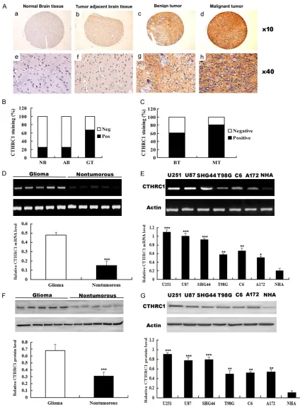
Figure 1. Expression of CTHRC1 in human glioma samples and glioma cell lines. A. Representative images depict CTHRC1 immunohistochemical staining. (a, e) Negative CTHRC1 staining in normal brain tissue (NB); (b, f) Nega-tive CTHRC1 staining in adjacent normal brain tissue (AB); (c, g) PosiNega-tive CTHRC1 staining in benign tumor (BT); (d,
h) Positive CTHRC1 staining in malignant tumor (MT). B. A significant difference in CTHRC1 staining was observed
between normal brain tissue and glioma tissue (GT) (P<0.01, χ2 test) and between tumor adjacent normal brain
tissue and glioma tumor (P<0.01, χ2 test). C. CTHRC1 staining was dramatically increased in malignant tumor
across the center of each well to produce a wound of ~0.5 mm in width. The wounded mo- nolayers were washed twice to remove nonad-herent cells, and fresh medium was added. The photo-graphs were taken at the same position of the wound at various time intervals. The
starting wound edges were defined in each
photo by white lines according to the scratch at 0 hour time point. The wound-healing percent-age was determined by the ratio of healing width at each time point to the wound width at 0 hour. Experiments were carried out in
tripli-cate, and three random fields of each well were
recorded.
Migration assay
Cell migration was determined by using a
modi-fied two chamber migration assay with a pore size of 8 μm. For migration assay, 1 × 10⁵ cells were seeded in serum-free medium in the upper chamber. After 12 hours incubation at 37°C, cells in the upper chamber were carefully removed with a cotton swab and the cells that
had traversed the membrane were fixed in
methanol, stained with Giemsa and
photo-graphed in five independent × 100 fields for
each well. Three independent experiments we- re done and used to calculate fold migration relative to control.
Cell attachment assay
96-well plates were coated with 1.25 μg/ml collagen type IV (Sigma) in 100 μl PBS over -night at 4°C. Wells coated with bovine serum albumin (BSA) served as negative control. The plates were blocked with 2.5 mg/ml BSA for 2 hours in DMEM at 37°C. Cells were trypsin-ized and 2 × 10⁴ cells were seeded in each well for 1 hour at 37°C, and then the cell adhesion assay was performed as previously described [19].
Gelatin zymography
2 × 10⁶ cells were seeded in 100 mm plate for 24 hours, cells were transfected with
nonspe-cific control siRNA or CTHRC1 siRNA. 48 hours
after transfection, serum-free medium was applied to the cells overnight and the proteins in the conditioned medium were concentrated
with Ultracel-30k centrifugal filters (Millipore,
Billerica, MA) at 5,000 × g for 20 min at 4°C.
Proteins (50 μg) were loaded on a 10% poly -acrylamide gel containing 0.1% gelatin (Sigma). After electrophoresis, gel was incubated in
Triton X-100 exchange buffer (20 mM Tris-HCl
[pH 8.0], 150 mM NaCl, 5 mM CaCl2 and 2.5%
Triton X-100) for 30 min followed by 10 min
wash with incubation buffer (same buffer
with-out Triton X-100) thrice. The gel was then incu -bated in incubation buffer overnight at 37°C, stained with 0.5% Coomassie blue R250 (Si- gma) for 4 hours and destained with 30% meth-anol and 10% glacial acetic acid for 2 hours. Gelatinolytic activity was shown as clear areas in the gel.
Statistical analysis
Statistical analysis was performed with SPSS 16.0 software (SPSS, Chicago, IL), Data are expressed as the means ± SD. The association between CTHRC1 staining and the clinicopa- thologic parameters of the glioma patients, including age, gender, WHO grade and
histo-logic type, was evaluated by χ2 test. For MTT cell proliferation assays, Student’s t test was
used. Differences were considered significant when P<0.05.
Results
CTHRC1 expression is up-regulated in glioma tissues and glioma cell lines
In order to investigate whether CTHRC1 expres-sion is changed in glioma, we utilized a TMA to evaluate the CTHRC1 expression in normal brain tissue, tumor adjacent normal brain tis-sue, benign tumor (Grade I and II) and
malig-nant (Grade III and IV). The representative pic -tures presented in Figure 1A showed that CTHRC1 protein in cytoplasm was stained in brown. CTHRC1 positive staining was observed (five samples shown) and nontumorous brain (five samples shown). Actin served as the internal control. E. RT-PCR
analysis of CTHRC1 expression in normal human astrocytes NHA and glioma cell lines, including U251, U87, SHG44,
in 2 of 8 (25%) normal brain tissues, 2 of 8 (25%) tumor adjacent normal brain tissues,
127 of 190 (67%) glioma tissues. A significant
difference in CTHRC1 staining was observed between normal brain tissues and glioma tis-sues (P<0.01, χ2 test) and between tumor adja-cent normal brain tissues and glioma tissues (P<0.01, χ2 test) (Figure 1B). To further confirm these observations, RT-PCR and Western blot
assay were done using five human glioma tis
-sues and five nontumorous brain tis-sues. It was
clear that the glioma tissue had a drastic increase of CTHRC1 expression as compared with the nontumorous tissues (Figure 1D, 1F), which was consistent with the level of CTHRC1 protein expression determined by immunohis-tochemical staining. In addition, RT-PCR and Western blot analyses showed that expression of CTHRC1 was markedly higher in all 6 ana-lyzed glioma cell lines, including SHG44, C6, U251, T98G, U87, A172 as compared with that in normal human astrocytes (NHA) (Figure 1E,
1G). Collectively, our results suggest that CT- HRC1 is up-regulated in gliomas.
Correlation of CTHRC1 expression with clinico-pathological parameters
The clinicopathologic features of 190 glioma biopsies were summarized in Table 1. WHO grade and histologic type are known to be important prognostic markers for patients with glioma. We studied whether CTHRC1
expres-Since CTHRC1 expression is drastically incr- eased in glioma compared with tumor adjacent normal brain tissue, we investigated the involvement of CTHRC1 in glioma cells
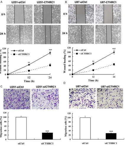
prolifer-ation and invasion. We first transiently trans -fected U251 and U87 glioma cells with CTHRC1 siRNA or control siRNA. Western blot indicated at least 60% knockdown of CTHRC1 protein expression in both U251 and U87 cells trans-fected with CTHRC1 siRNA compared with those transfected with control siRNA (Figure 2A, 2B). 36 hours after transfection, cells were subjected to cell proliferation and invasion assay. Our data suggested that the cell prolif-eration rates were similar between control group and the CTHRC1-siRNA group in both U251 and U87 cells (Figure 2C, 2D). However, in cell invasion assay, CTHRC1-siRNA inhibits cell invasive ability of U251 and U87 cells in matrigel-coated Boyden chamber by 60 and 65%, respectively (Figure 2E, 2F).
Since MMPs play a crucial role in cell invasion, we carried out the western blotting and gelatin zymography to measure the MMPs expression and activities in glioma cells. As shown in
Figure 2G, MMP-2 gelatinolytic activity was dramatically decreased by 47% and 53% in CTHRC1-siRNA U251 and U87 cells compared with the control cells, respectively. Then, we performed western blot to examine the MMP-2 expression in glioma cells. Western blot results showed that MMP-2 protein level was sharply
Table 1. CTHRC1 staining and clinicopathological characteris-tics of 190 glioma patients
Variables CTHRC1 staining
Negative, No. (%) Positive, No. (%) Total P*
Age
<45 years 17 (18.9%) 73 (81.1%) 90 0.719
≥45 years 22 (22.0%) 78 (78.0%) 100 Gender
Male 25 (21.7%) 90 (78.3%) 115 0.487 Female 20 (26.7%) 55 (73.3%) 75 WHO Grade
Benign (I-II) 52 (39%) 81 (61%) 133 0.011
Malignant (III-IV) 11 (19.3%) 46 (80.7%) 57 Histologic type
Astrocytoma 32 (23.9%) 102 (76.1%) 134 0.980 Glioblastoma 9 (25.7%) 26 (74.3%) 35 Oligoastrocytoma 3 (21.4%) 11 (78.6%) 14 Ependymoma 2 (28.6%) 5 (71.4%) 7
*x2 test.
sion correlates with these mark-ers. We found CTHRC1 positive staining in 81 of 133 (61%) be- nign tumor and 46 of 57 (80.7%) malignant tumor. Therefore, CTH- RC1 staining was dramatically
increased in WHO stages III-IV
compared with stages I-II (P< 0.05, χ2 test, Figure 1C). However,
we did not find significant
corre-lations between CTHRC1 expres-sion and histologic type (Table 1).
There is also no significant corre -lations between CTHRC1 expres-sion with other clinicopathologic variables, including patient age and gender (Table 1).
decreased after CTHRC1-siRNA in U251 and U87 cells (Figure 2H).
Knockdown of CTHRC1 in glioma cell lines inhibits cell migration, adhesion and Src, FAK expression in vitro
Tumor cell adhesion to the extracellular matrix is implicated in tumor cell motility, invasion and metastasis [20]. We then used the migration and adhesion assay to detect if CTHRC1 affect cell motility and adhesiveness. First, we investi-gated the role of CTHRC1 in migration of glioma cells by wound-healing assay and migration
assays. We found that there was significant
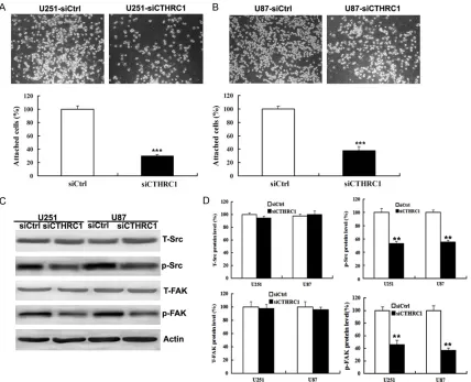
delay in wound closure following knockdown of CTHRC1 (Figure 3A, 3B). In addition, the cell migration assay revealed that U251-siCTHRC1 and U87-siCTHRC1 cells exhibited the ability to migrate through the Boyden chamber by 70 and 72%, respectively, compared with the con-trol cells (Figure 3C, 3D). Moreover, CTHRC1 knockdown decreased cell attachment ability of glioma cells by 70 and 62%, respectively (Figure 4A, 4B).
Convincing evidence exists that the activities of Src family kinases and focal adhesion kinase (FAK) control cell migration and adhesion pro-cesses, and increased expression of these kinases has been associated with more adhe-sive and aggresadhe-sive phenotypes [21, 22]. In order to elucidate the molecular mechanisms responsible for the induction of cell migration and adhesion by CTHRC1, we examined the protein levels of total Src and FAK, phosphory-lated Src and FAK by western blotting. Our results showed that CTHRC1 knockdown down-regulated phosphorylated Src and FAK expres-sion in U251 and U87 glioma cell lines, but not affect total Src and FAK expression (Figure 4C,
4D).
Discussion
The possible involvement of CTHRC1 in human
carcinogenesis was first suggested by a report
that some stromal cells in breast cancer ex-
pressed CTHRC1 mRNA as determined by cDNA microarray analysis and in situ hybridization [23]. Tang et al. then observed that CTHRC1 expression was upregulated in invasive and metastatic melanoma compared with non-met-astatic melanoma [16]. Furthermore, in a sur-vey of 310 human tumor-derived tissues repre-senting 19 types of human solid cancers, CTHRC1 was found to be increased within 16 of these 19 cancer types [16]. Immunohis- tochemical analysis also revealed that CTHRC1 is highly overexpressed in pancreatic cancer
[12, 24], gastric cancer [13] and dermatofibro -sarcoma protuberans (a locally aggressive neo-plasm that frequently recurs and metastasize [15]. The relevance of CTHRC1 in cancer was
reinforced by the identification that CTHRC1
upregulation was correlated with poor patient survival in metastatic breast [11] and colorec-tal cancer [25]. Therefore, CTHRC1 may play a role in general human cancer pathogenesis. However, the role of CTHRC1 in glioma has not
been clearly studied. In this study, our data first demonstrated that CTHRC1 was significantly
increased in glioma compared with tumor ad- jacent normal brain tissue by TMA and Im- munohistochemical technique (Figure 1A, 1B). In addition, CTHRC1 expression is upregulated
in high grade (III-IV) glioma in comparison to
low grade (I-II) glioma (Figure 1C). To further
confirm these observations, RT-PCR and
Western blot analyses showed that expres- sion of CTHRC1 was markedly higher in 5 clini-cal glioma tissues and all 6 glioma cell lines, (Figure 1D-G). Our results are consistent with
previous findings in other tumors [16]. This indi -cated that CTHRC1 may play an important role in the glioma development and progression. In the in vitro assays, we found that knockdown
of CTHRC1 did not significantly regulate cell
proliferation (Figure 2C, 2D), but the cell inva-sion ability is dramatically suppressed in glio-ma cell lines (Figure 3E, 3F). Invasion is the most characteristic biological phenotype of glioblastoma, which is thought to involve aber-rant interactions between tumor cells and ECM [26]. Previous studies found that CTHRC1 has
MTT cell proliferation assay was performed following knockdown of CTHRC1 in U251 and U87cells. E, F. Matrigel cell invasion assay was performed following knockdown of CTHRC1 in U251 and U87 cells. G. Gelatin zymography analysis of the relative enzyme activities of MMP-2 in CTHRC1 knockdown and siRNA control for both U251 and U87 cells. H. Western blot analysis of the relative protein levels of MMP-2 in CTHRC1 knockdown and control group of U251 and U87 cells. All experiments were carried out in triplicate. Data are shown as mean ± SD. **P<0.01;
been shown to be high active and potent degrading ECM proteins in melanoma cell lines [16]. High levels of MMP2 in tissues are associ-ated with tumor cell invasion [27, 28]. MMP2 is thought to be key enzymes involved in the
deg-radation of type IV collagen, which is a compo -nent of the ECM [27]. In this study, our data
suggested that down-regulation of CTHRC1
expression by short interfering RNA significant -ly inhibited MMP-2 protein expression and enzyme activity in glioma cell lines (Figure 2G,
[image:9.612.98.521.74.568.2]2H). Therefore, knockdown of CTHRC1 expres-sion can suppress glioma cell invasiveness through MMP2 down-regulation.
A multistep model of invasion suggests that
cancer cells must first adhere to the ECM, pro
-teolytically degrade the matrix, and finally
mig-rate through this barrier to surrounding tissue [29, 30]. Consequently, it is possible that CTHRC1 contributes to glioma tissue invasion by increasing tumor cell migration and adhe-sion. A previous study suggested that increased
CTHRC1 expression caused the fibroblast cells
to have increased migration in rat cells [6]. Thus, it might be possible that CTHRC1 has a promigratory role in glioma cells. Indeed, we found that knockdown of CTHRC1 decreased cell migration and adhesion abilities of glioma cells through down-regulating both phosphory-lated Src and FAK (Figures 3 and 4). Our finding
is consistent with another study in pancreatic
[image:10.612.94.522.73.420.2]cancer cell lines [12]. The Src-FAK signaling cascade has multiple cellular functions, and modulation of their activities can alter cellular responses that are often perturbed in cancer cells, such as adhesion, migration, and inva-sion [31-33]. FAK promotes cancer cell migra-tion and adhesion by regulating focal adhesion formation and turnover, which involve activa-tion of Src [34]. The constitutively activated Src-FAK pathway is capable of inducing malig-nant transformation of a variety of cell types, including glioma [35]. Inhibition of Src-FAK pathway decreased glioma migration and inva-sion [36]. Park et al. found that CTHRC1 pro-mote pancreatic cancer cell migration through activation of Wnt5a signaling cascade, which induce the activation of phosphorylated Src
and FAK [12]. However, the more exact molecu-lar mechanism of how CTHRC1 regulates Src and FAK expression need us further inves- tigation.
In summary, we found that CTHRC1 expression
is significantly increased in glioma compared
with tumor adjacent normal brain tissue and increased CTHRC1 staining is associated with WHO stages. The mRNA and protein levels of
CTHRC1 were significantly upregulated in
human primary glioma tissues. We also found
that CTHRC1 was significantly upregulated in
glioma cell lines compared to normal human astrocytes. Furthermore, we found that knock-down of CTHRC1 inhibited glioma cell invasion by suppressing MMP-2 protein expression and enzyme activity. Moreover, our data showed that knockdown of CTHRC1 inhibited glioma cell migration and adhesion capacity compared with the control and CTHRC1-siRNA reduced the levels of phosphorylated Src and FAK pro-tein expression. Our results imply that targeting of the CTHRC1 pathway may constitute a poten-tial treatment modality for glioma.
Acknowledgements
This project is supported by grants from the National Natural Science Foundation of China (No.81502160), and Jiangsu Provincial Medical Youth Talent (No.QNRC2016785).
Disclosure of conflict of interest
None.
Address correspondence to: Yue-Chao Fan, Depart-
ment of Neurosurgery, The Affiliated Hospital of Xuzhou Medical University, 99 West Huai-Hai Ro-ad, Xuzhou 221002, Jiangsu, China. Tel:
+86-516-85802319; E-mail: [email protected] ; Jun-Nian Zheng, Jiangsu Key Laboratory of Biological Cancer Therapy, Xuzhou Medical University, 84 West Huai-Hai Road, Xuzhou 221002, Jiangsu, China. Tel: +86-516-8558-2513; E-mail: [email protected]
References
[1] Ortega-Aznar A, Jimenez-Leon P, Martinez E
and Romero-Vidal FJ. [Clinico-pathological and
molecular aspects of diagnostic and progn- ostic value in gliomas]. Rev Neurol 2013; 56: 161-170.
[2] Chen W, Zheng R, Baade PD, Zhang S, Zeng H,
Bray F, Jemal A, Yu XQ and He J. Cancer statis
-tics in China, 2015. CA Cancer J Clin 2016; 66: 115-132.
[3] Mei PJ, Chen YS, Du Y, Bai J and Zheng JN.
PinX1 inhibits cell proliferation, migration and
invasion in glioma cells. Med Oncol 2015; 32: 73.
[4] Ding T, Ma Y, Li W, Liu X, Ying G, Fu L and Gu F.
Role of aquaporin-4 in the regulation of migra-tion and invasion of human glioma cells. Int J Oncol 2011; 38: 1521-1531.
[5] Rao JS. Molecular mechanisms of glioma inva-siveness: the role of proteases. Nat Rev Can-cer 2003; 3: 489-501.
[6] Pyagay P, Heroult M, Wang Q, Lehnert W,
Belden J, Liaw L, Friesel RE and Lindner V. Col -lagen triple helix repeat containing 1, a novel secreted protein in injured and diseased arter-ies, inhibits collagen expression and promotes cell migration. Circ Res 2005; 96: 261-268. [7] Kimura H, Kwan KM, Zhang Z, Deng JM,
Dar-nay BG, Behringer RR, Nakamura T, de Crom-brugghe B and Akiyama H. Cthrc1 is a positive regulator of osteoblastic bone formation. PLoS One 2008; 3: e3174.
[8] Beachy PA, Karhadkar SS and Berman DM. Tis-sue repair and stem cell renewal in carcino-genesis. Nature 2004; 432: 324-331.
[9] Coussens LM and Werb Z. Inflammation and
cancer. Nature 2002; 420: 860-867.
[10] Jiang N, Cui Y, Liu J, Zhu X, Wu H, Yang Z and
Ke Z. Multidimensional roles of collagen triple helix repeat containing 1 (CTHRC1) in malig-nant cancers. J Cancer 2016; 7: 2213-2220. [11] Kharaishvili G, Cizkova M, Bouchalova K,
Mge-brishvili G, Kolar Z and Bouchal J. Collagen tri-ple helix repeat containing 1 protein, periostin and versican in primary and metastatic breast cancer: an immunohistochemical study. J Clin Pathol 2011; 64: 977-982.
[12] Park EH, Kim S, Jo JY, Kim SJ, Hwang Y, Kim JM, Song SY, Lee DK and Koh SS. Collagen triple helix repeat containing-1 promotes pan-creatic cancer progression by regulating mig- ration and adhesion of tumor cells. Carcino-genesis 2013; 34: 694-702.
[13] Wang P, Wang YC, Chen XY, Shen ZY, Cao H,
Zhang YJ, Yu J, Zhu JD, Lu YY and Fang JY. CTHRC1 is upregulated by promoter demethyl-ation and transforming growth factor-beta1 and may be associated with metastasis in hu-man gastric cancer. Cancer Sci 2012; 103: 1327-1333.
[14] Yang XM, You HY, Li Q, Ma H, Wang YH, Zhang YL, Zhu L, Nie HZ, Qin WX, Zhang ZG and Li J.
[15] Wang L, Xiang YN, Zhang YH, Tu YT and Chen HX. Collagen triple helix repeat containing-1 in the differential diagnosis of dermatofibrosar
-coma protuberans and dermatofibroma. Br J
Dermatol 2011; 164: 135-140.
[16] Tang L, Dai DL, Su M, Martinka M, Li G and Zhou Y. Aberrant expression of collagen triple helix repeat containing 1 in human solid can-cers. Clin Cancer Res 2006; 12: 3716-3722. [17] Fan YC, Mei PJ, Chen C, Miao FA, Zhang H and
Li ZL. MiR-29c inhibits glioma cell proliferation, migration, invasion and angiogenesis. J Neu-rooncol 2013; 115: 179-188.
[18] Bai J, Mei PJ, Liu H, Li C, Li W, Wu YP, Yu ZQ and Zheng JN. BRG1 expression is increased in hu-man glioma and controls glioma cell prolifera-tion, migration and invasion in vitro. J Cancer Res Clin Oncol 2012; 138: 991-998.
[19] Jin XB, Mei HF, Pu QH, Shen J, Lu XM, Chu FJ
and Zhu JY. Effects of Musca domestica cecro-pin on the adhesion and migration of human hepatocellular carcinoma BEL-7402 cells. Biol Pharm Bull 2013; 36: 938-943.
[20] Lester BR and McCarthy JB. Tumor cell adhe-sion to the extracellular matrix and signal tran- sduction mechanisms implicated in tumor cell motility, invasion and metastasis. Cancer Me-tastasis Rev 1992; 11: 31-44.
[21] Chikara S, Lindsey K, Borowicz P,
Christofidou-Solomidou M and Reindl KM. Enterolactone alters FAK-Src signaling and suppresses mig- ration and invasion of lung cancer cell lines. BMC Complement Altern Med 2017; 17: 30. [22] Gabarra-Niecko V, Schaller MD and Dunty JM.
FAK regulates biological processes important for the pathogenesis of cancer. Cancer Metas-tasis Rev 2003; 22: 359-374.
[23] Allinen M, Beroukhim R, Cai L, Brennan C, La-hti-Domenici J, Huang H, Porter D, Hu M, Chin L, Richardson A, Schnitt S, Sellers WR and Polyak K. Molecular characterization of the tu-mor microenvironment in breast cancer. Can-cer Cell 2004; 6: 17-32.
[24] Liu W, Fu XL, Yang JY, Yang MW, Tao LY, Liu DJ,
Huo YM, Zhang JF, Hua R and Sun YW. Elevat-ed expression of CTHRC1 prElevat-edicts unfavorable prognosis in patients with pancreatic ductal adenocarcinoma. Am J Cancer Res 2016; 6: 1820-1827.
[25] Tan F, Liu F, Liu H, Hu Y, Liu D and Li G. CTHRC1 is associated with peritoneal carcinomatosis in colorectal cancer: a new predictor for progno-sis. Med Oncol 2013; 30: 473.
[26] Ulrich TA, de Juan Pardo EM and Kumar S. The mechanical rigidity of the extracellular matrix regulates the structure, motility, and prolifera-tion of glioma cells. Cancer Res 2009; 69: 4167-4174.
[27] Badiga AV, Chetty C, Kesanakurti D, Are D, Gu -jrati M, Klopfenstein JD, Dinh DH and Rao JS. MMP-2 siRNA inhibits radiation-enhanced in-vasiveness in glioma cells. PLoS One 2011; 6: e20614.
[28] Xiao LJ, Lin P, Lin F, Liu X, Qin W, Zou HF, Guo L, Liu W, Wang SJ and Yu XG. ADAM17 targets
MMP-2 and MMP-9 via EGFR-MEK-ERK path-way activation to promote prostate cancer cell invasion. Int J Oncol 2012; 40: 1714-1724. [29] Albini A. Tumor and endothelial cell invasion of
basement membranes. The matrigel chemoin-vasion assay as a tool for dissecting molecular mechanisms. Pathol Oncol Res 1998; 4: 230-241.
[30] Albini A, Benelli R, Noonan DM and Brigati C. The “chemoinvasion assay”: a tool to study tu-mor and endothelial cell invasion of basement membranes. Int J Dev Biol 2004; 48: 563-571. [31] Bianchi-Smiraglia A, Paesante S and Bakin AV.
Integrin beta5 contributes to the tumorigenic potential of breast cancer cells through the Src-FAK and MEK-ERK signaling pathways. On-cogene 2013; 32: 3049-3058.
[32] Slanina H, Hebling S, Hauck CR and Schubert-Unkmeir A. Cell invasion by Neisseria menin-gitidis requires a functional interplay between the focal adhesion kinase, Src and cortactin. PLoS One 2012; 7: e39613.
[33] Tomkiewicz C, Herry L, Bui LC, Metayer C,
Bour-deloux M, Barouki R and Coumoul X. The aryl
hydrocarbon receptor regulates focal adhesion sites through a non-genomic FAK/Src pathway. Oncogene 2013; 32: 1811-1820.
[34] Mitra SK and Schlaepfer DD. Integrin-regulat-ed FAK-Src signaling in normal and cancer cells. Curr Opin Cell Biol 2006; 18: 516-523. [35] Hecker TP, Grammer JR, Gillespie GY, Stewart J
Jr and Gladson CL. Focal adhesion kinase en-hances signaling through the Shc/extracellular signal-regulated kinase pathway in anaplastic astrocytoma tumor biopsy samples. Cancer Res 2002; 62: 2699-2707.