Original Article
Berberine improves motor function recovery by
inhibiting endoplasmic reticulum stress-induced
neuronal apoptosis via AMPK activation in rats
with spinal cord injury
Jiquan Wang1, Mingchao Zhang2, Haotian Li2, Gang Li2, Zhiqiang Jia2, Ping Sun2, Xingzhang Zhao2, Gang Lv2,
Zhongkai Fan2
1Department of Orthopaedics, Suizhou Hospital, Hubei University of Medicine, Suizhou, Hubei Province, People’s Republic of China; 2Department of Orthopaedics, The First Affiliated Hospital of Jinzhou Medical University, Jinzhou, Liaoning Province, People’s Republic of China
Received October 19, 2016; Accepted December 22, 2016; Epub April 1, 2017; Published April 15, 2017
Abstract: Evidences have supported the neuroprotective potential of berberine (BBR) in brain ischemia and injury. This study aimed to investigating whether BBR has neuroprotective effects on SCI and its mechanisms. Adult male Sprague-Dawley rats (220-250 g) were randomly divided into Sham, SCI, SCI + BBR (40 mg/kg/day), and SCI + BBR (40 mg/kg/day) + Compound C groups. The BBB score showed BBR improved functional recovery obviously 7 days later after SCI. Spinal cord tissue samples were harvested three days after SCI. Apoptotic neurons were assessed by TUNEL assay, and the expression levels of p-AMPK, AMPK, CHOP, cleaved caspase-12, Bcl-2, Bax, and cleaved
caspase-3 were determined via Western blot. Furthermore, the influence of BBR on p-AMPK, caspase-12 and CHOP expression was determined via immunofluorescence. Neuronal cell apoptosis after SCI was significantly attenuated
by BBR (P<0.01). Moreover, Western blot demonstrated that the expressions of cleaved caspase-12, CHOP, Bax, and caspase-3, which were linked to endoplasmic reticulum stress (ERS) associated apoptosis pathways, were
significantly increased after SCI and were inhibited by BBR (P<0.01). However, the expressions of p-AMPK and Bcl-2
were significantly decreased after SCI and ameliorated by BBR (P<0.01). Immunofluorescence analysis indicated
that BBR increased p-AMPK positive neurons number, reduced caspase-12 and CHOP positive neurons number following SCI (P<0.01). However, the protective effect of BBR on ERS-related protein expression was abolished by Compound C (P>0.05). Thus, BBR attenuates neuronal apoptosis and improves functional recovery in rats with SCI, and these neuroprotective effects may be associated with ERS inhibition and AMPK activation.
Keywords: Spinal cord injury, apoptosis, berberine, endoplasmic reticulum stress, AMPK, neuron
Introduction
SCI is a serious clinical problem linked to high morbidity worldwide, which often leads to dev-astating and catastrophic dysfunction [1].
Permanent neurological deficits result from
massive cell death through immediate cell death by necrosis and prolonged apoptotic cell death, which results from secondary injury after SCI [2]. Secondary injury includes distur-bances in ionic homeostasis, local edema, fo- cal hemorrhage, excitotoxicity, oxidative stress,
endoplasmic reticulum stress (ERS), and inflam -mation [3]. Neuronal apoptosis is a critical mechanism of secondary injury after SCI [4] and is triggered by various complex
mecha-nisms. Recent concerns have been raised re- garding the role of ERS, neuronal apoptosis,
and their interactions in pathological deficits
Berberine (BBR) is an alkaloid extract in tradi-tional medicine herbs. It is an AMP-activated protein kinase (AMPK) activator that is widely used in oriental medicine to treat diarrhea [12, 13]. BBR also exerts neuroprotective effects in brain ischemia [14-16], traumatic brain injury [17], and diabetic neuropathy in rats [18, 19]. Furthermore, previous studies have demon-strated that BBR attenuates ERS-induced cell apoptosis in several cell types [20, 21]. In addi-tion, AMPK activation has been demonstrated to inhibit ERS-mediated neuroblastoma cell apoptosis [22]. However, whether BBR has a neuroprotective effect on SCI and the neuro-protective molecular mechanisms of BBR in the nervous system remain unknown. This study aimed to determine the effect of BBR on neuro-nal apoptosis and ERS in a rat model of SCI
using behavioral, biochemical, and immunoflu -orescent assessments.
Materials and methods
Animals and experimental design
All animal experiments were conducted in accordance with the National Institutes of He- alth’s Guide for the Care and Use of Laboratory Animals. Adult male Sprague-Dawley rats (220-250 g) were purchased from the Experimental Animals Center of Liaoning Medical University. The animals were randomly divided into four groups: the sham group, SCI group, SCI + BBR group, and SCI + BBR + Compound C group. Berberine hydrochloride (BBR) (Nanjing Chunqiu Biology Engineering Company, 141433-60-5) was administered via intraperitoneal injection at a dose of 20 mg/kg every 12 hours per day following SCI. To investigate whether AMPK activation inhibits ERS-mediated neuronal ap-
optosis after SCI in rats, a specific inhibitor of
AMPK, Compound C (Sigma-Aldrich, St. Louis, MO, USA), was administered. Compound C was dissolved in 10% DMSO (Dimethyl sulfoxide; Sigma-Aldrich, St. Louis, MO, USA), and aliquots were stored at -20°C until use. Aliquots of
Compound C at a concentration of 5 μg/μl were
prepared in normal saline for single
intrave-nous (5 μl) injection 30 minutes prior to BBR treatment (the final concentration of DMSO
<0.1%).
Induction of SCI in rats
All rats were injured at the thoracic level 10
(T10) following a modified weight-drop model,
which was performed to induce SCI [23]. Briefly,
the rats were anaesthetized (10% chloral hydrate 0.03 ml/kg, i.p.), and the model was induced under sterile conditions. The skin and muscle were incised, and a laminectomy was performed at T10, leaving the dura intact. The impact of a 10-g weight at a height of 20 mm was subsequently applied to the dorsal surface of the spinal cord. The force was implemented by a steel rod (2-mm diameter tip), which was vertically dropped through a ruler tube perpen-dicular to the spinal cord. Following removal of the device, the muscle and skin were sutured. Treatments were subsequently administered as indicated for each group.
Behavioral tests
To investigate the functional recovery of the rats’ hind limbs after SCI, the Basso Beattie Bresnahan (BBB) behavioral test in an
open-field was performed at four time points after
SCI [24]. The scale used to measure hind limb function after SCI ranged from zero, which indi-cates no spontaneous movement, to a maxi-mum score of 21, which indicates normal motor function. The inclined plane test was performed
by a rubber mat secured to a flat board. The maximum angle of the flat board position was
increased from 0° horizontally in 5° increments until the rat was unable to maintain its position
on the board for five seconds without falling.
Each rat was scored by two observers blinded to the experimental groups.
Collection of spinal cord tissues
Seventy-two hours following sham surgery or injury, a subgroup of animals in each group
were sacrificed by cervical dislocation. For
epicen-ter, and caudal regions were harvested and stored in 4% paraformaldehyde for subsequent
analysis. The formalin-fixed spinal cord tissue
samples were subsequently embedded in OCT media and coronally sectioned at 5-µm thick-ness at two spinal cord levels (2-mm rostral and caudal to the epicenter of the injury) with a freezing microtome (Leica CM3050S, Heidel- berg, Germany).
TUNEL assay
To determine the ratio of cell apoptosis in the two tissue sections (2-mm rostral and caudal to the epicenter of the injury) of each spinal cord tissue sample, an in situ cell death detection kit (Roche, Mannheim, Germany, 10711900) was applied according to the manufacturer’s instructions. Images were captured by a confo-cal microscope (Olympus IX71, Tokyo, Japan). Positive apoptotic neurons were counted in six
randomly selected fields per sections in the spi -nal anterior horn by Image J 1.42q a-nalyzer software (Liaoning Medical University, Jinzhou, Liaoning, China). The average number of TUNEL-positive neurons (red dots neurons) and all neu-rons for the two sections were counted respec-tively as the number in a sample, at 400 ×
magnification (Scale bars = 20 μm) [25]. The
percentage of apoptotic neurons in a sample
was defined as follows: the percentage of apop
-totic cells (%) = 100 × (total apop-totic neurons/
total neurons).
Immunofluorescence staining
The two frozen sections of each spinal cord tis-sue sample from the 2-mm rostral and caudal
to the epicenter of the injury were rewarmed at room temperature for 20 minutes and then rinsed three times in PBS for 15 minutes. The sections were subsequently blocked with 5% normal donkey sera (Jackson Immuno Resear- ch, West Grove, PA, USA; 118727), 0.05% Triton X-100 for one hour. The sections were then incubated with a suitable dilution of primary IgG antibody, such as p-AMPK(1:50; Santa Cruz, sc-101630), Mouse mAb CHOP (1:3,200; CST, USA; L63F7, #2895S) or Rabbit mAb Caspa- se-12 (1:1000; Abcam, ab18766) mixture, ov- ernight at 4°C and then rinsed three times in PBS for 15 minutes. The sections were
subse-quently incubated with IFKine Red AffiniPure
Donkey Anti-Rabbit IgG (1:1,000; Abbkine, Re-
dlands, CA, USA; 142401B, 100 μl/500 μl) or IFKine Green AffiniPure Donkey Anti-Mouse IgG
(1:1,000; Abbkine, Redlands, CA, USA; 1337-
02A, 100 μl/500 μl) for one hour at room tem -perature. After washing in PBS for 15 minutes, the nuclei were marked with 4’6-diamidino-2-phenylindole (DAPI) according to the manu-facturer’s instructions (Sigma-Aldrich, St Louis, MO, USA; #033M4064V, 1 mg). Images were captured by a confocal microscope (Olympus
IX71, Tokyo, Japan) at a high magnification (Scale bars = 20 μm). The positive cell numbers
of CHOP and caspase-12 were counted in six
randomly selected fields per sections in the spi -nal anterior horn by Image J 1.42q a-nalyzer software (Liaoning Medical University, Jinzhou, Liaoning, China). The average positive cell num-ber for the two sections (2-mm rostral and cau-dal to the epicenter of the injury) was then cal-culated as the number of positive cells in a sample.
Western blot
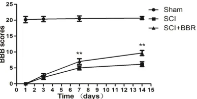
[image:3.612.93.287.72.170.2]The prepared spinal cord tissues were minced using eye scissors on ice, homogenized in lysis buffer that contained 1 mmol/L of PMSF, 1 µg/ ml of aprotinin, 1 µg/ml pepstatin A, 50 mM of NaF and centrifuged at 12,000 revolutions per minute (rpm) at 4°C for 20 minutes to collect the supernatant. BCA protein assay was used to determine the protein concentrations. Pro- teins were separated via SDS-polyacrylamide gel electrophoresis (SDS-PAGE) and transferred onto a PVDF membrane. The membrane was blocked with 0.1% BSA in Tris-buffered saline (TBS) for two h at room temperature and subse-quently incubated overnight at 4°C with anti-bodies for p-AMPK (1:1000; Santa Cruz, sc-10- Figure 1. BBR improved locomotor function after
spi-nal cord injury. Locomotor function was assessed by the BBB scale. Rats treated with BBR demonstrated
a significant improvement in locomotor function from
days 7 to 14 post-injury compared with the SCI group (**P<0.01). **P<0.01 vs. the SCI group. Data are presented as means ± standard deviations (SDs), n
1630), AMPK (1:1,000; Santa Cruz, sc-74461), CHOP (1:1,000; Cell Signaling, USA, L63F7, #2895S), Bcl-2 (1:1000; Abcam, ab183656), Bax (1:1000; Abcam, ab199613), caspase-12 (1:500; Abcam, ab18766), cleaved-caspase-3 (1:500; Abcam, Cambridge; ab32042), and
β-actin (1:500; Santa Cruz Biotechnology USA,
#D1713). The membrane was then incubat- ed with HRP-conjugated secondary antibody
(1:2,000; Santa Cruz, #L3902, 200 μg/0.5 ml)
in TBS-T for one hour at room temperature and visualized using a chemiluminescence system (ECL kit; Beyotime Institute of Biotechnology, Nanjing, Jiangsu, China). Optical density (OD) was determined using Image J 1.42q analyzer software (Liaoning Medical University, Jinzhou, Liaoning, China).
Statistical analysis
Data were analyzed using SPSS version 16.0 statistical software package (SPSS Inc., Chica- go, IL, USA). All data are expressed as means ± standard deviations (SDs). Comparisons bet- ween groups were performed via one-way anal-ysis of variance (ANOVA) and Dunnett’s post-hoc test for the statistical analysis. P<0.05 was
considered as statistically significance.
Results
Effects of BBR on neural function recovery after SCI
[image:4.612.92.523.72.513.2]To identify the effects of BBR on neural func-tion recovery during the two weeks after SCI,
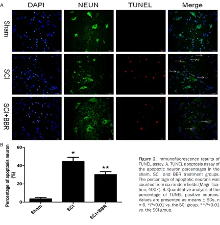
Figure 2. Immunofluorescence results of
TUNEL assay. A. TUNEL apoptosis assay of the apoptotic neuron percentages in the sham, SCI, and BBR treatment groups. The percentage of apoptotic neurons was
counted from six random fields (Magnifica -tion, 400×). B. Quantitative analysis of the percentage of TUNEL positive neurons. Values are presented as means ± SDs, n
= 6. *P<0.01 vs. the SCI group, **P<0.01
the evaluation of BBB scoring at four time points [23] post-surgery was used to quantify locomotor activity. In the sham group, the BBB scores were 20.17 ± 0.983, 20.33 ± 0.816, 20.5 ± 0.837, and 20.83 ± 0.408 at the four time points post-surgery, respectively. The rats in the two experimental groups exhibited grad-ual function improvement during the subse-quent seven days. In contrast, the rats in the
BBR group exhibited significantly increased
scores at days 7 (P<0.01) and 14 (P<0.01) compared with the SCI group. Two weeks after
SCI, the final BBB scores in the SCI (n = 6) and BBR (n = 6) groups were 6.17 ± 0.753 and 9.67
± 0.816, respectively (P<0.01, Figure 1).
BBR attenuates neuronal apoptosis in spinal cord tissues after SCI
Neuron survival was examined via an
immuno-fluorescence technique using a specific anti -body for neuron (NeuN) and TUNEL analysis (Figure 2). The number of TUNEL positive neu-rons in each group was counted with a color image analyzer (Image-ProPlus, Liaoning Medi- cal University, Jinzhou, Liaoning, China). The percentages of apoptotic spinal cord neurons
and cleaved caspase-12 in spinal cord tissues
Western blotting was used to determine the effects of BBR on the expressions of p-AMPK, CHOP, and cleaved caspase-12 in each group.
The SCI group exhibited significantly decreased p-AMPK expression and significantly increased
CHOP and cleaved caspase-12 expressions compared with the sham group (P<0.05). In
contrast, the BBR group exhibited a significant
-ly increased p-AMPK expression and signifi -cantly decreased CHOP and cleaved cas-pase-12 expressions compared with the SCI group (P<0.05, Figure 4).
Immunofluorescence staining of p-AMPK, cas -pase-12 and CHOP in spinal cord neurons
Immunofluorescence staining was used to
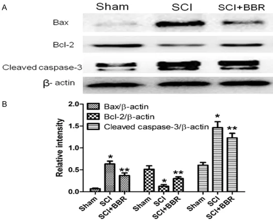
[image:5.612.91.373.72.301.2]identify the effects of BBR administration on the expressions of p-AMPK, caspase-12 and CHOP in spinal cord tissues three days after SCI in each group (Figure 5A-F). The number of p-AMPK, caspase-12 and CHOP positive neu-rons in each group was counted with a color image analyzer (Image-ProPlus, Liaoning Me- dical University, Jinzhou, Liaoning, China). The Figure 3. BBR ameliorates the level of apoptosis associated with Bax, Bcl-2,
and cleaved caspase-3 proteins in spinal cord lesions three days after SCI. A. Protein expressions of Bax, Bcl-2, and cleaved caspase-3 in the sham,
SCI, and BBR treated groups. β-actin was used as the loading control and
for band density normalization. B. Optical density analysis of Bax, Bcl-2, and cleaved caspase-3 proteins. *P<0.01 vs. the sham group, **P<0.01 vs. the
SCI group. Data are presented as means ± SDs, n = 6.
in the sham, SCI, and SCI + BBR groups were 3.56 ± 1.53%, 44.58 ± 4.52%, and 30.26 ± 3.22%, respectively.
These findings indicate that
BBR reduced spinal cord neu-ronal death after SCI.
Effects of BBR on Bcl-2, Bax, and cleaved caspase-3 expression in spinal cord tissues
The SCI group exhibited dec- reased Bcl-2 expression and increased Bax and cleaved caspase-3 expression com-pared with the sham group. In contrast, the BBR group exhi-
bited a significantly increased Bcl-2 expression and signifi -cantly decreased Bax and cl- eaved caspase-3 expression compared with the SCI group (P<0.01; Figure 3A, 3B).
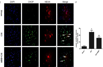
average numbers of p-AMPK, caspase-12 and CHOP positive neurons per section in each group were as follows (Figure 5B, 5D, 5F): sham group 25.67 ± 2.73, 8.33 ± 1.63 and 3.32 ± 1.42/mm2, respectively; SCI group 6 ± 1.78,
31.17 ± 3.19 and 24.51 ± 2.73/mm2,
respec-tively; and BBR group 17.5 ± 2.59, 20.51 ± 2.88 and 15.83 ± 1.72/mm2, respectively.
Immunofluorescence staining indicated
incre-ased CHOP and caspase-12 but decreincre-ased p-AMPK positive neuron numbers in the spinal cord tissues in the SCI group compared with the sham group (P<0.01). However, the spinal
cord tissues in the BBR group exhibited signifi -cantly higher p-AMPK but lower CHOP and cas-pase-12 positive neuron numbers compared with the SCI group (P<0.01).
Effects of compound C on the expressions of p-AMPK, CHOP, and cleaved caspase-12 in SCI with BBR administration
To investigate the molecular mechanism of the
influence of BBR on ERS-induced neuronal
apoptosis in the SCI model, the AMPK inhibitor Compound C was administered. The activations of AMPK, CHOP, and cleaved caspase-12 were
Following SCI, the pathological changes that ensue are a result of direct damage at the injured location and include disturbances in ionic homeostasis, local edema, focal
hemor-rhage, excitotoxicity, oxidative stress, inflam -mation, and ERS. These changes have been referred to as secondary damages [3]. Although the damage initiated by an original injury is irre-versible, secondary injury is an active process that occurs at molecular and cellular levels and
[image:6.612.92.375.72.289.2]is thus reversible and modifiable [26]. This sug -gests that therapeutic treatments to limit the pathological process and improve neural func-tion recovery are possible. Although the precise mechanisms of secondary spinal cord damage remain elusive, neuronal and glial cell apopto-sis plays a role in SCI, and the inhibition of neu-ronal and oligodendroglial apoptosis may be critical for improving neural function recovery [4, 27].
BBR is an alkaloid extracted from plants of the berberidaceae family and has a long history in Chinese medicines [28]. BBR can penetrate the blood-brain barrier via intraperitoneal adminis-tration [29]. However, to our knowledge, it has not been previously reported whether BBR has Figure 4. BBR administration activates AMPK and inhibits the protein
expres-sions of ERS-induced apoptosis response proteins, CHOP, and caspase-12. Western blot analysis of the protein expressions of CHOP and caspase-12 for
the sham, SCI, and BBR treatment groups is shown. β-actin was used as the
loading control and for band density normalization (A). Graphs indicate the
relative band intensities compared with β-actin (B). *P<0.05 vs. the sham group, **P<0.05 vs. the SCI group. Data are presented as means ± SDs, n
= 6.
determined by Western blot analysis. The expression lev-els of CHOP and caspase-12
were significantly reduced, but p-AMPK was significantly
increased in the SCI + BBR group compared with the SCI group (P<0.01). However, the expression levels of CHOP and cleaved caspase-12 were
significantly increased, but p-AMPK was significantly
de-creased in the SCI + BBR + Compound C group compared with the SCI + BBR group (P< 0.01). The expressions of CH- OP and cleaved caspase-12 in the SCI + BBR + Compound
C group was not significantly
different from the SCI group (P>0.05). Together, these find -ings indicate that the protec-tive role of BBR in ERS-induced apoptosis in SCI is related to AMPK activation (Figure 6A, 6B).
Figure 5. Immunofluorescence staining of the proteins: p-AMPK (A), caspase-12 (C) and CHOP (E) in rat spinal cord
tissues at 72 h after injury. There were lots of p-AMPK positive neurons but few caspase-12 and CHOP positive
neurons in the sham group. After SCI the number of p-AMPK positive neurons decreased significantly. What more,
Injury resulted in a substantial number of caspase-12 and CHOP positive neurons in the SCI group. This increase
was significantly attenuated by BBR treatment. *P<0.05 vs. the sham group, **P<0.05 vs. the SCI group. (B, D, F) The quantitative analysis of P-AMPK, caspase-12 and CHOP positive neurons per longitudinal section in both groups between 5 mm rostral and caudal to the injury epicenter (six views per section) is shown. Values are presented as
means ± SDs, n = 6.
neuroprotective effects in SCI. To assess the effect of BBR on neurological function in SCI rats, an extensively used BBB scoring method
was implemented in the current study. The find -ings indicated that BBR administration after
SCI was associated with significant improve -ment in locomotion at one week post-treat-ment (Figure 1). This finding suggests that BBR ame
-liorates deficits in locomotor function in rats with SCI. Furthermore, BBR not only significant -ly decreased the percentage of TUNEL positive neurons but also decreased the Bax/Bcl-2 ratio and suppressed cleaved caspase-3 expression (Figures 2 and 3). Together, these findings indi -cate that BBR has neuroprotective effects in SCI.
Early research demonstrated that BBR, an
AMPK activator, suppresses neuroinflammato -ry responses via AMPK activation in BV-2
microglia [30]. Other research has also demon-strated that AMPK activation plays a protective role in brain ischemia in rats [31, 32]. AMPK is a protein kinase that maintains the energy bal-ance in an organism [33, 34]. Disturbbal-ances in energy metabolism after SCI have been dem-onstrated in previous research [35]. The cen-tral nervous system has a high metabolic rate and a poor capacity for nutrient storage. How- ever, the changes in p-AMPK protein expres-sion in rats after SCI remain unknown. The-
refore, it is important to investigate the influ -ence of BBR on AMPK and the role of AMPK in neuronal survival and death following SCI. Our research demonstrates that the p-AMPK expression after SCI is lower than in normal spi-nal cord tissues; however, BBR administration after SCI promotes AMPK activation (Figures 4 and 5A, 5B). These findings indicate that the
which suggests that the energy metabolism in SCI is turbulent. Nevertheless, BBR administra-tion activated AMPK in rats after SCI. Together,
the current findings suggest that AMPK activa -tion may be associated with the effect of BBR inhibition of neuronal apoptosis following SCI. Nevertheless, the exact mechanism of the interaction between apoptosis and AMPK acti-vation remains unclear. Several studies have demonstrated that the protein expression of CHOP and caspase-12 progressively increased to a maximum at three days post-injury [5, 7]. The protein expression levels of CHOP and cas-pase-12 at three days after SCI were deter-mined to investigate the molecular mechanism of BBR in SCI in this study. The results demon-strated that ERS was involved in SCI, and the
expressions of CHOP and caspase-12 signifi -cantly increased, which is similar to a previous study [5]. Furthermore, the current study dem-onstrated that ERS was inhibited by BBR admin-istration three days after SCI in rats (Figures 4 and 5C-F), which suggests that the
neuropro-Despite these promising findings, several study
limitations must be considered in the interpre-tation of these results. First, our research sug-gests that the administration of BBR (40 mg/ kg/day) has the effect to inhibit ER stress-induced apoptosis in rats after SCI, while a pre-vious study indicated that a relatively high con-centration of berberine induces apoptosis via ER stress through the elevation of ROS and the mitochondrial-dependent pathway in human glioblastoma T98G cells [40]. It is necessary to clarify whether the different concentrations of berberine is associated with its different drug effects in cell apoptosis and investigate the optimal therapeutic dose for SCI. Several stud-ies have demonstrated that BBR has the poten-tial to promote axonal regeneration in injured nerves of the peripheral nervous system [41] and promote Nrf2-related neurite outgrowth [18]. In addition, BBR also attenuates axonal transport impairment and axonopathy in neuro-blastoma-2a cells [42]. Nevertheless, whether the positive effect of BBR on functional recov-ery after SCI is related to its potential to pro-Figure 6. Blocking AMPK activation facilitated ERS-induced apoptosis. A.
Ex-pression of p-AMPK, CHOP, and cleaved caspase-12 proteins in the sham, SCI, SCI + BBR, and SCI + BBR + compound C groups. B. The optical density analysis of p-AMPK, CHOP, and cleaved caspase-12 in each group are shown
as histograms. β-actin was used as the loading control and for band den -sity normalization. *P<0.01 vs. the sham group; **P<0.01 vs. the SCI group; ##P>0.05 vs. the SCI group. Data are presented as means ± SDs, n = 6.
tective effect of BBR in SCI may be relevant to ERS inhibi-tion. It has been reported that AMPK activation has neuro-protective effects [36] and that this is an important de- fensive response to stress [37]. AMPK activation pro-tects cardiomyocytes against hypoxic injury via the attenua-tion of ERS [38]. Moreover, AMPK activation may function against ERS-mediated neuro-toxicity [39]. To investigate whether AMPK activation in- hibits ERS-mediated neuronal apoptosis following SCI in
rats, a specific inhibitor of
[image:9.612.92.369.73.327.2]mote axonal regeneration requires further investigation.
In summary, the main findings in this study indi -cate that BBR treatment inhibits the activation of the ERS-induced apoptotic pathway in a rat SCI model, and this protective effect may occur via AMPK activation.
Acknowledgements
This study was supported by the National Natural Science Foundation of China (No. 81- 272074), the Program for Liaoning Excellent Talents in University (No. 2014091), and the President Foundation of Liaoning Medical University (No. QM2014011).
Disclosure of conflict of interest
None.
Address correspondence to: Zhong-Kai Fan, Depart-
ment of Orthopaedics, The First Affiliated Hospital of
Jinzhou Medical University, 5-2 Renmin Street, Guta District, Jinzhou 121000, Liaoning Province, Peo- ple’s Republic of China. Fax: +86 4164673679; E-mail: fanzk_ln@163.com
References
[1] Di Paola R, Impellizzeri D, Salinaro AT, Mazzon E, Bellia F, Cavallaro M, Cornelius C, Vecchio G, Calabrese V, Rizzarelli E, Cuzzocrea S. Admin-istration of carnosine in the treatment of acute spinal cord injury. Biochem Pharmacol 2011; 82: 1478-89.
[2] Emery E, Aldana P, Bunge MB, Puckett W, Srini-vasan A, Keane RW, Bethea J, Levi AD. Apopto-sis after traumatic human spinal cord injury. J Neurosurg 1998; 89: 911-20.
[3] Lee JY, Maeng S, Kang SR, Choi HY, Oh TH, Ju BG, Yune TY. Valproic acid protects motor neu-ron death by inhibiting oxidative stress and en-doplasmic reticulum stress-mediated cyto-chrome C release after spinal cord injury. J Neurotrauma 2014; 31: 582-94.
[4] Liu C, Shi Z, Fan L, Zhang C, Wang K, Wang B. Resveratrol improves neuron protection and functional recovery in rat model of spinal cord injury. Brain Res 2011; 1374: 100-9.
[5] Zhang H, Wu F, Kong X, Yang J, Chen H, Deng L, Cheng Y, Ye L, Zhu S, Zhang X, Wang Z, Shi H, Fu X, Li X, Xu H, Lin L, Xiao J. Nerve growth fac-tor improves functional recovery by inhibiting endoplasmic reticulum stress-induced neuro-nal apoptosis in rats with spineuro-nal cord injury. J Transl Med 2014; 12: 130.
[6] Zhang HY, Zhang X, Wang ZG, Shi HX, Wu FZ, Lin BB, Xu XL, Wang XJ, Fu XB, Li ZY, Shen CJ,
Li XK, Xiao J. Exogenous basic fibroblast growth
factor inhibits ER Stress-induced apoptosis and improves recovery from spinal cord injury. CNS Neurosci Ther 2013; 19: 20-9.
[7] Penas C, Guzmán MS, Verdú E, Forés J, Navar-ro X, Casas C. Spinal cord injury induces endo-plasmic reticulum stress with different cell-type dependent response. J Neurochem 2007; 102: 1242-55.
[8] Oyadomari S, Koizumi A, Takeda K, Gotoh T, Akira S, Araki E, Mori M. Targeted disruption of the Chop gene delays endoplasmic reticulum stress-mediated diabetes. J Clin Invest 2002; 109: 525-32.
[9] Zinszner H, Kuroda M, Wang X, Batchvarova N, Lightfoot RT, Remotti H, Stevens JL, Ron D. CHOP is implicated in programmed cell death in response to impaired function of the endo-plasmic reticulum. Genes Dev 1998; 12: 982-95.
[10] Nakagawa T, Yuan J. Cross-talk between two cysteine protease families. Activation of cas-pase-12 by calpain in apoptosis. J Cell Biol 2000; 150: 887-94.
[11] Nakagawa T, Zhu H, Morishima N, Li E, Xu J, Yankner BA, Yuan J. Caspase-12 mediates
en-doplasmic reticulum specific apoptosis and
cytotoxicity by amyloid-beta. Nature 2000; 403: 98-103.
[12] Mo C, Wang L, Zhang J, Numazawa S, Tang H, Tang X, Han X, Li J, Yang M, Wang Z, Wei D, Xiao H. The crosstalk between Nrf2 and AMPK
sig-nal pathways is important for the anti-inflam -matory effect of berberine in LPS-stimulated macrophages and endotoxin-shocked mice. Antioxid Redox Signal 2014; 20: 574-88. [13] Kim WS, Lee YS, Cha SH, Jeong HW, Choe SS,
Lee MR, Oh GT, Park HS, Lee KU, Lane MD, Kim JB. Berberine improves lipid dysregulation in obesity by controlling central and peripheral AMPK activity. Am J Physiol Endocrinol Metab 2009; 296: E812-9.
[14] Hong JS, Chu YK, Lee H, Ahn BH, Park JH, Kim MJ, Lee S, Ryoo HS, Jang JH, Lee SR, Park JW. Effects of berberine on hippocampal neuronal damage and matrix metalloproteinase-9 activ-ity following transient global cerebral ischemia. J Neurosci Res 2012; 90: 489-97.
[15] Chai YS, Yuan ZY, Lei F, Wang YG, Hu J, Du F, Lu X, Jiang JF, Xing DM, Du LJ. Inhibition of retino-blastoma mRNA degradation through poly (A) involved in the neuroprotective effect of ber-berine against cerebral ischemia. PLoS One 2014; 9: e90850.
model of transient global cerebral ischemia. Eur J Pharmacol 2013; 720: 98-106.
[17] Chen CC, Hung TH, Lee CY, Wang LF, Wu CH, Ke CH, Chen SF. Berberine protects against neuro-nal damage via suppression of glia-mediated
inflammation in traumatic brain injury. PLoS
One 2014; 9: e115694.
[18] Hsu YY, Tseng YT, Lo YC. Berberine, a natural antidiabetes drug, attenuates glucose neuro-toxicity and promotes Nrf2-related neurite out-growth. Toxicol Appl Pharmacol 2013; 272: 787-96.
[19] Kim SO, Kim HJ. Berberine ameliorates cold and mechanical allodynia in a rat model of dia-betic neuropathy. J Med Food 2013; 16: 511-7. [20] Yu W, Sheng M, Xu R, Yu J, Cui K, Tong J, Shi L,
Ren H, Du H. Berberine protects human renal proximal tubular cells from hypoxia/reoxygen-ation injury via inhibiting endoplasmic reticu-lum and mitochondrial stress pathways. J Transl Med 2013; 11: 24.
[21] Hao X, Yao A, Gong J, Zhu W, Li N, Li J.
Berber-ine Ameliorates pro-inflammatory cytokBerber-ine-in -duced endoplasmic reticulum stress in human
intestinal epithelial cells in vitro. Inflammation
2012; 35: 841-9.
[22] Kim J, Park YJ, Jang Y, Kwon YH. AMPK activa-tion inhibits apoptosis and tau hyperphosphor-ylation mediated by palmitate in SH-SY5Y cells. Brain Res 2011; 1418: 42-51.
[23] Allen AR. Surgery of experimental lesion of spi-nal cord equivalent to crush injury of fracture dislocation of spinal column. J Am Med Assoc 1911; 57: 878-880.
[24] Basso DM, Beattie MS, Bresnahan JC. Graded histological and locomotor outcomes after spi-nal cord contusion using the NYU weight drop device versus transection. Exp Neurol 1996; 139: 244-56.
[25] Li G, Jia Z, Cao Y, Wang Y, Li H, Zhang Z, Bi J, Lv G, Fan Z. Mitochondrial division inhibitor 1 ameliorates mitochondrial injury, apoptosis, and motor dysfunction after acute spinal cord injury in rats. Neurochem Res 2015; 40: 1379-92.
[26] Jin W, Ming X, Hou X, Zhu T, Yuan B, Wang J, Ni H, Jiang J, Wang H, Liang W. Protective effects of erythropoietin in traumatic spinal cord injury by inducing the Nrf2 signaling pathway activa-tion. J Trauma Acute Care Surg 2014; 76: 1228-34.
[27] Chen KB, Uchida K, Nakajima H, Yayama T, Hi-rai T, Watanabe S, Guerrero AR, Kobayashi S, Ma WY, Liu SY, Baba H. Tumor necrosis factor-alpha antagonist reduces apoptosis of neu-rons and oligodendroglia in rat spinal cord in-jury. Spine (Phila Pa 1976) 2011; 36: 1350-8. [28] Vuddanda PR, Chakraborty S, Singh S.
Berber-ine: a potential phytochemical with
multispec-trum therapeutic activities. Expert Opin Inves-tig Drugs 2010; 19: 1297-307.
[29] Wang X, Wang R, Xing D, Su H, Ma C, Ding Y, Du L. Kinetic difference of berberine between hip-pocampus and plasma in rat after intravenous administration of Coptidis rhizoma extract. Life Sci 2005; 77: 3058-67.
[30] Lu DY, Tang CH, Chen YH, Wei IH. Berberine
suppresses neuroinflammatory responses
through AMP-activated protein kinase activa-tion in BV-2 microglia. J Cell Biochem 2010; 110: 697-705.
[31] Ashabi G, Khodagholi F, Khalaj L, Goudarzvand M, Nasiri M. Activation of AMP-activated pro-tein kinase by metformin protects against glob-al cerebrglob-al ischemia in mglob-ale rats: interference
of AMPK/PGC-1α pathway. Metab Brain Dis
2014; 29: 47-58.
[32] Culmsee C, Monnig J, Kemp BE, Mattson MP. AMP-activated protein kinase is highly ex-pressed in neurons in the developing rat brain and promotes neuronal survival following glu-cose deprivation. J Mol Neurosci 2001; 17: 45-58.
[33] Ramamurthy S, Ronnett GV. Developing a head for energy sensing: AMP-activated protein ki-nase as a multifunctional metabolic sensor in the brain. J Physiol 2006; 574: 85-93.
[34] Zhang Y, Hillered L, Olsson Y, Holtz A. Time course of energy perturbation after compres-sion trauma to the spinal cord: an experimen-tal study in the rat using microdialysis. Surg Neurol 1993; 39: 297-304.
[35] Okon EB, Streijger F, Lee JH, Anderson LM, Russell AK, Kwon BK. Intraparenchymal micro-dialysis after acute spinal cord injury reveals differential metabolic responses to contusive versus compressive mechanisms of injury. J Neurotrauma 2013; 30: 1564-76.
[36] Kuramoto N, Wilkins ME, Fairfax BP, Revilla-Sanchez R, Terunuma M, Tamaki K, Iemata M, Warren N, Couve A, Calver A, Horvath Z, Free-man K, Carling D, Huang L, Gonzales C, Cooper E, Smart TG, Pangalos MN, Moss SJ. Phospho-dependent functional modulation of GABA (B) receptors by the metabolic sensor AMP-depen-dent protein kinase. Neuron 2007; 53: 233-47. [37] Hardie DG. Roles of the AMP-activated/SNF1
protein kinase family in the response to cellu-lar stress. Biochem Soc Symp 1999; 64: 13-27..
[38] Terai K, Hiramoto Y, Masaki M, Sugiyama S, Kuroda T, Hori M, Kawase I, Hirota H. AMP-acti-vated protein kinase protects cardiomyocytes against hypoxic injury through attenuation of endoplasmic reticulum stress. Mol Cell Biol 2005; 25: 9554-75.
homo-cysteine-mediated neurotoxicity in SH-SY5Y cells. Neurochem Res 2013; 38: 1561-71. [40] Eom KS, Kim HJ, Hong-Seob SO, Park R, Kim
TY. Berberine-induced apoptosis in human glioblastoma T98G cells is mediated by endo-plasmic reticulum stress accompanying reac-tive oxygen species and mitochondrial dys-function. Biol Pharm Bull 2010; 33: 1644-9. [41] Han AM, Heo H, Kwon YK. Berberine promotes
axonal regeneration in injured nerves of the peripheral nervous system. J Med Food 2012; 15: 413-7.