Original citation:
Johnson, D. W., Langford, C. R., Didsbury, M. P., Lipp, B., Przyborski, S. A. and Cameron, Neil
R.. (2015) Fully biodegradable and biocompatible emulsion templated polymer scaffolds by
thiol-acrylate polymerization of polycaprolactone macromonomers. Polymer Chemistry, 6
(41). pp. 7256-7263.
Permanent WRAP URL:
http://wrap.warwick.ac.uk/85217
Copyright and reuse:
The Warwick Research Archive Portal (WRAP) makes this work of researchers of the
University of Warwick available open access under the following conditions. Copyright ©
and all moral rights to the version of the paper presented here belong to the individual
author(s) and/or other copyright owners. To the extent reasonable and practicable the
material made available in WRAP has been checked for eligibility before being made
available.
Copies of full items can be used for personal research or study, educational, or not-for-profit
purposes without prior permission or charge. Provided that the authors, title and full
bibliographic details are credited, a hyperlink and/or URL is given for the original metadata
page and the content is not changed in any way.
Publisher statement:
First published by Royal Society of Chemistry 2015
http://dx.doi.org/10.1039/C5PY00721F
A note on versions:
The version presented here may differ from the published version or, version of record, if
you wish to cite this item you are advised to consult the publisher’s version. Please see the
‘permanent WRAP URL’ above for details on accessing the published version and note that
access may require a subscription.
Journal Name
ARTICLE
Received 00th January 20xx, Accepted 00th January 20xx
DOI: 10.1039/x0xx00000x
www.rsc.org/
Fully Biodegradable and Biocompatible Emulsion Templated
Polymer Scaffolds by Thiol-‐Acrylate Polymerization of
Polycaprolactone Macromonomers
D. W. Johnson,a C. R. Langforda,b, M. P. Didsburya, B. Lippa, S. A. Przyborskic* and N. R.
Camerona,b,d**
The emulsion templating process offers a route to highly porous polymers with well-‐defined morphologies. This study describes the preparation of such porous polymers (polyHIPEs) via the photopolymerization of a multi-‐functional thiol and polycaprolactone macromonomer. The resulting materials have nominal porosities of 90% and 95%, and are seen to have an interconnected pore morphology, with an average pore diameter of approximately 60 µm. Initial biocompatibility assessments with fibroblast cells (L929) have shown that the polymers are capable of supporting cell growth over 7 days and degradation products are non-‐toxic to cells up to a concentration of 0.1 mg ml-‐1.
Introduction
Highly porous polymeric materials with well-‐defined morphology can be prepared by the emulsion templating process, whereby the continuous phase of a high internal phase emulsion (HIPE) is solidified1. With this method the internal droplet phase templates the pores, and hence, the porosity is defined by the internal volume phase fraction (φ).
Upon polymerization, the continuous phase of the HIPE contracts allowing the droplets to connect with neighbouring droplets. This results in a fully interconnected, low density polymer foam, referred to as a polyHIPE. PolyHIPE polymers have been prepared from a wide range of chemistries, notably styrene2, 3 and its derivatives4, 5, (meth)acrylates6-‐8, polysaccharides9 and nobornenes10, and have found a wide
range of applications11-‐16. Most commonly, polyHIPEs are
produced from styrene with a divinylbenzene comonomer via a thermally induced free radical polymerization. PolyHIPEs fabricated in this way have found application as scaffolds for 3-‐ dimensional cell culture in the form of Alvetex®17. However, these scaffolds are limited in their range of applications as they are non-‐biodegradable, and are thus unsuitable for in vivo applications. As a result, there is great interest in producing polyHIPE polymers that are fully degradable. There are two
approaches that have been explored in order to produce biodegradable polyHIPEs: the first is the use of biodegradable
macromers such as poly(ε-‐caprolactone)18 (PCL) and poly(lactic
acid)19 (PLA); the second is to crosslink the polymers in such a way that can be degraded, using chemistries including thiol-‐ ene “click” chemistry7.
Degradable polyHIPE materials can be prepared from thiol and acrylate monomers via photoinitiated thiol-‐ene “click”
chemistry7. Although the thermal curing of thiol-‐ene polyHIPE
materials has also been described20, the rapid cure times associated with photocuring offers a distinct advantage as it allows polyHIPE materials to be obtained from monomers which produce highly unstable emulsions, such as multifunctional thiols. Such materials can be produced with nominal porosities up to 90% and are inherently biodegradable due to their aliphatic ester crosslinks16. Evaluation of these materials as scaffolds for tissue engineering has shown promise as the materials are capable of supporting the growth of keratinocytes over a culture period of 11 days16.
Partially degradable styrene-‐based polyHIPE polymers have previously been prepared via thermally initiated free radical polymerization with up to 50 wt% of the total organic phase
replaced by PCL-‐diacrylate macromonomers18, 21. Polymer
ARTICLE Journal Name
In an attempt to increase the material’s suitability for tissue engineering applications, the styrene comonomer was replaced with methyl methacrylate (MMA). Poly(methyl methacrylate) (PMMA) is an accepted biomaterial commonly used as bone cement in hip replacement surgery. As with styrene/PCL polyHIPEs, the upper limit of PCL content was 40 wt% before organic phase viscosity became too great. The HIPEs were also observed to display reduced stability than their styrene-‐containing counterparts, limiting the porosity to 84% and resulting in materials that appear to have undergone emulsion collapse during the curing process.
In this paper we describe the synthesis of thiol-‐ene polyHIPEs from PCL-‐triacrylate (PCL-‐TA) and a trithiol, trimethylolpropane tris(3-‐mercaptopropionate), via a photoinitiated thiol-‐acrylate “click” reaction. Replacing the comonomer with a thiol results in a fully degradable material which is capable of supporting the growth of murine fibroblasts (L929s) over a culture period of up to 7 days.
Experimental Section
Materials
Polycaprolactonetriol (PCL-‐triol) (900 Da), trimethylolpropane
tris(3-‐mercaptopropionate) (trithiol), trimethylolpropane
triacrylate (TMPTA), acryloyl chloride, and dipentaerythritol penta/hexa-‐acrylate (DPEHA), the photo initiator (a blend of diphenyl(2,4,6-‐trimethylbenzoyl)phosphine oxide/2-‐hydroxy-‐2-‐ methylpropiophenone), dichloroethane, and Hyflo® filter aid were obtained from Sigma Aldrich and used as supplied. The surfactant Hypermer B246, obtained from Croda, is a triblock copolymer of polyhydroxystearic acid and polyethylene glycol and was used as supplied. Alvetex® 3D cell culture scaffolds were obtained from Reinnervate Ltd. Histoclear® and VectorShield® were obtained from Fisher Scientific. Deuteroxide, deuterated chloroform and sodium deuteroxide (1 M in D2O) were purchased from Apollo Scientific.
Instrumentation
Scanning electron microscopy. SEM images were obtained using a Philips/FEI XL30 SEM operating at 20 kV. Samples of fractured polyHIPEs were mounted on carbon fibre pads attached to aluminium stubs and coated with gold using an Edwards Pirani 502 sputter coater.
Fluorescence microscopy. Fluorescence microscopy was carried out using a Leica DMI3000 B inverted microscope.
FT-‐IR. FT-‐IR spectra were obtained using a Perkin Elmer 1600 Series FT-‐IR spectrometer fitted with a Golden Gate ATR element.
MALDI-‐TOF. MALDI-‐TOF mass spectra were obtained using a Bruker Autoflex II ToF/ToF mass spectrometer. Mass spectra
were analysed using the software MestReNova 6 by MESTRE-‐
LAB RESEARCH.
Synthetic Procedures
Synthesis of PCL-‐Triacrylate. Preparation of polycaprolactone triacrylate (PCL-‐TA) followed a modified version of an earlier procedure18. Briefly, PCL-‐triol (20.0 g, 900 Da, 0.022 mol) was dissolved in anhydrous dichloromethane (100 mL) with triethylamine (15.17 g, 0.15 mol) under a flow of nitrogen and cooled to 0 oC in an ice bath. To this, acryloyl chloride (22.33 g, 0.25 mol), dissolved in 60 mL of DCM and cooled to 0 oC, was added dropwise with vigorous stirring. The PCL solution turned a deep orange colour, and was allowed to warm to room temperature (18h). The solution was observed to contain solid, triethylamine hydrochloride, which was removed by filtering over Hyflo® celite. The filtrate was then concentrated by rotary evaporation. The solution was
extracted with KOH(aq) (3%w) until the washings were
colourless and then the HCl(aq) (1%v) until the washings were
colourless. During the extraction process a thick, cream-‐like emulsion formed which was very slow to separate. To aid separation the emulsion was centrifuged (8000 rpm, 60-‐ 180min). The extracted organic phase was then dried over magnesium sulphate, filtered, and the DCM removed by rotary evaporation. This yielded a waxy yellow solid which was then further dried on a high vacuum line. The final product was obtained in 84% yield and stored at -‐4 oC. MALDI-‐tof MS, m/z (Da) = 774, 888, 1003, 1117, 1231, 1345, 1460, 1574, 1689, 1803, 1916 [(M)n + Na]+.
PCL-‐TA PolyHIPE Preparation. The procedure was based on the earlier work of Lovelady et al7. Trithiol, PCL-‐TA (Figure 1), 1,2-‐dicholoroethane (DCE), surfactant Hypermer B246 and the photoinitiator were combined to form an oil phase in a two necked round bottom flask wrapped in aluminium foil to exclude light. An initial test determined that a DCE content of 2 mL gave an oil phase with a suitable viscosity (free flowing at a stirrer speed of 300 rpm), however the precise volume of DCE had to be varied between batches. Best results were achieved by adding additional DCE once the organic phase was mixed until the solution was free flowing. Once this was achieved the volume of the organic phase was then measured with a measuring cylinder.
Table 1: PCL-‐TA polyHIPE formulations
Sample PCL-‐TA
(g) Thiol (g) (mL) DCE Hypermer (g) Initiator (g) Volume
a
(mL) Water (mL) Taq
b Porosityc
(%) <D>SEM
d
(µm) <D>FM
e
(µm)
PCL-‐TA 90 2.0 0.64 4 0.27 0.4 6.9 62 Ambient 90 54 68
PCL-‐TA 95 1.0 0.31 2.2 0.14 0.2 4.5 86 Ambient 95 41 62
PCL-‐TA
40Cf 2.0 0.65 4.5 0.31 0.4 7.2 65 40
oC 90 N/A 82
(a) Total measured organic phase volume, (b) temperature of the water (droplet) phase, (c) nominal porosity, (d) average void diameter by SEM, (e) average void diameter by FM, (f) aqueous phase heated to 40oC .
Figure 1: Schematic of crosslinkers used in polyHIPE formation
The HIPE was then transferred to a 1 mm deep square-‐shaped mould mounted on a glass plate. The mould with then closed with a second glass plate and passed under a UV irradiator (Fusion UV Systems Inc. Light Hammer® 6 variable power UV curing system with LC6E bench top conveyor) six times on each side of the emulsion. The resulting monolithic sheet was then carefully removed and immersed in an ethanol bath for 48 h (with four ethanol changes), then in chloroform 24 h, then in ethanol 24 h, before transferring and storing in deionized water.
Samples were then either dried by freeze drying thin sections (<1 mm in at least one dimension) or otherwise used without further purification.
Hexa-‐Acrylate and Triacrylate PolyHIPE Preparation. For comparison two biodegradable polyHIPEs reported by Caldwell
et al. based on a hexa-‐functional acrylate (DPEHA) and a second on a trifunctional acrylate (TMPTA) were also produced
according to the previously published procedure16. The
nominal porosity in each case was 85%.
PolyHIPE Morphology
Scanning electron microscopy. Samples were mounted on carbon fibre pads attached to aluminum stubs and coated with gold using an Edwards Pirani 502 sputter coater. Where possible, the image analysis software Image J was used to calculate the average void diameter. 130 voids were measured in a random walk of connecting voids across the scanning electron microscopy (SEM) image of the sample and the diameters measured. Void diameters measured in this way
underestimate the true value as the voids are unlikely to be exactly bisected. Therefore a statistical correction factor was used to account for this underestimate22.
Optical microscopy. Cross section samples of polyHIPE were dehydrated through alcohol, then transferred into Histoclear® for 15 minutes, after which an equal volume of molten wax (60
oC) was added and the sample left for a further 15 minutes
before being transferred to molten wax and incubated (60 oC,
60 min). The sample was then removed and mounted in wax in a histology cartridge. After hardening, the sample was sectioned using a microtome (10-‐20 µm thick). These samples were then mounted on a glass microscope slide from a section bath. The wax was then removed by Histoclear® (3 min), and the sections hydrated through water-‐ethanol mixtures including 100%, 95%, 90%, 70%, 50%, 30%, 10%v ethanol (1 minute in each) and finally distilled water for 2 minutes. The section was then mounted with VectorShield® and the coverslip held in place using nail varnish. Samples were then imaged by fluorescence microscopy and the images analyzed in the same manner as SEM images.
Cell Culture
Culture of L929 Fibroblasts. Unless otherwise stated all cells were cultured in DMEM media supplemented per 500 mL of media with 10% horse serum, 5 mL L-‐glutamine, and pen-‐step antibiotic. The polyHIPEs were used engorged with water to prevent their collapse from drying as detailed in the polyHIPE section. For each scaffold, a disc (ɸ 20 mm x 1 mm) was cut using a coring device. A thin layer of the planar surfaces of each disc was carefully shaved off using a razor blade to expose the bulk porous structure beneath. The discs were then mounted in a well insert and inserted into a 6 well plate. Seven discs for each polyHIPE were prepared alongside an additional 4 discs of Alvetex® control scaffolds.
[image:4.595.64.541.116.194.2] [image:4.595.39.295.251.357.2]ARTICLE Journal Name
A 100 µL suspension of L929 fibroblasts in DMEM (passage 151; 500,000 cells per 100 µL) was placed on each scaffold and left for 15 minutes in an incubator (37.5 oC, 5.0 %CO2) to allow
cell attachment as per the manufacturer’s recommendations for Alvetex®. After incubation 6 mL of DMEM was then placed in each well and the cells returned to the incubator for 3 and 7 days with medium changes every two days or as required.
MTT Assay. For each scaffold type an MTT assay was carried out at 3 and 7 days with three repeats for each scaffold. Briefly the scaffolds were removed from the cell inserts and washed with PBS (37.5 oC) and then transferred to a clean well with 1 mL MTT reagent, 3-‐(4,5-‐dimethylthiazoyl-‐2-‐yl)-‐2,5-‐ diphenyltetrazolium bromide (1 mg.mL-‐1 in phenol red-‐free DMEM, 37.5 oC) and incubated for 1 hour under darkness. The excess MTT solution was then removed and the scaffolds rinsed with PBS, and then washed with acidified isopropanol (1 mL) on a plate stirrer (100 rpm, 10 minutes). 20 µL of each solution was then transferred to a 96 well plate and diluted with 180 µL of isopropanol. The plate was then placed in a spectrophotometer plate reader and the absorbance recorded at 570 nm.
Histology. For histology, 7 day cultured scaffolds were washed three times with PBS (3 mL) and fixed using para-‐formaldehyde solution (4% in PBS, 24h, 4 oC). The fixed scaffolds were then washed with PBS and dehydrated through 50%, 70%, 80%, 90%, 95% and 100% ethanol with 60 minutes in each solution. The ethanol was then removed and each scaffold was placed in 3 mL of Histoclear for 15 minutes, after which approx. 3 mL of molten paraffin wax was added (60 oC) and the solution allowed to stand for 15 minutes. The Histoclear solution was then removed and replaced with molten wax (60 oC) and incubated for at least 60 minutes. The scaffold was then bisected with scissors and transferred to a plastic embedding mould, mounting the scaffold such that sectioning would result in visualisation of the scaffold cross section. The wax blocks were then allowed to harden over 18h before then microtoming the sections to (10-‐20 µm). The sections were then transferred to a slide bath and mounted on a microscope slide. The slides were then dried overnight.
To stain, the slides were deparaffinised in Histoclear for 5 minutes before being transferred to 100% ethanol. The slides were rehydrated through 95%, 70%, 50%, 30%, 10% ethanol and distilled water for 1 minute each time. The sections were stained in Mayer’s Haematoxylin solution for 5 minutes and washed in distilled water. The nuclei were stained blue with alkaline alcohol (ammonia: 70% ethanol, 3: 97) and dehydrated in 70% and 90% ethanol leaving for 30 seconds each time. The slides were then stained in Eosin (30 seconds) and further dehydrated in 95% and 100% ethanol. The slides were then placed twice in Histoclear® for 3 minutes before being mounted with DPX. All staining was done ‘on the flat’ i.e. the microscope slides were placed flat on a surface and the aforementioned solutions carefully placed on to the slides surface, this prevented the scaffold from being washed off the
microscope slide. Sections were then mounted with DPX and viewed using a light microscope.
Degradation of PolyHIPEs. Product degradation was achieved by adding 50 mg of polyHIPE to 10 mL of 0.01 M NaOH(aq)
solution in a 20 mL sealed microwave reaction vessel. The sample was then placed in a microwave synthesiser and heated to 120 oC for 24h. The resulting solutions were then used in toxicity assays.
For analysis, 25 mg of polyHIPE was added to 5 mL of 0.01 M NaOD (in deuterium oxide), decomposed by microwave irradiation and analysed by NMR spectroscopy.
Toxicity Assays. The materials to be tested were diluted into DMEM to a concentration of 0.1 mg/mL and then serially diluted with DMEM to give a range of concentration between 10-‐1 and 10-‐8 mg/mL. This resulted in media containing the degradation products and sodium hydroxide. Test solutions included: the polyHIPE degradation products of PCL-‐TA-‐90 polyHIPE, DPEHA polyHIPE, TMPTA polyHIPE, the degradation product of PCL (900 Da) and Triton X-‐100 (positive control). In addition to this, DMEM containing an equivalent quantity of sodium hydroxide to the degradation solutions was tested, in order to discriminate between the effects of sodium hydroxide and the degradation products.
The toxicity of the degradation products was studied in the following way. Briefly, mouse fibroblast cells (L929, passage 114) were grown to confluence in a 48 well plate. The media was then removed, the cells washed with PBS, and then exposed to the solutions of degradation products for 24h (5% CO2, 37.5 oC). The cell viability was then tested by MTT assay
and live dead cell counts using a haemocytometer and trypan blue stain.
Preparation of PCL-‐TA PolyHIPEs for Microscopy After Cell Culture. Cross section samples of polyHIPE were dehydrated through alcohol, then transferred into Histoclear® for 15 minutes, after which an equal volume of molten wax (60 oC) was added and the sample left for a further 15 minutes before being transferred to molten wax and incubated (60 oC, 60 min). The sample was then removed and mounted in wax in a histology cartridge. After hardening, the sample was sectioned using a microtome (10-‐20 µm thick). These samples were then mounted on a glass microscope slide from a section bath. The wax was then removed by Histoclear® (3 min), and the sections hydrated through water-‐ethanol mixtures including 100%, 95%, 90%, 70%, 50%, 30%, 10%v ethanol (1 minute in each) and finally distilled water for 2 minutes. The section was then mounted with VectorShield® and the coverslip held in place using nail varnish. Samples were then imaged by fluorescence microscopy and the images analyzed in the same manner as SEM images.
Results and Discussion
PCL-‐TA PolyHIPE Preparation
In forming a polyHIPE it is clear that a stable structure will be dependent on efficient crosslinking. It is also clear that a high crosslink density will slow the rate of biodegradation, as large numbers of bonds must be cleaved to break down the network. Previous attempts to crosslink biodegradable scaffolds from PCL have encountered the problem of low functional group density. That is to say, the number of acrylate-‐bearing PCL chain ends is low relative to the number of crosslinking vinyl bonds. As Busby et al. observed, the crosslinker has a tendency to react preferentially with itself thereby limiting the number of crosslinked PCL chains18. To overcome this we have prepared a PCL triacrylate (PCL-‐TA)
crosslinker using a literature procedure18 from commercially
available PCL triol and acryloyl choride. The structure of the product was confirmed by NMR spectroscopy and MALDI-‐tof mass spectrometry. Detailed analysis of the NMR spectra revealed that the extent of acrylation was 84%. The number of caprolactone units in the PCL triol and in PCL-‐TA was found to
be between 6 and 7, by both 1H NMR spectroscopy and mass
spectrometry.
The thiol-‐ene crosslinking reaction proceeds via a step growth mechanism, due to the inability of the thiol crosslinker to
homopolymerise7. This overcomes the problems encountered
by Busby et al.18 in which the vinyl crosslinker tended to homopolymerize and exclude PCL chain ends, leading to a largely non-‐degradable material. The acrylate groups are able both to homopolymerise via radical chain growth and to participate in a radical thiol-‐ene reaction. Previous work has revealed that an initial 1:1 ratio of thiols to acrylates leads to a residual excess of thiols in the polymerised material. Consequently, we decided to use a 0.9 mole equivalent of thiol relative to acrylate to prevent the thiol from being in excess of the acrylate. A system in which the thiol is in excess would lead to incomplete network formation as the PCL-‐TA chain ends would become entirely consumed. By contrast, acrylate chain ends in excess are able to homo-‐polymerize by radical
chain growth and therefore are still able to participate in network formation (Figure 2). A further important parameter appears to be emulsion viscosity, which was modified using DCE. Too little DCE resulted in an extremely thick paste-‐like HIPE in which it was difficult to incorporate water, however too much DCE led to a polyHIPE which was weak and prone to collapse.
Figure 2: Schematic of the crosslinked network showing a mixture of chain and step growth polymerization products.
The resulting polyHIPEs were found to be elastic and collapsed upon drying under vacuum; whilst porosity could be observed in the material, it was evidently the result of a collapsed porous structure. Freeze drying thin sections of the polyHIPE did allow the open porous network to be observed (Figure 3). However, the polyHIPE was subject to buckling and contraction during drying.
[image:6.595.304.554.157.395.2]
ARTICLE Journal Name
Figure 3: SEM and FM micrographs of PCL-‐TA polyHIPEs. (A) SEM of PCL-‐TA-‐90 scale bar 500µm, (B) SEM of PCL-‐TA-‐90 scale bar 200µm, (C) FM of PCL-‐TA-‐90 scale bar 200µm, (D) SEM of PCL-‐TA-‐40C scale bar 500µm, (E) SEM of PCL-‐TA-‐40C scale bar 200µm, (F) FM of PCL-‐TA-‐40C scale bar 500µm, (G) SEM of PCL-‐TA-‐95 scale bar 500µm, (H) SEM of PCL-‐TA-‐ 95C scale bar 200µm, (I) FM of PCL-‐TA-‐95C scale bar 500µm
It was realised that the polyHIPEs could maintain their structure provided they were not dried, the liquid-‐filled pores providing structural support. As a result the polymers were embedded in wax for sectioning and mounted wet (in VectorShield®) under a glass coverslip. The polymer auto-‐ fluorescence was then exploited to image the cross section and obtain an average void diameter. Where sufficient numbers of voids were visible in the SEM micrograph a comparison between the average void diameter from SEM and
fluorescence microscopy (FM) was made (table 1). Values from SEM and FM were found to be approximately the same, but the FM images were always more consistent and showed no evidence of collapsed structure.
The distribution of void diameters was wide in all cases, but within the range which has previously been used for cell culture. In common with earlier work we observe that heating the droplet phase (40 oC) increases both the average void size and distribution16, 22 (Table 1, figure 4).
Figure 4: Corrected void size histogram based on measurement of 130 voids by FM
Cell Culture
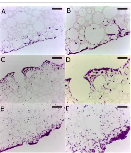
Cell culture experiments with L929 cells showed that cells readily penetrated into and migrated through the structure as would be required for 3D cell growth (Figure 5, 6). The slight decrease in cell viability seen between days 3 and 7 for the Alvetex® control is likely to be due to the high cell seeding density (500,000 cells). The Alvetex® product being a thinner section is likely to reach cell confluence at an earlier time point. High seeding densities were used due to the large thickness of the PCL-‐TA polyHIPEs.
[image:7.595.46.555.88.417.2] [image:7.595.43.292.598.731.2]Figure 5: MTT assay of L929 cultured cells on PCL-‐TA scaffolds vs an Alvetex® control.
Figure 6: 7 day time point micrographs of H&E stained sections of scaffold grown L929 cultures: (A) PCL-‐TA-‐40C scale bar 200 µm, (B) PCL-‐TA-‐40C scale bar 100 µm, (C) PCL-‐TA-‐90 scale bar 200 µm, (D) PCL-‐TA-‐90 scale bar 100 µm, (E) PCL-‐TA-‐ 95 scale bar 200 µm, (F) PCL-‐TA-‐40C scale bar 100 µm
Degradation of PCL-‐TA PolyHIPEs
The degradability of scaffolds was demonstrated by heating in 0.01 M NaOH(aq) to hydrolyse the ester bonds within the
network which would reasonably be expected to be the pathway for any biodegradation of the scaffold either in vivo
or in vitro. Analysis of the degradation products gave 1H NMR
spectra which were consistent with the hypothesised ultimate degradation products (figure 7). Similarly the degradation products of DPEHA and TMPTA scaffolds were also found to be consistent with their proposed hydrolysis products and these were tested alongside the PCL-‐TA products. For comparison PCL was also degraded in the same manner. In addition sodium hydroxide DMEM solutions (to the same concentration of sodium hydroxide in the degradation solutions) were also
prepared, as was a cell membrane disrupting surfactant (Triton X-‐100) as a positive control.
Figure 7: Proposed ultimate degradation products of PCL-‐TA scaffolds and previously reported DPEHA and TMPTA scaffolds.
Results indicate that up to a concentration of 0.1 mg.mL-‐1 the degradation products do not cause significant cell death relative to control. This compares favourably with the degradation products of other biodegradable scaffolds DPEHA and TMPTA, as well as PCL (figure 8 and 9). By contrast, the Triton X-‐100 positive control sees a large drop in cell viability at 10-‐4 mg/mL before reaching near zero cell viability at 10-‐1 mg.mL-‐1. The spike in the MTT absorbance in the presence of Triton X-‐100 at 10-‐3 mg.mL-‐1 should be interpreted as the onset of apoptosis. Tests at higher concentrations were limited by the foul odour of the degraded scaffold solution (presence of sulphur compounds) and the apparent acidity of the decomposition products of PCL-‐TA, DPEHA, TMPTA. Specifically, at 1 mg.mL-‐1 the media became too foul-‐smelling to place in the incubator and at 10 mg.mL-‐1 the phenol red indicator in DMEM was observed to change colour from red to yellow indicating pH <6.8.
[image:8.595.51.289.69.212.2] [image:8.595.305.552.111.266.2] [image:8.595.44.293.234.526.2] [image:8.595.306.550.535.699.2]
ARTICLE Journal Name
Figure 9: Live-‐dead cell count by the trypan blue haemocytometer method of L929 cultures after 24h exposure to PCLA-‐TA-‐90, DPEHA, TMPTA polyHIPE degradation products. Also shown are PCL degradation products and a sodium hydroxide control.
Conclusions
PCL-‐triol was end-‐functionalized with acryloyl chloride to create a macro cross-‐linker. This was then used to prepare a polyHIPE by photochemical radical reaction with a trithiol co-‐ monomer. The resulting structures were shown to be macroporous and suitable for cell culture. The material’s elasticity meant that the scaffold was prepared and stored engorged with liquid to prevent collapse. When applied to the
in vitro culture of L929 cells the scaffold was found to be biocompatible and compared favourably with the commercial product Alvetex®. The scaffold was found to readily hydrolyse in NaOH(aq). The degradation products were found to be non-‐
cytotoxic up to a concentration of 0.1mg.mL-‐1 in media, again
comparing well with other degradable scaffolds.
Acknowledgements
Support from the “Precision Polymer Materials (P2M)” RNP programme from the European Science Foundation is acknowledged. The European Commission (for DWJ: FP7-‐SME-‐ 2008-‐2-‐243542-‐HIP), EPSRC (studentships for CRL and MPD) and the Erasmus Training Program (for BL) are also thanked for funding.
Notes and references
1. N. R. Cameron, Polymer, 2005, 46, 1439-‐1449.
2. N. R. Cameron, D. C. Sherrington, I. Ando and H. Kurosu, J. Mater. Chem., 1996, 6, 719-‐726.
3. J. M. Williams and D. A. Wrobleski, Langmuir, 1988, 4, 656-‐662.
4. A. Barbetta, N. R. Cameron and S. J. Cooper, Chem. Commun., 2000, 221-‐222.
5. D. Štefanec and P. Krajnc, React. Funct. Polym., 2005, 65, 37-‐45.
6. S. D. Kimmins, P. Wyman and N. R. Cameron, React. Funct. Polym., 2012, 72, 947-‐954.
7. E. Lovelady, S. D. Kimmins, J. J. Wu and N. R. Cameron,
Polym. Chem., 2011, 2, 559-‐562.
8. N. Leber, J. D. B. Fay, N. R. Cameron and P. Krajnc, J. Polym. Sci. Pt. A: Polym. Chem., 2007, 45, 4043-‐4053. 9. A. Barbetta, M. Dentini, M. S. De Vecchis, P. Filippini, G.
Formisano and S. Caiazza, Adv. Funct. Mater., 2005, 15, 118-‐124.
10. H. Deleuze, R. Faivre and V. Herroguez, Chem. Commun.,
2002, 2822-‐2823.
11. S. Kovačič and P. Krajnc, J. Polym. Sci. Pt. A: Polym. Chem., 2009, 47, 6726-‐6734.
12. M. Ottens, G. Leene, A. Beenackers, N. Cameron and D. C. Sherrington, Ind. Eng. Chem. Res., 2000, 39, 259-‐266. 13. M. W. Hayman, K. H. Smith, N. R. Cameron and S. A.
Przyborski, J. Biochem. Biophys. Methods, 2005, 62, 231-‐ 240.
14. S. D. Kimmins, P. Wyman and N. R. Cameron, Polymer, 2014, 55, 416-‐425.
15. M. G. Schwab, I. Senkovska, M. Rose, N. Klein, M. Koch, J. Pahnke, G. Jonschker, B. Schmitz, M. Hirscher and S. Kaskel, Soft Matter, 2009, 5, 1055-‐1059.
16. S. Caldwell, D. W. Johnson, M. P. Didsbury, B. A. Murray, J. J. Wu, S. A. Przyborski and N. R. Cameron, Soft Matter, 2012, 8, 10344-‐10351.
17. E. Knight, B. Murray, R. Carnachan and S. Przyborski, in 3D Cell Culture, ed. J. W. Haycock, Humana Press, Editon edn., 2011, vol. 695, pp. 323-‐340.
18. W. Busby, N. R. Cameron and C. A. B. Jahoda,
Biomacromolecules, 2001, 2, 154-‐164.
19. W. Busby, N. R. Cameron and A. B. C. Jahoda, Polym. Int., 2002, 51, 871-‐881.
20. B. Sergent, M. Birot and H. Deleuze, React. Funct. Polym., 2012, 72, 962-‐966.
21. Y. Lumelsky and M. S. Silverstein, Macromolecules, 2009,
42, 1627-‐1633.
22. R. J. Carnachan, M. Bokhari, S. A. Przyborski and N. R. Cameron, Soft Matter, 2006, 2, 608-‐616.
[image:9.595.47.315.65.201.2]