Original Article
Hypertensive venous changes and Eph-B4/caveolin-1
pathways: an exploratory study
Qian Wang1, Min Zhou2, Yang Li3, Bo Zhang4, Na Li5, Zhen Xu6, Ling Li6
1Special Medical Service Center, 6Department of Neurology, Guangdong Key Laboratory for Diagnosis and Treat-ment of Major Neurological Diseases, National Key Clinical DepartTreat-ment, National Key Discipline, The First Affili -ated Hospital, Sun Yat-Sen University, Guangzhou, China; 2Medical Affairs, Glaxosmithkline, Guangzhou, China; 3Department of Geriatrics, Zhujiang Hospital, Southern Medical University, Guangzhou, China; 4Department of
Neurology, The Eighth People’s Hospital of Guangzhou, Guangzhou, China; 5Department of Rehabilitation, The Third Affiliated Hospital, Sun Yat-Sen University, Guangzhou, China
Received March 22, 2018; Accepted July 7, 2018; Epub October 15, 2018; Published October 30, 2018
Abstract: Hypertension is a major risk factor for cerebral vascular disease (CVD). Previous studies have only focused on cerebral artery disease, failing to attach importance to the role of the cerebral venous system in CVD. The aim of this study was to investigate the structural change of jugular veins and to explore whether Eph-B4/caveolin-1 (Cav-1) pathways plays a role in jugular venous changes caused by arterial hypertension. A stroke-prone renovascular hypertensive rat (RHRSP) model was established. Expression of MMP-9, EphB4, Cav-1, and p-Cav-1 was detected through Western blot, PCR, immunohistochemistry, or immunofluorescence. Jugular veins in the RHRSP group were thicker than those in the sham group, as were the intima of the jugular veins, mainly composed of type I collagen. Compared to the sham group, expression of Eph-B4 was significantly decreased and levels of Caveolin-1 (Cav-1) and phosphorylated Caveolin-1 (p-Cav-1) were significantly increased in jugular veins of the RHRSP group. Furthermore, endothelial p-Cav-1 and Eph-B4 were found to be diminished in jugular veins of the RHRSP group. Arterial hyper-tension caused venous collagenosis in the jugular veins. These changes may be strongly related to Eph-B4 and its downstream factor Cav-1/p-Cav-1.
Keywords: Hypertension venous remodeling, venous collagenosis, Eph-B4/caveolin-1 pathways
Introduction
Cerebrovascular disease remains the leading cause of death, worldwide, and hypertension is one of the primary causes of cerebrovascular disease. At the same time, vascular remodeling is the most primary pathological mechanism caused by hypertension. A previous study [1] observed changes in the cerebral veins of stroke-prone renovascular hypertensive rats (RHRSPs) using susceptibility-weighted imag-ing (SWI) and histopathological methods, find-ing that long-term hypertension in RHRSPs leads to increased visibility of cerebral veins on SWI and thickened cerebral venous walls (venous collagenosis). Thickened walls of the affected veins in RHRSPs may be ascribed to several conditions that result from hyperten-sion. However, how arterial hypertension affects the venous system, effects on venous
remodeling, and mechanisms responsible for these changes remain unknown.
To resolve this issue, further research was performed on jugular veins using an RHRSP model, examining the effects and mechanisms of altered Eph-B4 signaling.
Materials and methods
Animal treatment
All animal procedures were approved by the Sun Yat-Sen University (Guangzhou, China) Committee for the Care and Use of Animals. A total of 80 male Sprague-Dawley rats, weighing 80-100 g, were obtained from the Experimental Animal Center of Guangdong Province and were randomly divided into the following 2 groups: sham-clipped group (sham, n=28) and RHRSP model group (RHRSP, n=52).
These male Sprague-Dawley rats were fed ad libitum and housed in conventional conditions with controlled temperature (23±2°C), humidity (55±10%), and light (12-hour light/12-hour da- rkness). All animal were treated in strict accor-dance with International Ethical Guidelines and the National Institutes of Health Guide for the Care and Use of Laboratory Animals.
An RHRSP rat model was used to induce reno-vascular hypertension [4-6]. Under anesthesia with 3% sodium pentobarbital (36 mg/kg body wt IP), a midline laparotomy was used for bilat-eral placement of partially occlusive silver clips (0.3 mm internal diameter) on the renal arter-ies of RHRSP rats. The ring part of the clip was placed around the root of each artery and the outer gap of the clip was then shut. Rats in the
tracted protein were loaded onto 10% gels for sodium dodecyl sulfate polyacrylamide gel electrophoresis (SDS-PAGE). Membranes were blotted using the following primary antibodies: caveolin-1 (R&D Systems, Minneapolis, USA), EphB4 (Santa Cruz Biotechnology, California, USA), and phosphorylated caveolin-1 (p-Cav-1, Santa Cruz Biotechnology, California, USA). Real-time quantitative PCR
The cDNA sequence of rat EphB4 was pur-chased from Open Biosystems. The following primers were used for real-time PCR: GAPDH forward, 5’-GGCCTCCAAGGAGTAAGAAA-3’ and GAPDH reverse, 5’-GCCCCTCCTGTTATTATGG-3’; and EPHB4 forward, 5-GCTCGGAACATCTTG- GTCAA-3’ and EPHB4 reverse, 5’-CCCAGGG- AACTTGTGTAGGT-3’. PCR conditions were as follows: one cycle at 94°C for 4 minutes, fol-lowed by 30 cycles at 94°C for 30 seconds, and 60°C for 30 seconds. Amplification was quantified using miScript SYBR Green PCR Kit (Qiagen, Hilden, Germany). Quantified results for individual cDNAs were normalized to GAPDH using the ΔΔct method. Purities of the amplified products were examined using dissociation curves.
Immunohistochemistry and immunofluores -cence
[image:2.612.90.374.74.252.2]Samples were fixed with 4% paraformaldehyde, embedded in paraffin, and cut into 5-μm cross sections. Hematoxylin and eosin (H&E) and Masson trichrome staining were performed for all samples.
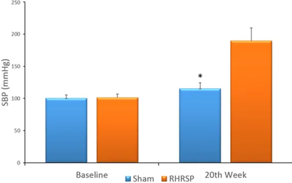
Figure 1. SBPs before and after surgery in the two groups (n=28). Baseline: before surgery; 20th week: 20 weeks after surgery. *P < 0.05 vs SBP of the RHRSP group in 20th week. Error bars denote SEM.
sham surgery group under-went laparotomies and iso- lation of the bilateral renal arteries was without clip pl- acement. After 4 weeks of recovery from surgery, systol-ic blood pressure (SBP) was measured with a tail cuff once per week.
Western blot analysis
Primary antibody treatments were performed, according to manufacturer instructions. Expe- riments for antibody validation and document-ed controls were carridocument-ed out (Figure 1). Co- ncentrations of all antibodies were optimized as needed. Primary antibodies included Cav-1, p-Cav-1, and Eph-B4. Antigen retrieval was performed using 10-mmol/L citrate buffer at pH 6.0. For immunohistochemistry, secondary detection was performed using DAB. Sections were counterstained with Mayer’s hematoxylin. For immunofluorescence, secondary detection was performed using Alexa Fluor 488 and 568 and DAPI counterstain (Sigma, St. Louis, MO, USA). Images were captured with a fluorescent microscope under identical conditions.
Statistical analysis
SPSS 19.0 was used for statistical analyses. Data are expressed as mean ± SD. Student’s t-tests were used to compare pairs of groups. P values < 0.05 are considered statistically significant.
Results
Of the 80 Sprague-Dawley rats, 4 rats died dur-ing the experiment in the RHRSP group. The sham group consisted of 28 normal healthy rats, used as a reference control group.
RHRSP model was established successfully
SBP increased gradually in all RHRSP rats but did not significantly increase in the sham gro- up. Compared to baseline and sham group val-ues, SBPs of the RHRSP group were significant-ly increased 4 weeks after surgery. Twelve we- eks after the RHRSP rats underwent surgery, increased SBPs were maintained at a relatively higher and stable level (Figure 1, p < 0.05). Jugular veins thickened in the RHRSP group and walls were mainly composed of collagen I
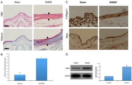
[image:3.612.89.516.70.347.2]H&E staining demonstrated normal thin walls of jugular veins in the sham group and thick-ened walls of jugular veins in the RHRSP group
(Figure 2A). Masson trichrome staining rev- ealed that these thickened walls were com-posed of collagen (Figure 2A). Intimal thickn- ess of jugular veins was measured for both groups. Differences between the two groups were significant (Figure 2B).
To explore the main constituent of the thick-ened walls of jugular veins, this study dealt with jugular veins of both groups using immunohis-tochemistry staining (Figure 2C). It was found that the thickened walls mainly consisted of type I collagen, however, no significant differ-ences were detected concerning expression of smooth muscle actin (SMA) in the jugular veins between the sham group and RHRSP group. MMP9 expression increased in jugular veins of the RHRSP group compared to that of the sham group
Protein expression of MMP9 was examined in jugular veins via Western blotting, finding that it
was significantly increased in the RHRSP group, compared to the sham group (Figure 2D). Eph-B4 expression decreased and Cav-1 and p-Cav-1 expression increased in jugular veins of the RHRSP group
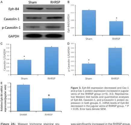
Levels of Eph-B4, Cav-1, and p-Cav-1 proteins were detected by Western blot in the jugular veins of sham and RHRSP groups. Compared to the sham group, protein expression of Eph-B4 was significantly decreased and levels of Cav-1 and p-Cav-1 were significantly increased in jugular veins of the RHRSP group (Figure 3A-D).
[image:4.612.89.524.70.483.2]Next, mRNA levels of Eph-B4 were examined in the jugular veins of both groups using quantita-tive real-time PCR. The RHRSP group exhibited less detectable Eph-B4 than the sham group, suggesting that expression of Eph-B4 tran-scripts was downregulated in the hypertensive rats (Figure 3E).
Eph-B4 and p-Cav-1 expression diminished in endothelial cells of jugular veins of the RHRSPs
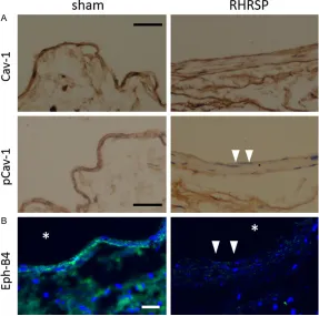
Immunohistochemistry demonstrated that en- dothelial Cav-1 was detectable in jugular veins of both groups, but endothelial p-Cav-1 was strongly diminished in the RHRSP group (Figure 4A). Immunofluorescence showed that Eph-B4 was diminished in jugular veins of the RHRSP group (Figure 4B).
Discussion
In a previous study [1], it was proven that arte-rial hypertension affects the venous system of the brain. Increased visibility of cerebral veins on SWI and thickened cerebral venous walls (venous collagenosis) were both consequenc- es of long-term hypertension in RHRSPs. Fu- rthermore, it was validated that stenosis in these affected veins led to slower velocities, resulting in disordered venous drainage. The present study investigated venous remodeling in jugular veins of the RHRSP group, examining
tension (CWT), an important determinant of vascular remodeling. Moreover, it was found that expression of MMP9 was increased in the jugular veins of hypertensive rats. Thus, it was hypothesized that elevated blood pressure leads to overload capacity of the body, increas-ing CWT. This, on one hand, stimulates the pro-liferation of vascular smooth muscle cells and collagen. On the other hand, it promotes MMP9 secretion which reduces the elasticity of blood vessels. The pathologic changes above, in turn, increase CWT, forming a vicious circle and lead-ing to venous remodellead-ing.
Remodeling of jugular veins in hypertensive rats (i.e., venous collagenosis) is substantially different from the corresponding process in arteries. This elicits the question, through which signaling pathways does venous remod-eling occur?
[image:5.612.90.377.73.358.2]During embryonic development, Eph receptors and their membrane-bound ephrin ligands play essential roles in the formation of func-tional vascular networks [7-9]. Eph-B4 is an
Figure 4. Endothelial p-Cav-1 and Eph-B4 was diminished in jugular veins of RHRSP group (n=5). A. Immunohistochemistry against Cav-1 and p-Cav-1, Bar, 200 μm. Arrowheads denote loss of p-Cav-1 signal; B. Immunofluores -cence against Eph-B4, Bar, 100 μm. Arrowheads denote loss of Eph-B4 sig -nal. *, lumen.
possible mechanisms. It was found these changes may be strongly related to Eph-B4 and its downstream factor Cav-1/p-Cav-1.
active determinant of embryonic venous devel-opment [2, 10-12]. It is also present in adult veins where it provides a marker of venous identity. Kudo [3] used immunofluorescence to confirm that Eph-B4 is expressed in aged rat jugular veins in both the endothelium and α-actin-positive medial SMCs. He also con-firmed that Eph-B4 is strongly diminished, but not eliminated in the intima media of venous grafts placed in the relatively hypertensive environment of arterial circulation. The present study obtained similar results. Eph-B4 signal-ing was reduced in jugular veins of the hyper-tensive rats, as was signaling in the intima. Thus, it was questioned whether reduced Eph-B4 expression is responsible for venous re- molding in hypertensive rats.
A study in 2011 [13] demonstrated that the loss of Eph-B4 during venous transposition to the arterial circulation was directly responsible for the loss of normal venous structures and excessive venous wall thickening and remo- deling. Lynn S. Model et al. [14] examined changes in vessel identities of human saphe-nous veins in a flow circuit in which shear stress could be precisely controlled. They found that venous Eph-B4 expression was diminished and expression of osteopontin was increased with exposure to arterial magnitudes of shear st- ress. These authors concluded that arterial magnitudes of shear stress cause the loss of venous identity. To increase knowledge of the function of Eph-B4, a study in 2013 found that diminished venous endothelial Eph-B4 expression was associated with an angioge- nic and mitogenic phenotype that is charact- erized by increased secretion of smooth mus-cle cell mitogens and reduced nitric oxide pro-duction [15]. This result suggests that de- creased Eph-B4 expression is related to vascu-lar remodeling.
Cav-1 is a major structural protein of the caveo-lae in endothelial cells. It is thought to be involved in the mechanotransduction of dynam-ic shear stress changes via interactions with several signaling protein families, including Eph receptors [16, 17]. Forrester’s research [18] found that Cav1(-/-) mice, infused with AngII, showed attenuation of medial thickness and perivascular fibrosis in the thoracic aorta, in- dicating that dysfunction of Cav-1 leads to vas-cular remodeling. Moreover, a study by Muto [13] suggested that Cav-1 is downstream of
Eph-B4 signaling during venous adaptation. This author examined vein graft adaptation in WT and Cav-1 KO mice, finding that vein grafts derived from Cav-1 KO mice exhibit sig-nificantly increased wall thickness, like that observed in vein grafts with reduced Eph-B4 function. However, vein grafts derived from Cav-1 RC mice, such as Cav-1 KO mice with an EC-specific Cav-1 transgenic reconstitution, exhibited greatly reduced thickness compar- ed with vein grafts derived from Cav-1 KO mi- ce. These results suggest that the thickening response is a result of endothelial Cav-1, not global Cav-1, dysfunction and that endotheli- al Cav-1 is essential to the Eph-B4 signaling that limits venous wall thickness. The present study further observed increased Cav-1 and p-Cav-1 in the thickened jugular veins of RHRSPs at both protein and mRNA levels. This seemed to contradict the findings that Eph-B4 stimulated phosphorylation of Cav-1 in the endothelial cells [13] and that Eph-B4 was greatly reduced in RHRSPs. Furthermore, im- munohistochemistry was used to detect endo-thelial Cav-1 and p-Cav-1. It was found that endothelial Cav-1 was detectable in both gro- ups, but endothelial p-Cav-1 was strongly dimin-ished in the RHRSP group, in accord with Mito’s results. Results also suggest that probably only p-Cav-1 in the endothelial cells is functional active and plays a role in vascular remodeling. Decreased p-Cav-1 in the endothelium may have been caused by the reduction in Eph-B4 in the RHRSP group.
Acknowledgements
This work was supported by the National Na- tural Science Foundation of China (NSFC) (No. 81671153) and the Natural Science Fo- undation of Guangdong Province, China (No. 2016A030313203).
Disclosure of conflict of interest
None.
Address correspondence to: Ling Li, Department of Neurology, Guangdong Key Laboratory for Dia- gnosis and Treatment of Major Neurological Dis- eases, National Key Clinical Department, National Key Discipline, The First Affiliated Hospital of Sun Yat-Sen University, 58 Zhongshan Er Road, Guang- zhou 510080, China. Tel: +86 20-87332200-8291; E-mail: [email protected]
References
[1] Zhou M, Mao L, Wang Y, Wang Q, Yang Z, Li S and Li L. Morphologic changes of cerebral veins in hypertensive rats: venous collagenosis is associated with hypertension. J Stroke Cere-brovasc Dis 2015; 24: 530-536.
[2] Adams RH, Wilkinson GA, Weiss C, Diella F, Gale NW, Deutsch U, Risau W and Klein R. Roles of ephrinB ligands and EphB receptors in cardiovascular development: demarcation of arterial/venous domains, vascular morpho-genesis, and sprouting angiogenesis. Genes Dev 1999; 13: 295-306.
[3] Kudo FA, Muto A, Maloney SP, Pimiento JM, Bergaya S, Fitzgerald TN, Westvik TS, Frattini JC, Breuer CK, Cha CH, Nishibe T, Tellides G, Sessa WC and Dardik A. Venous identity is lost but arterial identity is not gained during vein graft adaptation. Arterioscler Thromb Vasc Biol 2007; 27: 1562-1571.
[4] Zeng J, Huang R and Su Z. Stroke-prone reno-vascular hypertensive rats. Chin Med J (Engl) 1998; 111: 741-744.
[5] Zeng J, Zhang Y, Mo J, Su Z and Huang R. Two-kidney, two clip renovascular hypertensive rats can be used as stroke-prone rats. Stroke 1998; 29: 1708-1713; discussion 1713-1704. [6] Liao SJ, Huang RX, Su ZP, Zeng JS, Mo JW, Pei
Z, Li L, Fang YN, Hong H and Huang HW. Stroke-prone renovascular hypertensive rat as an ani-mal model for stroke studies: from artery to brain. J Neurol Sci 2013; 334: 1-5.
[7] Adams RH and Klein R. Eph receptors and eph-rin ligands. essential mediators of vascular de-velopment. Trends Cardiovasc Med 2000; 10: 183-188.
[8] Kullander K and Klein R. Mechanisms and functions of Eph and ephrin signalling. Nat Rev Mol Cell Biol 2002; 3: 475-486.
[9] Lawson ND, Scheer N, Pham VN, Kim CH, Chit-nis AB, Campos-Ortega JA and Weinstein BM. Notch signaling is required for arterial-venous differentiation during embryonic vascular de-velopment. Development 2001; 128: 3675-3683.
[10] Wang HU, Chen ZF and Anderson DJ. Molecu -lar distinction and angiogenic interaction be-tween embryonic arteries and veins revealed by ephrin-B2 and its receptor Eph-B4. Cell 1998; 93: 741-753.
[11] Gerety SS, Wang HU, Chen ZF and Anderson DJ. Symmetrical mutant phenotypes of the re-ceptor EphB4 and its specific transmembrane ligand ephrin-B2 in cardiovascular develop-ment. Mol Cell 1999; 4: 403-414.
[12] Shin D, Garcia-Cardena G, Hayashi S, Gerety S, Asahara T, Stavrakis G, Isner J, Folkman J, Gim -brone MA Jr and Anderson DJ. Expression of ephrinB2 identifies a stable genetic difference between arterial and venous vascular smooth muscle as well as endothelial cells, and marks subsets of microvessels at sites of adult neo-vascularization. Dev Biol 2001; 230: 139-150. [13] Muto A, Yi T, Harrison KD, Davalos A, Fancher
TT, Ziegler KR, Feigel A, Kondo Y, Nishibe T, Sessa WC and Dardik A. Eph-B4 prevents ve-nous adaptive remodeling in the adult arterial environment. J Exp Med 2011; 208: 561-575. [14] Model LS, Hall MR, Wong DJ, Muto A, Kondo Y,
Ziegler KR, Feigel A, Quint C, Niklason L and Dardik A. Arterial shear stress reduces eph-b4 expression in adult human veins. Yale J Biol Med 2014; 87: 359-371.
[15] Jadlowiec CC, Feigel A, Yang C, Feinstein AJ, Kim ST, Collins MJ, Kondo Y, Muto A and Dardik A. Reduced adult endothelial cell EphB4 func-tion promotes venous remodeling. Am J Physiol Cell Physiol 2013; 304: C627-635.
[16] Gratton JP, Bernatchez P and Sessa WC. Cave-olae and caveolins in the cardiovascular sys-tem. Circ Res 2004; 94: 1408-1417.
[17] Yu J, Bergaya S, Murata T, Alp IF, Bauer MP, Lin MI, Drab M, Kurzchalia TV, Stan RV and Sessa WC. Direct evidence for the role of caveolin-1 and caveolae in mechanotransduction and re-modeling of blood vessels. J Clin Invest 2006; 116: 1284-1291.
[18] Forrester SJ, Elliott KJ, Kawai T, Obama T, Boy -er MJ, Preston KJ, Yan Z, Eguchi S and Rizzo V. Caveolin-1 deletion prevents hypertensive vas-cular remodeling induced by angiotensin II. Hy-pertension 2017; 69: 79-86.