Original Article
Quantification of clinical parameters in patients with
vertical food impaction treated by occlusal adjustment:
cone-beam computed tomography analysis
Feng Wu1, Yue Wang2, Lu Wang1, Fusong Yuan3, Bin Zhao1, Yuchun Sun3
1Department of Stomatology, Shanxi Medical University, Taiyuan 030001, China; 2Department of Stomatology,
People’s Hospital of Beijing Daxing District, Beijing, China; 3Center of Digital Dentistry, Faculty of
Prosthodon-tics, Peking University School and Hospital of Stomatology and National Engineering Laboratory for Digital and Material Technology of Stomatology and Research Center of Engineering and Technology for Digital Dentistry of Ministry of Health, Beijing 100081, China
Received March 9, 2017; Accepted February 10, 2018; Epub April 15, 2018; Published April 30, 2018
Abstract: Objectives: This study aimed to determine the effectiveness of occlusal adjustment for vertical food im-paction, and evaluate the quantitative standards of vertical food impaction treated by occlusal adjustment. Meth-ods: A total of 51 patients who suffered from vertical food impaction without anatomical structure destruction and presented to the Dental Hospital of Shanxi Medical University were selected for this study. These patients were treated by occlusal adjustment and the dental impressions were made before and after treatment. Cone-beam computed tomography (CBCT) was used to scan and measure six clinical parameters, including the length of the interproximal contact area in the food impaction zone, angle of the occlusal buccal-lingual embrasure, and occlu-sal height and width. Results: Therapeutic results were followed up, and the relevance between each parameter and the treatment outcome was investigated through statistical analysis. Results indicated that the length of the
interproximal contact and occlusal height significantly increased after adjustment (P < 0.001), and the difference
in occlusal buccal-lingual embrasure angle before and after treatment was not statistically significant (P>0.05).
Conclusions: The length of the interproximal contact and occlusal height are critical factors for curing vertical food impaction. The best therapeutic effect could be achieved when occlusal height and width measurements reach 1.0-1.6 mm and 0.9-1.2 mm, respectively.
Keywords: Food impaction, quantitive standards, occlusal adjustment, cone-beam computed tomography
Introduction
Food impaction is defined as the forceful we- dging of food through occlusal pressure into the interproximal spaces, and characterizes the typical phenomenon that food particles or fibers is embedded in the gap of adjacent teeth during the process of chewing [1, 2]. Frequent food impaction can increase the risk of oral and dental diseases such as halitosis, dental car-ies, gingivitis, periodontitis, and even tooth loss [3, 4]. According to the different directions of food dregs between teeth, food impaction can be subdivided into three forms: vertical, hori-zontal and mixing [5]. Clinically, patients who suffer from vertical and mixing food impactions are the key group, accounting for 90.3% of the
total cases [6]. Vertical food impaction with anatomical structure destruction can be cured through methods of tooth filling, inlay and cro- wn prosthesis [7-11], while occlusal adjustment is an effective treatment for vertical food imp- action without dental anatomical structure de- struction [12].
vertical food impaction with anatomical struc-ture destruction [13]. It has been reported that occlusal adjustment is an effective treatment for food impaction, which does not cause ana-tomical structure destruction and is performed by acquiring sufficient mesial movement [14]. In addition, occlusal adjustment was performed by using diamond and polishing stones to cre-ate buccal and lingual food escape grooves that would allow food to escape buccally and lingually from occlusal surfaces, preventing food impaction [15].
In particular, the means of occlusal adjustme- nt to treat food impaction involves the adju- stment of filling cusps, reduction of the inter-proximal contact area, opening of dental em- brasures, and the expansion or reconstruction of a food spillway [15]. Unfortunately, no previ-ous studies have systematically investigated occlusal adjustment for clinical application and further established quantitive standards. This has brought difficulties and unpredictabilities to clinical treatment and practice. In the pres-ent study, cone-beam computed tomography (CBCT) was used to measure six parameters in the food impaction zone before and after occlu-sal adjustment. Compared with previous stud-ies, the present study aimed to investigate the influence of each parameter on the pathogen-esis and curative effect of vertical food impac-tion, and to ultimately provide quantitative standards for clinical treatment.
Study subjects selection
A total of 51 patients who suffered from food impaction and presented to the Dental Hos- pital of Shanxi Medical University between 2014 and 2015 were selected for this study. Among these patients, 28 patients were male and 23 patients were female; and the age of these patients ranged between 26 and 58 years. These patients were included into the study if they frequently underwent food impac-tion between the maxillary first molar and sec-ond molar, with duration of at least four weeks. In addition, the adjacent teeth in the impacted zone of these patients should be complete without looseness, dentin hypersensitivity and severe wear. Furthermore, the dental interproxi-mal system should be norinterproxi-mal without caries. Patients who had their designated impacted zone restored by fillings, patients who have inlays and full crowns, or patients who suffered from buccoversion or linguoversion and uncon-trolled periodontitis were excluded from this study.
Clinical treatment
[image:2.612.90.377.72.188.2]Ultrasonic teeth cleaners were first used for scaling impacted teeth, in order to remove embedded food particles, debris and dental calculus. The maxillary and mandibular denti-tion was recorded with a silicon-rubber impres-sion (DMG, Germany) before treatment (Figure 1A).
Figure 1. A. Making impressions. B. Perfusing target teeth position by die stone.
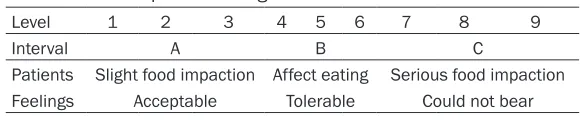
Table 1. Food impaction rating scale
Level 1 2 3 4 5 6 7 8 9
Interval A B C
Patients Slight food impaction Affect eating Serious food impaction Feelings Acceptable Tolerable Could not bear
Materials and methods
Ethic statement
[image:2.612.87.378.246.306.2]Then, the position of the impacted tooth and the other tooth was perfused with die ston- es (Heraeus, Germany) and self-curing resins (Shanghai New Century Dental Materials Co. Ltd.) (Figure 1B). The prepared plaster models were fixed according to the original occlusal relationship.
The extent of food impaction before occlusal adjustment was determined using a table (Table 1). Specifically, the feel of patients on the food impaction was divided into three inter-vals and nine levels, according to the severity of the food impaction.
Then, occlusal adjustment was applied to th- ese patients. A TF-21 diamond bur was used to grind the cusp and marginal ridge of the maxillary first molar and second molar, as well as the paired jaw teeth. The TF-14 diamond bur was employed to prepare a spillway for food and deepen the occlusal embrasure. Furthe- rmore, the occlusal adjustment also expand- ed the buccal-lingual embrasure angle and reduced the length of the interproximal con- tact.
The curative effects were evaluated through the difference (d) between the level of food impaction, which the patients selected from Table 1 before and after treatment. Specifically, d≥5 indicates that the food impaction symptom was cured, 3≤d≤4 indicates that the food impaction symptom improved, and d≤2 indi-cates that the treatment was ineffective. After treatment, the grinded teeth should be pol-ished and covered with a desensitizer or fluor protector, in order to prevent tooth sensitivity. Similarly, silicon-rubber impressions and plas-ter-resin models were made after occlusal adjustment.
edge of the maxillary central incisor, the sagit-tal position line was aligned with the contact zone of maxillary central incisor, and the coro-nal position line was leveled with the contact region of the maxillary first molar and second molar (Figure 2B). The scanning region was lim-ited to a 30-mm area on each side of the three position lines. Scanning conditions: tube volt-age and current was 60 kV and 2 mA, respec-tively; and scan thickness and layer spacing was 1.0 mm.
Parameter definition
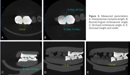
The first layer cross-section that appeared from the occlusion to the gingiva in the horizontal position was the base plane on which the length of the interproximal contact and the buc-cal-lingual angle were measured: (1) Length of the interproximal contact: the line between adjacent points close to the buccal and lingu- al orifices, respectively (Figure 3A); (2) Buccal-lingual orifice angle: the angle formed with the adjacent point near to the buccal or lingual side, and the food spillway between the ma- xillary first molar and second molar on the sa- me cross section (Figure 3B). Measurements of the occlusal embrasure and occlusal height and width were performed on the coronal plane where the adjacent point firstly appeared: (1) Occlusal embrasure angle: the angle formed with this adjacent point and the food spillway between the maxillary first molar and second molar on the same cross section (Figure 3C); (2) Occlusal height: the minimum vertical dis-tance measured from the bottom of the occlu-sal embrasure on the selected cross-section to the cusp or ridge of the antagonistic teeth (Figure 3D); (3) Occlusal width: the horizontal distance of the occlusal embrasure bottom after occlusal adjustment (Figure 3E). All data mentioned above were completed by the same Figure 2. A. Positioned upper lower jaw model. B. Scanning position lines.
Measurement
person. For each sample, at least three mea-surements were carried out, and the average values were reported.
Statistical analysis
Statistical comparisons were performed using SPSS 16.0 software. Parameters before and after occlusal adjustment were compared us-
[image:4.612.89.521.74.325.2]ing the Wilcoxon paired test. Parameters vie- wed from different curative effects were com-pared using the Wilcoxon rank sum test. Chi-square test was used to analyze the difference between group and logistic regression analysis was utilized to determine the relationship between certain key parameters and treatme- nt outcomes after occlusal adjustment. When Chi-square test was used to test the difference Figure 3. Measured parameters: A. Interproximal contacts length; B. Buccal/lingual embrasures angle; C. Occlusal embrasure angle; D, E. Occlusal height and width.
Table 2. Comparisons of the parameters before and after treament (median (interquartile range))
Parameter Case number Before treatment After treatment Z P
Interproximal contacts length (mm) 51 4.190±1.56 3.630±1.695 -0.516 < 0.001* The angle of buccal embrasure (°) 51 54.64±19.26 58.24±14.51 -0.66 0.948 The angle of lingual embrasure (°) 51 52.25±13.95 51.34±14.16 -0.019 0.985 The angle of occlusal embrasure (°) 51 59.75±42.34 69.20±20.74 -4.181 0.606 Occlusal height (mm) 51 0.670±0.213 1.043±0.376 -5.785 < 0.001*
Note: According to the inspection level of α=0.05, “*” represents statistically significant difference before and after treatment.
Table 3. Comparisons of parameters from the view of curative effects (median (interquartile range))
Parameter Treatment effects Z P
Improved (n=14) Cured (n=37)
Interproximal contacts length (mm) 4.26±1.61 3.51±1.51 -1.46 0.144* The angle of buccal embrasure (°) 49.64±13.62 59.30±14.51 -0.676 0.499 The angle of lingual embrasure (°) 58.24±19.28 51.21±11.1 -1.352 0.276 The angle of occlusal embrasure (°) 67.12±5.11 73.45±20.74 -1.204 0.329
Occlusal height (mm) 0.960±0.170 1.003±0.389 -1.291 0.197*
Occlusal width (mm) 0.712±0.250 0.793±0.250 -1.987 0.047**
[image:4.612.90.525.362.442.2] [image:4.612.92.523.489.595.2]between three groups, the P value was correct-ed and 0.05/3 was the new inspection which means that P < 0.0168 indicated statistical significance. For more free variables entering the regression equation, more than 80% power of test was acceptable which means P < 0.20. Unless stated otherwise, statistical signifi-cance was set at a P value of < 0.05.
Results
Comparisons of the parameters before and after occlusal adjustment
As shown in Table 2, the difference in occlusal buccal-lingual embrasure angle was not statis-tically significant before and after treatment (P>0.05). This indicates that there was no ob- vious change in these three parameters. The length of the interproximal contact and occlu-sal height significantly increased after adjust-ment (P < 0.001).
Comparisons of various parameters in view of different curative effects
The results presented in Table 3 revealed that the difference in occlusal width was
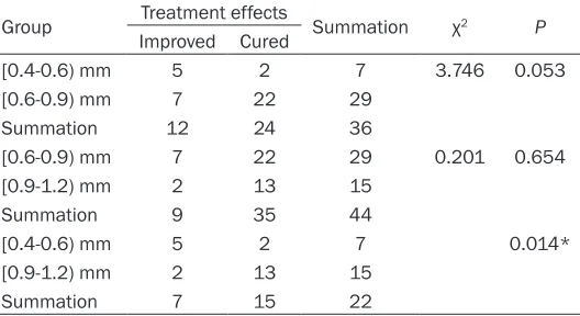
statistical-Occlusal width is critically important for solv- ing food impaction problems. As shown in Tab- le 6, the difference in occlusal width among groups was statistically significant (P < 0.05). Treatment effects in the 0.4-0.6 mm group was inferior to that of the 0.9-1.2 mm group, and the results were statistically significant (Tab- le 7, P < 0.0168). However, the difference in curative effects between the 0.4-0.6 mm and 0.6-0.9 mm groups or 0.6-0.9 mm and 0.9-1.2 mm groups was not statistically significant (P>0.0168).
The relationship between key parameters and treatment outcomes after occlusal adjustment
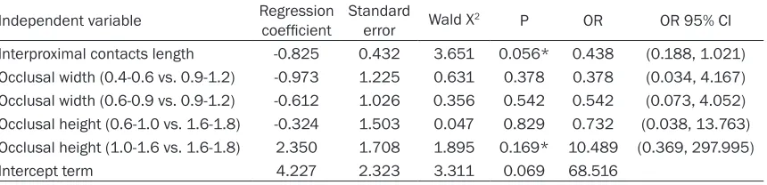
[image:5.612.90.340.96.177.2]According to logistic regression analysis resul- ts (Table 8), the difference in the length of the interproximal contact and occlusal height was statistically significant in terms of evaluating the therapeutic effects. Quantitatively, the pos-sibility of healing would decrease by 0.562 times for every 1 mm increase in length of the interproximal contact, while patients with an occlusal height of 1.0-1.6 mm might achieve a cure rate 10.489 times higher than that in the 1.6-1.8 mm group.
Table 4. Comparisons of curative effects resulted from different occlusal height (%)
Group Treatment effects Summation χ2 P Improved Cured
[0.6-1.0) mm 10 15 25 7.095 0.020*
[1.0-1.6) mm 2 20 22
[1.6-1.8) mm 2 2 4
Summation 14 37 51
Note: “*” means P < 0.05.
ly significant from the perspective of treatment outcomes (P < 0.05). Occ- lusal width increased in the cured group, compared with the improved group.
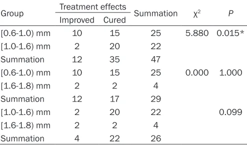
Comparison of curative effects that re-sulted from different occlusal height
[image:5.612.92.339.236.382.2]Adjustment of the occlusal height co- uld lead to various therapeutic results, and the difference in occlusal height between these groups was statistically significant (Table 4, P < 0.05).
Table 5 shows the paired comparisons of various occlusal height groups. The 1.0-1.6 mm group revealed a signifi-cant improvement in curative effect, compared with the 0.6-1.0 mm group. However, the difference in treatment outcomes that resulted from the 0.6-1.0 mm and 1.6-1.8 mm groups, or 1.0-1.6 mm and 1.6-1.8 mm groups were not statistically significant.
Comparison of curative effects that resulted from different occlusal width Table 5. The paired comparisons of various occlusal
height groups (%)
Group Treatment effects Summation χ2 P Improved Cured
[0.6-1.0) mm 10 15 25 5.880 0.015*
[1.0-1.6) mm 2 20 22
Summation 12 35 47
[0.6-1.0) mm 10 15 25 0.000 1.000
[1.6-1.8) mm 2 2 4
Summation 12 17 29
[1.0-1.6) mm 2 20 22 0.099
[1.6-1.8) mm 2 2 4
Summation 4 22 26
Discussion
Vertical food impaction is a common and fre-quently-occurring disease in clinic [16], and refers to the phenomenon that food is pushed into the tooth clearance from the vertical direc-tion by external forces during the chewing pro-cess [5]. Clinically, occlusal adjustment is an effective tool for vertical food impaction, which includes grinding the filling cusps and occlusal marginal ridge, expanding embrasures, recon-structing the spillway of food [13], and adjust-ing the bite force of the impacted teeth [17]. However, commonly used treatments had ne- ver attracted enough academic attention, and had not been systematically and quantitative- ly investigated. In this study, food impaction was treated by occlusal adjustment, and rele-vant parameters were studied quantitatively for further guidance in more accurate clinical practices.
In the present study, patients who suffered from food impaction between the maxillary first molar and second molar were selected. The reason for this choice was that food impaction was prone to occurring in the clearance of the
The data presented in Table 3 indicates that there was some correlation between occlusal width and its curative effects, but no evidence revealed that the occlusal width was an impor-tant factor to evaluate treatment outcomes by multi-factor logistic regression analysis. The reason for such result is that the measurement of the occlusal width was established on the occlusal height after treatment. Actually, it was more appropriate to observe the width as an assistant indicator to affect therapeutic effica-cy. Nonetheless, the research of this parameter was necessary, because it can provide a quan-titative standard correlated to the width for bet-ter treatment results.
In general, occlusal height and width provides synergistic effects for treating food impaction. On the basis of statistical analysis, the best therapeutic effect could be achieved when occlusal height and width reaches 1.0-1.6 mm and 0.9-1.2 mm, respectively.
[image:6.612.89.356.98.177.2]In addition to occlusal height, there was evi-dence that the length of the interproximal con-tact was also critically important to gain good curative effects (Table 8). According to logistic Table 6. Comparisons of curative effects resulted from
differ-ent occlusal width (%)
Group Treatment effects Summation χ2 P Improved Cured
[0.4-0.6) mm 5 2 7 7.319 0.02*
[0.6-0.9) mm 7 22 29
[0.9-1.2) mm 2 13 15
Summation 14 37 51
Note: “*” means P < 0.05.
Table 7. The paired comparisons of various occlusal width groups (%)
Group Treatment effects Summation χ2 P Improved Cured
[0.4-0.6) mm 5 2 7 3.746 0.053
[0.6-0.9) mm 7 22 29
Summation 12 24 36
[0.6-0.9) mm 7 22 29 0.201 0.654
[0.9-1.2) mm 2 13 15
Summation 9 35 44
[0.4-0.6) mm 5 2 7 0.014*
[0.9-1.2) mm 2 13 15
Summation 7 15 22
Note: “*” means P < 0.0168.
first molar and second molar, and symptoms that occur in the maxi- lla and under jaw are almost id- entical.
[image:6.612.92.356.237.381.2]regression analysis results, the possibility of healing would decreased by 0.562 times for every 1 mm increase in length of the interproxi-mal contact. The decrease of this parameter could lead to the expansion of the food spillway, and consequently relieve these food impaction symptoms. Furthermore, the method of adjust-ing the length of the interproximal contact to treat food impaction was simple in clinical prac-tice; because it could be reduced accordingly during the process of increasing the occlusal height without any special attention.
The remaining relevant parameter in this study was the occlusal buccal-lingual embrasure angle. Statistical analysis revealed that the dif-ference in occlusal buccal-lingual embrasure angle before and after treatment was not sta-tistically significant. However, clinical experi-ence has proven that embrasure grinding was beneficial to the therapeutic effects. Actually, the buccal-lingual embrasure angle changed along with the occlusal height and length of the interproximal contactin clinical treatment. In most situations, the embrasure angle could only obtain a small change or even remain con-sistent after occlusal adjustment. Although the change in embrasure angle was not obvious, the area of the food spillway significantly in- creased after treatment, which would be effec-tive to improve the food impaction.
Compared with a previous study, the occlusal adjustment used in this study was more moder-ate and the grinding degree was milder, which was more safe and effective for treating verti-cal food impaction. It should be noted that occlusal adjustment is irreversible, and could lead to permanent change in teeth morphology and occlusal relationship [18]. Therefore, doc-tors should pay attention to the following aspects during the course of treatment: grasp
the indications rigidly and perform frequent but small amounts of adjustment, do not reduce the height of the functional tooth cusp to avoid the decrease in occlusal vertical dimension, and the occlusal force should tend to the axial direction after occlusal treatment [19].
Disclosure of conflict of interest
None.
Address correspondence to: Bin Zhao, Department of Stomatology, Shanxi Medical University, 63 New South Road, Yingze District, Taiyuan 030001, China. Tel: 0086-351-4690377; Fax: 0086-351-4690700; E-mail: [email protected]; Yuchun Sun, Cen- ter of Digital Dentistry, Faculty of Prosthodontics, Peking University School and Hospital of Stomato- logy and National Engineering Laboratory for Digital and Material Technology of Stomatology and Res- earch Center of Engineering and Technology for Di- gital Dentistry of Ministry of Health, 22 Zhongguan- cun South Street, Haidian District, Beijing 100081, China. Tel: 82195553; Fax: 0086-10-62142111; E-mail: [email protected]
References
[1] Clickman. Clinical Periodontology 1979, Phila-delphia: W.B Saunders Company.
[2] Isador H. Food impaction. Journal of the Amer-ican 1930; 17: 1504-1528.
[3] Colgan CM, Henry J, Napier SS and Cowan CG. Paradental cysts: a role for food impaction in the pathogenesis? A review of cases from Northern Ireland. Br J Oral Maxillofac Surg 2002; 40: 163-168.
[4] Du H, Gao M, Qi C, Liu S and Lin Y. Drug-in-duced gingival hyperplasia and scaffolds: they may be valuable for horizontal food impaction. Med Hypotheses 2010; 74: 984-985.
[image:7.612.94.524.86.190.2][5] Meng HX. Periodontics, 4th ed. Beijing: Peo-ple’s Medical Publishing House, 2012. Table 8. The results of logistic regression analysis
Independent variable Regression coefficient Standard error Wald X2 P OR OR 95% CI Interproximal contacts length -0.825 0.432 3.651 0.056* 0.438 (0.188, 1.021) Occlusal width (0.4-0.6 vs. 0.9-1.2) -0.973 1.225 0.631 0.378 0.378 (0.034, 4.167) Occlusal width (0.6-0.9 vs. 0.9-1.2) -0.612 1.026 0.356 0.542 0.542 (0.073, 4.052) Occlusal height (0.6-1.0 vs. 1.6-1.8) -0.324 1.503 0.047 0.829 0.732 (0.038, 13.763) Occlusal height (1.0-1.6 vs. 1.6-1.8) 2.350 1.708 1.895 0.169* 10.489 (0.369, 297.995)
Intercept term 4.227 2.323 3.311 0.069 68.516
[6] Peng M, Zhu ZM and Yang XM. An epidemio-logical investigation of food impaction in 283 patient. Chinese Journal of Conservative Den-tistry 2008; 18: 636-638.
[7] Creugers NH and Kayser AF. The use of adhe-sive metal partial crowns to restore attrition defects: a case report. Quintessence Int 1992; 23: 245-248.
[8] Crawford PJ and Aboush YE. The use of adhe-sively retained gold onlays in the management of dental erosion in a child: a 4-year case re-port. Br Dent J 1993; 175: 414-416.
[9] El-Badrawy WA, Leung BW, El-Mowafy O, Rubo JH and Rubo MH. Evaluation of proximal con-tacts of posterior composite restorations with 4 placement techniques. J Can Dent Assoc 2003; 69: 162-167.
[10] Salz U and Bock T. Testing adhesion of direct restoratives to dental hard tissue-a review. J Adhes Dent 2010; 12: 343-371.
[11] Burke FJ, Crisp RJ, James A, Mackenzie L, Pal A, Sands P, Thompson O and Palin WM. Two year clinical evaluation of a low-shrink resin composite material in UK general dental prac-tices. Dent Mater 2011; 27: 622-630.
[12] Günay H, Seeger A, Tschernitschek H and Geurtsen W. Tschemitschek placement of the
preparation fine and periodontal health apros -pective 2 year clinical study. Int J Periodontics Restorative Dent 2000; 20: 171-181.
[13] Xu J, Fang BS, Ma H and Sun XQ. [Clinical ob-servation of sequential occlusal adjustment for kinetic food impaction]. Hua Xi Kou Qiang Yi Xue Za Zhi 2009; 27: 626-628, 632.
[14] Xu J and Yu RY. A study on etiology of food im-paction without anatomical structure destruc-tion. Journal of Modern Stomatology 1998; 12: 33-35.
[15] Newell DH, John V and Kim SJ. A technique of occlusal adjustment for food impaction in the presence of tight proximal contacts. Oper Dent 2002; 27: 95-100.
[16] Soikkonen KT. Endodontically treated teeth
and periapical findings in the elderly. Int Endod
J 1995; 28: 200-203.
[17] Wright EF. Elimination of a food impaction problem in the posterior maxillary region. J Prosthet Dent 1993; 69: 540-541.
[18] Zhao YM. Prosthodontics, 6th ed. Beijing: Peo-ple’s Medical Publishing House, 2008.