REVIEW ARTICLE
Spinal Dural Arteriovenous Fistulas
T. Krings S. Geibprasert
SUMMARY:Spinal dural arteriovenous (AV) fistulas are the most commonly encountered vascular malformation of the spinal cord and a treatable cause for progressive para- or tetraplegia. They most commonly affect elderly men and are classically found in the thoracolumbar region. The AV shunt is located inside the dura mater close to the spinal nerve root where the arterial blood from a radiculo-meningeal artery enters a radicular vein. The increase in spinal venous pressure leads to decreased drainage of normal spinal veins, venous congestion, and the clinical findings of progressive myelopa-thy. On MR imaging, the combination of cord edema, perimedullary dilated vessels, and cord enhance-ment is characteristic. Therapy has to be aimed at occluding the shunting zone, either by superselec-tive embolization with a liquid embolic agent or by a neurosurgical approach. Following occlusion of the fistula, the progression of the disease can be stopped and improvement of symptoms is typically observed.
D
espite being the most commonly encountered spinal vas-cular malformation, spinal dural arteriovenous fistulas (SDAVFs) are rare and still underdiagnosed entities, which, if not treated properly, can lead to considerable morbidity with progressive spinal cord symptoms. Because presenting clinical symptoms are unspecific, the neuroradiologist is often the first clinician to raise the possibility of this diagnosis, which ini-tially rests mainly on MR imaging. For a thorough under-standing of the disease and for planning the therapeutic strat-egy, however, selective spinal digital subtraction angiography (DSA) still is necessary. The aim of the following article is to review the epidemiology, etiology, clinical and imaging fea-tures, and therapeutic approaches of this type of spinal vascu-lar malformation. Because an understanding of spinal vascuvascu-lar malformations both from an etiologic and pathophysiologic standpoint is based on the spinal vascular anatomy, we will start by briefly describing the salient features of the spine and spinal cord arterial supply and venous drainage followed by a classification of spinal vascular malformations in general and a classification of dural arteriovenous (AV) shunts in particular.Embryology and Anatomy of the Spinal Vasculature Development of the neural plate starts during the third gesta-tional week and is derived from the embryologic ectoderm. This process is induced by the underlying notochord and ad-jacent mesoderm, which regulate the development of the sur-rounding structures, including the nerves, blood vessels, and somites.1In this stage, the angioblasts initially form small cell clusters (blood islands) within the embryonic and extraem-bryonic mesoderm.2Formation of the neural tube begins early
in the fourth week (days 22–23) with closure of the rostral and caudal neuropore during days 25–27, which coincides with the establishment of the intrinsic blood vascular circulation
within the spinal cord.3Two longitudinal collector systems
form in the subarachnoid space at the dorsal and ventral sur-face of the cord, later joining the epidural space laterally through numerous bridging or radicular veins.4We propose the term “bridging veins” because these veins do not necessar-ily follow the spinal nerves as the arteries always do.5 This
adult-type venous drainage pattern is already seen by the 10th gestational week.6
In the adult, segmental arteries (ie, segmental feeders from the vertebral arteries; the deep and ascending cervical arteries for the cervical levels; intercostal or lumbar arteries at the tho-racic and lumbar levels; and, for the sacral levels, the ileolum-bar arteries) supply the spine (including the vertebral bodies, paraspinal muscles, dura, and nerve roots) and the spinal cord with blood.7The bony spine is supplied by anterior and
pos-terior central arteries that arise directly from the segmental and radicular arteries. A spinal radicular branch supplying the dura and the nerve root as a radiculomeningeal artery is present at each segment. From these radicular arteries, radicu-lomedullary arteries might branch, following the anterior or posterior nerve root to reach the anterior or posterior surface of the cord, where they form the anterior or posterior spinal artery.8
In the adult patient, not all lumbar or intercostal arteries have a radiculomedullary feeder, and their location for a given patient is not predictable. The anterior and posterior spinal arteries constitute a superficial longitudinal anastomosing sys-tem. The anterior spinal artery travels along the anterior sulcus and typically originates from the 2 vertebral arteries, whereas the typically paired posterolateral spinal arteries originate from the intradural part of the vertebral artery or from the posterior inferior cerebellar artery (PICA). These 3 arteries run from the cervical spine to the conus medullaris but are not capable of feeding the entire spinal cord.8Instead, they are
reinforced from the above-mentioned radiculomedullary ar-teries, which derive from various (and unpredictable) seg-mental levels. The best known anterior radiculomedullary ar-tery is the radiculomedullaris magna, (ie, the Adamkiewicz artery). The anterior radiculomedullary arteries branch in a very typical way to reach the spinal cord. The ascending branch continues along the direction of the radicular artery in the midline of the anterior surface. The descending branch, being the larger one at thoracolumbar levels, forms a hairpin curve as soon as it reaches the midline at the entrance of the anterior fissure.9
Received November 30, 2008; accepted December 2.
From the Division of Neuroradiology (T.K., S.G.), Department of Medical Imaging, University of Toronto, Toronto Western Hospital and Hospital for Sick Children, Toronto, Ontario, Canada; Clinic for Neuroradiology (T.K.), University Hospital Aachen, Aachen, Germany; Service de Neuroradiologie Diagnostique et The´rapeutique (T.K., S.G.), CHU Le Kremlin Bicetre, Paris, France; and Department of Radiology (S.G.), Ramathibodi Hospital, Mahidol University, Bangkok, Thailand.
Please address correspondence to T. Krings, MD, PhD, University of Toronto, Toronto Western Hospital, UHN, Division of Neuroradiology, 399 Bathurst St, 3MCL-429, Toronto, ON, M5T 2S8, Canada; email: [email protected]
Indicates open access to non-subscribers at www.ajnr.org
DOI 10.3174/ajnr.A1485
REVIEW
The intrinsic network of the spinal cord arteries can be divided into central or sulcal arteries from the anterior spinal artery on the one hand and, on the other, into the rami per-forantes of the vasocorona, which supplies the periphery of the spinal cord and is derived from both the anterior and the paired posterolateral arteries.10The venous drainage of the
cord is via radially symmetric intrinsic spinal cord veins and small superficial pial veins that open into the superficial lon-gitudinal median anastomosing spinal cord veins. These veins follow more or less the arteries (ie, the anterior and posterior median spinal veins) but have many anastomoses (including transmedullary anastomoses) creating a network with com-monly more than 1 anterior and posterior vein.8They may use the roots as a vehicle to reach the epidural plexus and the extraspinal veins and plexus, with a reflux-impeding mecha-nism within the dura mater.11The transition of a median vein
into a radicular vein shows the same hairpin-shape as the ar-tery. At the superior cervical part, these veins can run through the occipital foramen to connect the vertebral plexus to the inferior dural sinuses. Drainage of blood from the spine occurs through the valveless internal and external venous vertebral plexus, connected to the azygos and hemiazygos venous systems.
Classification of Spinal Vascular Malformations in General and Dural AV Shunts in Particular
Multiple different classification schemes have been proposed for spinal vascular malformations. The Bicetre group classified spinal cord AV malformations into 3 main groups: 1) Genetic hereditary lesions that are caused by a genetic disorder affect-ing the vascular germinal cells. Spinal cord malformations as-sociated with hereditary hemorrhagic telangiectasia (HHT) fall into this category.122) Genetic nonhereditary lesions that share metameric links such as the Cobb syndrome (or spinal AV metameric syndrome), which affects the whole my-elomere.13 These patients typically present with multiple
shunts of the spinal cord; nerve root; bone; and paraspinal, subcutaneous, and skin tissue. Klippel-Trenaunay and Parkes-Weber syndromes also belong to this group. 3) Single lesions that may reflect the incomplete expression of one of the pre-viously mentioned situations and include spinal cord, nerve root, and filum terminale lesions.14Because most spinal
vas-cular malformations fall into the last group, we use a classifi-cation that is based on the vascular anatomy of the spinal cord as described above. According to this classification, spinal vas-cular shunting malformations can be differentiated, similar to vascular malformations of the brain, into pial and dural AV shunting lesions depending on the vessels feeding the shunt.15
Spinal cord AV malformations are (like their cerebral counterpart) shunts, which are fed by arteries normally sup-plying the neural tissue (ie, the intrinsic arteries of the spinal cord), whereas SDAVFs (like their cranial counterparts, the dural AV fistulas [DAVFs]) are fed by radiculomeningeal ar-teries (which are, in fact, similar to meningeal arar-teries). We will not discuss the pial AV malformations in this review but rather will focus solely on the dural AV shunts, (ie, those shunts that are fed by radiculomeningeal arteries and that lead to a retrograde drainage into the radicular veins toward the perimedullary veins).
In a recent uniform classification of dural AV shunts,5both
cranial and spinal AV shunts can be categorized into 3 groups on the basis of the embryologic development of the venous drainage of the surrounding structures: the ventral, dorsal, and lateral epidural groups.
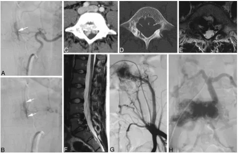
The ventral epidural group consists of shunts into those veins that normally drain structures developed from the notochord (ie, the vertebral body at the spinal level). These veins are known as the basivertebral venous plexus, which subsequently drains into the anterior internal vertebral ve-nous plexus, located at the ventral epidural space of the spinal canal, which joins the basilar venous plexus and cav-ernous sinus cranially. The previously called “epidural,” “osteodural,” or “paravertebral” AV shunts can be catego-rized into this group. Because the draining veins of these shunts do not drain the spine but the bone, these shunts will not become symptomatic due to venous congestion of the cord (Fig 1). Instead they may become symptomatic due to compression of the spinal cord or nerve roots by the en-larged epidural venous pouches.16-18There have been only
a few case reports describing associated perimedullary re-flux causing congestive myelopathy.11,19 A hypothesis about a possible defective valve-like mechanism normally impeding retrograde flow from the epidural plexus to peri-medullary veins has been put forward to explain this find-ing.11However, it may also be argued, that the reflux is due to an extensive thrombosis of the normal epidural outlets that leads to secondary retrograde drainage into the peri-medullary veins.5
The dorsal epidural group of AV shunts is related to veins that normally drain the spinous process and lamina at the spinal level. Although they are related to the major dural ve-nous sinuses (superior sagittal sinus and torcular and trans-verse sinuses) at the cranial level, the corresponding veins at the spinal level are poorly developed4and consist of a pair of
longitudinal channels (ie, the posterior internal venous plexus). Patients with dural AV shunts within this space typi-cally present with spontaneous epidural hematomas.20,21 These shunts are extremely rare.
The most common “classic” types of SDAVFs are the lateral epidural DAVFs. These AV shunts develop in the lateral epi-dural space at the junction of the bridging (or radicular) veins that connect the spinal cord drainage to the epidural venous system. Outflow obstruction of its adjacent venous outlet, ei-ther due to thrombosis or fibrosis related to aging, will then lead to immediate drainage into the perimedullary veins.5As a result, patients within this group present with aggressive clinical symptoms and at an older age. A strong male predom-inance is also observed,22which is similar to that in the
crani-ally located lateral epidural DAVFs, such as in the foramen magnum (medulla bridging vein) and tentorial (petrosal bridging vein) locations. Below, we will focus only on the latter “classic” form of dural AV shunt of the spine because it repre-sents⬎90% of all spinal dural AV shunts, whereas the remain-der are only seldom encountered and, if so, rarely present with clinically significant symptoms.
Epidemiology
ret-rospective series of the major German referral center for spinal vascular diseases (Prof Thron, Aachen, Germany), arrived at 5–10/million/year in the general population.25
However, the disease seems to be underdiagnosed.26,27 Usually, SDAVFs become symptomatic in elderly men. A recent meta-analysis22 of all series larger than 5 patients
concluded that men are affected 5 times more often than women and that the mean age at the time of diagnosis is 55– 60 years. Patients younger than 30 years of age consti-tuted less than 1% of patients with a DAVF, whereas, to our knowledge, no patient younger than 20 years of age has ever been reported. Most fistulas are solitary lesions and are found in the thoracolumbar region. In our experience, ⬎80% of all DAVFs are located between T6 and L2. Sacral lesions occur in approximately 4% of patients,28whereas high cervical lesions (at the level of the foramen magnum) occur in 2% of patients.29Low cervical DAVFs (below C2 and
above T1) are extremely rare.30,31In approximately 2% of pa-tients, double spinal DAVFs or an association of a spinal dural with a spinal pial AV shunt may be present, raising the possibility of a potential etiologic connection.32,33
Etiology and Pathophysiology
It is presumed that SDAVFs are acquired diseases, though their exact etiology is not known. The AV shunt is located inside the dura mater close to the spinal nerve root where the arterial blood from the radiculomeningeal artery (ie, the artery
that supplies the nerve root and meninges but not necessarily the spinal cord) enters a radicular vein, where the latter passes the dura at the dorsal surface of the dural root sleeve in the intervertebral foramen.22This transition is classically located directly underneath the pedicle of the vertebral body, which is supplied by the injected segmental artery. The increase in spi-nal venous pressure due to arterialization diminishes the AV pressure gradient and leads to a decreased drainage of normal spinal veins and a venous congestion with intramedullary edema because the intramedullary veins and the radicular vein share a common venous outflow.34,35This congestion, in turn,
leads to chronic hypoxia and progressive myelopathy.7Direct
intraoperative measurement of the vascular pressure of the fistula was found to be as high as 74% of the systemic arterial pressure.36,37This finding may explain why, in some patients,
symptoms become worse during physical activity with a con-comitant increase in arterial pressure.38,39Because the lower
thoracic region has relatively fewer venous outflow channels compared with the cervical region,40the venous congestive
edema is likely to be transmitted in a caudocranial direction throughout the spinal cord. This may explain why the first symptoms of myelopathy sometimes reflect dysfunction of the conus medullaris, even though the shunt is located remotely.41
Clinical Features
[image:3.594.52.537.41.354.2]more often, sensory symptoms such as paresthesias, diffuse or patchy sensory loss, but also radicular pain that may affect both lower limbs or initially only 1 limb.42Lower back pain
without radicular distribution is also frequently encountered. These neurologic symptoms are progressive with time and are often ascending.41Bowel and bladder incontinence, erectile dysfunction, and urinary retention are more often seen late in the course of the disease. Whereas classically the deficits are slowly progressive, an acute onset of the disease and a progres-sive development interrupted by intermediate remissions are also possible.43
A spinal hemorrhage has, in our experience, never oc-curred and, therefore, rather points toward a perimedullary (ie, pial) shunt rather than a true DAVF.15In rare cases, an SDAVF at the level of the foramen magnum with reflux toward the brain may cause a cranial subarachnoid hemorrhage.44
Because the course of the disease is slowly progressive, the
neurologic deficits at the time of diagnosis are often consider-ably worse: Two thirds of patients show a combination of gait difficulty, sensory disturbance, and involvement of sacral seg-ments (micturition, defecation, or sexual dysfunction).42
Up-per motoneuron involvement with clonus and positive Babin-ski signs and lower motoneuron involvement may coexist in the same patient, as has been described in the original obser-vations of Foix and Alajouanine.45,46
Imaging Features
In our experience, the diagnosis rests on MR imaging, is guided by MR angiography (MRA), and is confirmed by DSA.7
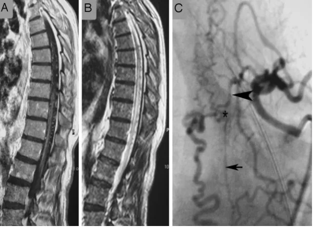
[image:4.594.132.453.42.476.2](Fig 2).47In the further course of the disease, the cord will
become atrophic.48The perimedullary vessels are dilated and
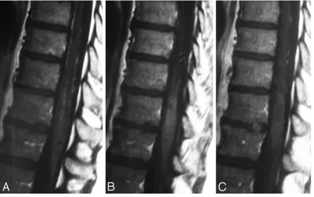
coiled and can be observed on the T2-weighted images as flow voids, which are often more pronounced on the dorsal surface compared with the ventral surface. However, if the shunt vol-ume is small, they might only be seen after contrast enhance-ment (Fig 2). The coiled or serpentine vascular structures may be better appreciated on heavily T2-weighted sequences (con-structive interference in steady state [CISS], fast imaging em-ploying steady-state acquisition [FIESTA], or 3D turbo
[image:5.594.54.286.44.323.2]spin-echo [3D-TSE]) compared with standard T2 TSE sequences (Fig 3).7In addition, these sequences may be useful to
differ-entiate pulsation artifacts, which are sometimes mistaken for flow voids, from true vascular tubular structures. Neither the location of pathologic vessels nor the intramedullary imaging findings seem to be related to the height of the fistula.30
On T1-weighted scans, the swollen cord is slightly hypoin-tense and enlarged. Following contrast administration, diffuse enhancement may be seen within the cord as a sign of chronic venous congestion with a breakdown of the blood⫺spinal cord barrier (Fig 4).49,50SDAVFs may occur anywhere from the level of the foramen magnum to the sacrum and localiza-tion of these lesions may be difficult and challenging, espe-cially in cases in which cord edema occurs distant from the AV shunt location.51 Thus, the noninvasive evaluation of the
shunt location is extremely helpful to guide the invasive con-ventional angiography.52Spinal contrast-enhanced MRA has
greatly contributed to localizing these lesions and helping to avoid unnecessary superselective injections of all possible ar-terial feeders. The technique of first-pass gadolinium-en-hanced MRA can clearly demonstrate the early venous filling, thereby confirming the shunt, and, in most cases, can also demonstrate the level of the shunt (Fig 5).53-56Spinal CT an-giography has also been shown to demonstrate the fistula lo-calization57; however, given the potential location from the foramen magnum to the sacrum, it may lead to a large radia-tion burden for the patient and, in our experience, does not constitute a practical approach to localizing SDAVFs.
On selective angiography, stasis of contrast material in the radiculomedullary arteries,7especially the anterior spinal ar-tery, can be seen. The delayed venous return following ASA injection indicates venous congestion and underlines the ne-cessity to search for a shunting lesion, whereas in most cases, a normal venous return following injection of the anterior spi-nal artery (ASA) will exclude the possibility of an SDAVF (Fig 6).30After injection into the segmental artery harboring the
AV fistula, the early venous filling and the retrograde contrast uptake of the radiculomedullary veins are visualized. Often an extensive network of dilated perimedullary veins is visible. Fig 3.When comparing a routine T2 TSE (A) sequence and a heavily T2-weighted (FIESTA,
3D T2 TSE, or CISS) sequence (B), the former depicts the cord edema better (arrow,A), whereas the latter is better suited to demonstrate the perimedullary flow voids (arrow,B), as seen in this patient.
[image:5.594.134.455.503.705.2]This network may even recruit supply from dural arteries that ascend or descend from neighboring radiculomeningeal arter-ies. In rare cases, the flow from the radiculomeningeal artery into the radicular vein may be slow; therefore, we classically perform spinal angiographies to search for DAVFs with a low frame rate (1 image/second) and hold for at least 4 seconds to exclude delayed retrograde filling of the radicular veins.
Differential Diagnosis
Clinical differential diagnosis of the rather unspecific neuro-logic symptoms is manifold, including polyneuropathy, tu-mor, or degenerative disk diseases.22It, therefore, is not sur-prising that patients with SDAVF see orthopedic surgeons, urologists (urinary retention being misinterpreted as being related to prostrate hypertrophy),58or psychologists (erectile
Fig 6.In patients with spinal AV shunts, the venous return after injection into the ASA is delayed due to the arterialized pressure in the spinal cord veins. Therefore, stasis following injection into the segmental artery from which the ASA originates is seen as in this patient.A, The arrow demonstrates the hairpin curve of the ASA visible in the early arterial phase.B, The late venous phase still demonstrates contrast material within the ASA, which should have been washed out by this time under physiologic conditions (arrowheads).C, The reason for the delayed washout is a DAVF at a different level, leading to massive venous congestion.
[image:6.594.136.452.42.295.2] [image:6.594.136.453.352.591.2]dysfunction)59before the neurologist. From an imaging point
of view, the MR imaging findings of cord edema together with perimedullary dilated vessels without any intramedullary ni-dus of vessels are typical for an SDAVF, and the only viable imaging differential diagnosis is another type of spinal vascu-lar malformation. An SDAVF that drains solely into the ante-rior spinal veins may go along with cord hypersignal on T2 only because the anterior spinal veins are located subpial and may, therefore, not be visualized as being dilated.60In these
cases, a glioma (especially when contrast uptake is present),61
an inflammatory lesion, or spinal ischemia should be in the differential diagnosis.7
Concerning the DSA appearance, the classic SDAVFs are typically of the slow-flow type, supplied by radiculomeningeal arteries and draining into radicular veins, which directly con-nect with either ascending or descending perimedullary veins; however, occasionally these angiographic characteristics may be difficult to differentiate from other simulating vascular shunting lesions (ie, radicular AV malformations [rAVMs]; epidural AV shunts, which belong to the ventral and dorsal epidural group of DAVFs; or perimedullary AV fistulas).30
rAVMs or AV malformations of the nerves usually have ab-normal vessels forming a nidus surrounding the nerve root,
whereas SDAVFs have an apparent shunt zone with converg-ing radicular feeders into the same drainconverg-ing vein. These pa-tients also often present with radicular pain and not direct symptoms of congestive venous myelopathy. The ventral and dorsal epidural DAVFs are located in the epidural space and will normally recruit a supply from the vertebral body and surrounding structures, with drainage directly into the epi-dural plexus and not a perimedullary vein.5
Symptoms are related to compression of the adjacent nerve root or cord rather than venous congestion, unless unusual cases of perimedullary reflux occur.51Perimedullary AV
fistu-las, including fistulas of the filum terminale, are pial shunts at the surface of the cord (or the filum) that are invariably sup-plied by arteries that, under normal circumstances, would supply the cord (ie, the ASA or the posterior spinal artery). In high-flow shunts, venous pouches are commonly encountered in those vascular malformations, especially in young patients with HHT.12,62
Treatment Modalities
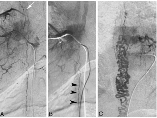
The aim of treatment in SDAVF is to occlude the shunting zone (ie, the most distal part of the artery together with the most proximal part of the draining vein, Fig 7).43,63A
[image:7.594.132.454.43.377.2]mal arterial occlusion will lead to a transient improvement of symptoms; however, owing to the good collateralization of the dura, the fistula is prone to recur within the following months. There are 2 options in the treatment of SDAVFs: surgical oc-clusion of the intradural vein that received the blood from the shunt zone, a relatively simple and safe intervention with the exception of sacral fistulas64; or endovascular therapy using a liquid embolic agent after superselective catheterization of the feeding radiculomeningeal artery.15 As pointed out
previ-ously, the embolic agent must pass the nidus and reach and occlude the proximal segment of the draining vein to prevent subsequent intradural collateral filling of the fistula (Fig 8). Therefore, proximal occlusions with coils or Gelfoam (Phadia, Uppsala, Sweden) are contraindicated. Embolization with particles is also prone to early recanalization and is, therefore, not indicated.65
The success rates of endovascular therapy have been re-ported to vary between 25% and 75%,43,66whereas a recent
meta-analysis suggested complete occlusion of the fistula fol-lowing surgery in 98%.67If the glue does not reach the venous
site, we strongly advocate early surgical intervention because a recent study has shown that patients in whom the endovascu-lar occlusion was incomplete and who required surgical inter-vention had a bad clinical outcome, which was likely due to the delay of the secondary intervention.68The treatment strategy
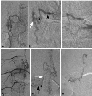
that is adopted by most centers currently includes a tentative embolization if this is believed to be a safe approach (ie, no spinal cord⫺supplying artery that may arise from the same pedicle as the feeder to the shunt as present in Fig 9). If the liquid embolic agent penetrates into the vein, long-term clin-ical follow-up has proved to result in complete obliteration of the fistula and good clinical outcome.69If the glue stays
intra-arterial, the liquid embolic agent may at least be used to label the feeding artery internally and thereby ease intraoperative
fluoroscopic localization of the exact height of the fistula. In the authors’ experience, a slow continuous injection of liquid glue (2 parts of iodized oil [Lipiodol; Guerbet, Aulnay-sous-Bois, France] and 1 part of glue) has a high chance to reach the draining vein and obliterate the fistula.
Prognosis
Treatment is aimed at halting the progression of the disease, and the prognosis is dependent on the duration of symptoms before treatment and the pretreatment disability. Following complete occlusion of the fistula, the progression of the dis-ease can be stopped in most instances70; however, only two
thirds of all patients have a regression of their motor symp-toms (including gait and strength) and only one third show an improvement of their sensory disturbances.43Impotence and sphincter disturbances are seldom reversible, and pain may persist. In rare cases of long-standing SDAVFs, patients may experience worsening despite complete occlusion.71Still, a
de-terioration of symptoms after initial improvement should raise the awareness of recanalization of the shunt or a second-ary shunt.33
Conclusions
[image:8.594.136.453.44.275.2]Acknowledgments
The authors are indebted to their clinical and neuroradiologic teachers, Drs Armin Thron (Aachen), Sirintara Pongpech (Bangkok) and Pierre Lasjaunias (Paris), to whom we dedicate this article posthumously.
References
1. Fleming A, Keynes RJ, Tannahill D.The role of the notochord in vertebral column formation.J Anat2001;199:177– 80
2. Eichmann A, Yuan L, Moyon D, et alVascular development: from precursor cells to branched arterial and venous networks.Int J Dev Biol2005;49:259 – 67 3. O’Rahilly R, Muller F.Neurulation in the normal human embryo.Ciba Found
Symp1994;181:70 – 82, discussion 82– 89
4. Groen RJ, Grobbelaar M, Muller CJ, et al.Morphology of the human internal vertebral venous plexus: a cadaver study after latex injection in the 21–25-week fetus.Clin Anat2005;18:397– 403
5. Geibprasert S, Pereira V, Krings T, et al.Dural arteriovenous shunts: a new classification of craniospinal epidural venous anatomical bases and clinical correlations.Stroke2008;39:2783–94
6. Zawilinski J, Litwin JA, Nowogrodzka-Zagorska M, et al.Vascular system of the human spinal cord in the prenatal period: a dye injection and corrosion cast-ing study.Ann Anat2001;183:331– 40
7. Krings T, Lasjaunias PL, Hans FJ, et al.Imaging in spinal vascular disease.
Neuroimaging Clin N Am2007;17:57–72
8. Lasjaunias PL, Berenstein A, terBrugge K.Surgical Neuroangiography:Clinical Vascular Anatomy and Variations.Vol 1. Berlin, Germany: Springer-Verlag; 2001
9. Krings T, Geibprasert S, Thron A.Spinal vascular anatomy.In: Naidich T, ed.
Neuroradiology of the Brain and Spine.New York: Elsevier; 2009. In press 10. Thron A.Vascular Anatomy of the Spinal Cord: Neuroradiological Investigations
and Clinical Syndromes.Berlin, Germany: Springer-Verlag; 1988
11. Krings T, Mull M, Bostroem A, et al.Spinal epidural AV fistula with perimed-ullary drainage: case report and pathomechanical considerations.J Neurosurg
2006;5:353–58
[image:9.594.132.456.43.519.2]12. Krings T, Ozanne A, Chng SM, et al.Neurovascular phenotypes in hereditary haemorrhagic telangiectasia patients according to age: review of 50 consecu-tive patients aged 1 day-60 years.Neuroradiology2005;47:711–20. Epub 2005 Sep 1
13. Rodesch G, Hurth M, Alvarez H, et al.Classification of spinal cord arterio-venous shunts: proposal for a reappraisal—the Bicetre experience with 155 consecutive patients treated between 1981 and 1999. Neurosurgery
2002;51:374 –79
14. Rodesch G, Hurth M, Alvarez H, et al.Angio-architecture of spinal cord arte-riovenous shunts at presentation: clinical correlations in adults and chil-dren—the Bicetre experience on 155 consecutive patients seen between 1981– 1999.Acta Neurochir (Wien) 2004;146:217–26
15. Krings T, Mull M, Gilsbach JM, et al.Spinal vascular malformations.Eur Radiol
2005;15:267–78
16. Alexander MJ, Grossi PM, Spetzler RF, et al.Extradural thoracic arteriovenous malformation in a patient with Klippel-Trenaunay-Weber syndrome: case report.Neurosurgery2002;51:1275–78, discussion 1278 –79
17. Kahara V, Lehto U, Sajanti J.Presacral arteriovenous fistula: case report. Neu-rosurgery2003;53:774 –76, discussion 776 –77
18. Schmidt C, Lonjon J, Costalat V, et al.Paraspinal arteriovenous malforma-tions with perimedullary venous drainage[in French].J Neuroradiol2008;35: 165–72. Epub 2008 May 16
19. Silva N Jr, Januel AC, Tall P, et al.Spinal epidural arteriovenous fistulas asso-ciated with progressive myelopathy: report of four cases.J Neurosurg Spine
2007;6:552–58
20. Asai J, Hayashi T, Fujimoto T, et al.Exclusively epidural arteriovenous fistula in the cervical spine with spinal cord symptoms: case report.Neurosurgery
2001;48:1372–75, discussion 1375–76
21. Chuang NA, Shroff MM, Willinsky RA, et al.Slow-flow spinal epidural AVF with venous ectasias: two pediatric case reports.AJNR Am J Neuroradiol
2003;24:1901– 05
22. Jellema K, Tijssen CC, van Gijn J.Spinal dural arteriovenous fistulas: a con-gestive myelopathy that initially mimics a peripheral nerve disorder.Brain
2006;129:3150 – 64
23. Kendall BE, Logue V.Spinal epidural angiomatous malformations draining into intrathecal veins.Neuroradiology1977;13:181– 89
24. Merland JJ, Riche MC, Chiras J.Intraspinal extramedullary arteriovenous fis-tulae draining into the medullary veins.J Neuroradiol1980;7:271–320 25. Thron A. Spinal dural arteriovenous fistulas [In German]. Radiologe
2001;41:955– 60
26. Grandin C, Duprez T, Stroobandt G, et al.Spinal dural arterio-venous fistula: an underdiagnosed disease?Acta Neurol Belg1997;97:17–21
27. Jellema K, Tijssen CC, Sluzewski M, et al.Spinal dural arteriovenous fistulas: an underdiagnosed disease—a review of patients admitted to the spinal unit of a rehabilitation center.J Neurol2006;253:159 – 62
28. Schaat TJ, Salzman KL, Stevens EA.Sacral origin of a spinal dural arterio-venous fistula: case report and review.Spine2002;27:893–97
29. Reinges MH, Thron A, Mull M, et al.Dural arteriovenous fistulae at the fora-men magnum.J Neurol2001;248:197–203
30. Geibprasert S, Jiarakongmun P, Krings T, et al.C5-cervical spinal dural arte-riovenous fistula presenting with congestive myelopathy of the cone.J Neu-rosurg Spine2009. In press
31. Willinsky R, TerBrugge K, Lasjaunias P, et al.The variable presentations of craniocervical and cervical dural arteriovenous malformations.Surg Neurol
1990;34:118 –23
32. Krings T, Coenen VA, Weinzierl M, et al.Spinal dural arteriovenous fistula associated with a spinal perimedullary fistula: case report.J Neurosurg Spine
2006;4:241– 45
33. Krings T, Mull M, Reinges MH, et al.Double spinal dural arteriovenous fistulas: case report and review of the literature. Neuroradiology
2004;46:238 – 42
34. Kataoka H, Miyamoto S, Nagata I, et al.Venous congestion is a major cause of neurological deterioration in spinal arteriovenous malformations. Neurosur-gery2001;48:1224 –29, discussion 1229 –30
35. Hurst RW, Kenyon LC, Lavi E, et al.Spinal dural arteriovenous fistula: the pathology of venous hypertensive myelopathy.Neurology1995;45:1309 –13 36. Hassler W, Thron A.Flow velocity and pressure measurements in spinal dural
arteriovenous fistulas.Neurosurg Rev1994;17:29 –36
37. Hassler W, Thron A, Grote EH.Hemodynamics of spinal dural arteriovenous fistulas: an intraoperative study.J Neurosurg1989;70:360 –70
38. Aminoff MJ, Barnard RO, Logue V.The pathophysiology of spinal vascular malformations.J Neurol Sci1974;23:255– 63
39. Khurana VG, Perez-Terzic CM, Petersen RC, et al.Singing paraplegia: a dis-tinctive manifestation of a spinal dural arteriovenous fistula.Neurology
2002;58:1279 – 81
40. Tadie M, Hemet J, Freger P, et al.Morphological and functional anatomy of spinal cord veins.J Neuroradiol1985;12:3–20
41. Koenig E, Thron A, Schrader V, et al.Spinal arteriovenous malformations and fistulae: clinical, neuroradiological and neurophysiological findings.J Neurol
1989;236:260 – 66
42. Jellema K, Canta LR, Tijssen CC, et al.Spinal dural arteriovenous fistulas: clinical features in 80 patients.J Neurol Neurosurg Psychiatry2003;74:1438 – 40 43. Van Dijk JM, TerBrugge KG, Willinsky RA, et al.Multidisciplinary manage-ment of spinal dural arteriovenous fistulas: clinical presentation and long-term follow-up in 49 patients.Stroke2002;33:1578 – 83
44. Kai Y, Hamada J, Morioka M, et al.Arteriovenous fistulas at the cervicomed-ullary junction presenting with subarachnoid hemorrhage: six case reports with special reference to the angiographic pattern of venous drainage.AJNR Am J Neuroradiol2005;26:1949 –54
45. Foix CH, Alajouanine T. La myelite necrotique subaigue. Rev Neurol
1926;46:1– 42
46. Schrader V, Koenig E, Thron A, et al.Neurophysiological characteristics of spinal arteriovenous malformations. Electromyogr Clin Neurophysiol
1989;29:169 –77
47. Gilbertson JR, Miller GM, Goldman MS, et al.Spinal dural arteriovenous fistulas: MR and myelographic findings. AJNR Am J Neuroradiol
1995;16:2049 –57
48. Huffmann BC, Spetzger U, Reinges M, et al.Treatment strategies and results in spinal vascular malformations.Neurol Med Chir (Tokyo) 1998;38(suppl):231–37 49. Chen CJ, Chen CM, Lin TK.Enhanced cervical MRI in identifying intracranial
dural arteriovenous fistulae with spinal perimedullary venous drainage. Neu-roradiology1998;40:393–97
50. Terwey B, Becker H, Thron AK, et al.Gadolinium-DTPA enhanced MR imag-ing of spinal dural arteriovenous fistulas. J Comput Assist Tomogr
1989;13:30 –37
51. Bostroem A, Thron A, Hans FJ, et al.Spinal vascular malformations: typical and atypical findings.Zentralbl Neurochir2007;68:205–13. Epub 2007 Oct 26 52. Bowen BC, Fraser K, Kochan JP, et al.Spinal dural arteriovenous fistulas:
evaluation with MR angiography.AJNR Am J Neuroradiol1995;16:2029 – 43 53. Farb RI, Kim JK, Willinsky RA, et al.Spinal dural arteriovenous fistula
local-ization with a technique of first-pass gadolinium-enhanced MR angiography: initial experience.Radiology2002;222:843–50
54. Mull M, Nijenhuis RJ, Backes WH, et al.Value and limitations of contrast-enhanced MR angiography in spinal arteriovenous malformations and dural arteriovenous fistulas.AJNR Am J Neuroradiol2007;28:1249 –58
55. Bowen BC, Pattany PM.Contrast-enhanced MR angiography of spinal vessels.
Magn Reson Imaging Clin N Am2000;8:597– 614
56. Bowen BC, Saraf-Lavi E, Pattany PM.MR angiography of the spine: update.
Magn Reson Imaging Clin N Am2003;11:559 – 84
57. Yamaguchi S, Eguchi K, Kiura Y, et al.Multi-detector-row CT angiography as a preoperative evaluation for spinal arteriovenous fistulae.Neurosurg Rev
2007;30:321–26, discussion 327
58. Sheikh SI, Busl KM, Ning M, et al.Spinal dural arteriovenous fistula mimick-ing prostate hyperplasia.J Emerg Med2008 Nov 18. [Epub ahead of print] 59. Jellema K, Tijssen CC, van Rooij WJ, et al.Spinal dural arteriovenous fistulas:
long-term follow-up of 44 treated patients.Neurology2004;62:1839 – 41 60. Thiex R, Mayfrank L, Krings T, et al.Delayed diagnosis of spinal dural
arterio-venous fistula in the absence of pathological vessels on MRI.Zentralbl Neuro-chir2006;67:94 –98
61. Roccatagliata L, Centanaro F, Castellan L.Venous congestive myelopathy in spinal dural arteriovenous fistula mimicking neoplasia. Neurol Sci
2007;28:212–15
62. Krings T, Chng SM, Ozanne A, et al.Hereditary hemorrhagic telangiectasia in children: endovascular treatment of neurovascular malformations—results in 31 patients.Neuroradiology2005;47:946 –54
63. Jellema K, Sluzewski M, van Rooij WJ, et al.Embolization of spinal dural arte-riovenous fistulas: importance of occlusion of the draining vein.J Neurosurg Spine2005;2:580 – 83
64. Huffmann BC, Gilsbach JM, Thron A.Spinal dural arteriovenous fistulas: a plea for neurosurgical treatment.Acta Neurochir (Wien) 1995;135:44 –51 65. Nichols DA, Rufenacht DA, Jack CR Jr, et al.Embolization of spinal dural
arteriovenous fistula with polyvinyl alcohol particles: experience in 14 pa-tients.AJNR Am J Neuroradiol1992;13:933– 40
66. Niimi Y, Berenstein A, Setton A, et al.Embolization of spinal dural arterio-venous fistulae: results and follow-up.Neurosurgery1997;40:675– 82, discus-sion 682– 83
67. Steinmetz MP, Chow MM, Krishnaney AA, et al.Outcome after the treatment of spinal dural arteriovenous fistulae: a contemporary single-institution se-ries and meta-analysis.Neurosurgery2004;55:77– 87, discussion 87– 88 68. Andres RH, Barth A, Guzman R, et al.Endovascular and surgical treatment of
spinal dural arteriovenous fistulas.Neuroradiology2008;50:869 –76 69. Sherif C, Gruber A, Bavinzski G, et al.Long-term outcome of a
multidisci-plinary concept of spinal dural arteriovenous fistulae treatment. Neuroradiol-ogy2008;50:67–74
70. Aghakhani N, Parker F, David P, et al.Curable cause of paraplegia: spinal dural arteriovenous fistulae.Stroke2008;39:2756 –59