ORIGINAL RESEARCH
ADULT BRAIN
Localized Marked Elongation of the Distal Internal Carotid
Artery with or without PHACE Syndrome: Segmental
Dolichoectasia of the Distal Internal Carotid Artery
XZ.Y. Jia,X L.B. Zhao, andXD.H. Lee
ABSTRACT
BACKGROUND AND PURPOSE: Segmental intracranial dolichoectasia of the distal ICA is a feature of PHACE syndrome or a sporadic phenomenon. We evaluated the relationship between intracranial dolichoectasia of the distal ICA and PHACE syndrome and illustrated the characteristic radiologic findings of the lesions.
MATERIALS AND METHODS: Intracranial dolichoectasia of the distal ICA was identified in 20 patients at our institution from 2005 to 2016 through a review of diagnostic cerebral angiography results. All radiologic images were reviewed to determine the vascular morphologic dispositions around the distal ICA, including dysplasia, mural calcification, vessel wall enhancement, lumen narrowing, and aneurysm formation. Medical records were reviewed to determine the symptoms of PHACE syndrome. Subsequently, the correlation between radiologic findings and PHACE syndrome was assessed.
RESULTS:In this cohort, which had a strong female predominance (male/female ratio⫽2:18), intracranial dolichoectasia had a more ipsilateral vascular morphologic disposition. Mural calcification was detected more frequently in elderly patients, whereas vessel wall enhancement was detected more frequently in younger patients. Follow-up images showed a slow progression of the lesions. However, no significant differences in the vascular morphologic disposition and brain structural changes were observed between patients with (n⫽ 11) and without (n⫽9) PHACE syndrome.
CONCLUSIONS: The striking elongation and tortuosity of the distal ICA generally appeared to be a type of congenital lesion occurring early in embryogenesis as either a sporadic phenomenon or an arterial change associated with PHACE syndrome. Imaging findings revealed various mural abnormalities with a benign clinical course.
ABBREVIATIONS:AchoA⫽anterior choroidal artery; BA⫽basilar artery; CS⫽communicating segment; ICDE⫽intracranial dolichoectasia; PCA⫽posterior cerebral artery; PcomA⫽posterior communicating artery; PHACE⫽posterior fossa malformations, hemangiomas, arterial anomolies, cardiac defects, and eye abnormalities; OA⫽ophthalmic artery
M
arked arterial elongation and tortuosity (intracranial dolicho-ectasia [ICDE]) have been reported previously as common features of PHACE syndrome1-4or as a sporadic phenomenon.5-7PHACE syndrome refers to conditions associated with posterior fossa malformations, hemangiomas, arterial anomolies, cardiac de-fects, and eye abnormalities.8ICDE of the intracranial arteries is one
of the various features of the arterial cerebrovascular abnormalities.
On cerebrovascular imaging, from time to time, we encounter striking arterial elongations, particularly the distal ICA elonga-tion, regardless of the PHACE syndrome status. This pathologic ar-terial elongation often co-occurs with marked tortuosity and may even appear as a conglomerated vascular mass to compensate for the limited length of the allocated arterial segment. It is often accompa-nied by conditions such as multiple stenoses, dilations, or aneurysms and usually features a variable amount of mural calcification.2
An-giographically, this vascular morphologic disposition exhibits a more congenital appearance because the unique features cannot be ex-plained by an acquired anatomic alteration. Although these types of arterial changes may present in any arterial segment of the body, we focused on cases of segmental involvement of the distal ICA because in our angiographic experience (limited to the cerebrovascular sys-tem), this site is the most frequently affected.
The easily recognizable symptoms and clinical significance of the above-mentioned vascular morphologic disposition completely dif-Received July 29, 2017; accepted after revision January 1, 2018.
From the Department of Radiology and Research Institute of Radiology (Z.Y.J., L.B.Z., D.H.L.), Asan Medical Center, University of Ulsan College of Medicine, Seoul, Korea; and Department of Radiology (Z.Y.J., L.B.Z.), The First Affiliated Hospital of Nanjing Medical University, Jiangsu Province, China.
Please address correspondence to Deok Hee Lee, MD, PhD, Department of Radiol-ogy and Research Institute of RadiolRadiol-ogy, Asan Medical Center, University of Ulsan College of Medicine, 88, Olympic-ro 43-gil, Songpa-gu, Seoul 138-736, Korea; e-mail: dhlee@amc.seoul.kr
Indicates article with supplemental on-line table.
fer from those of acquired nonsegmental dilative arteriopathy, which is frequently observed in the basilar artery (BA). Accordingly, this study describes angiographic and other imaging characteristics of this peculiar anatomic disposition, particularly in the distal ICA, by reviewing the radiologic findings and medical records of affected pa-tients. We aimed to evaluate the possible associations of the radio-logic findings with PHACE syndrome and illustrate the clinical fol-low-up results of the imaging morphologic abnormalities.
MATERIALS AND METHODS
Definition of ICDE and Segmentation of the ICA
Because pathologic arterial tortuosity cannot be quantified, we subjectively defined segmental ICDE by comparing other arterial segments, particularly those in the same segment on the contralat-eral side, as unusually marked areas of dolichosis with variable degrees of ectasia on the affected side. Notably, these areas could be easily demarcated from other adjacent segments with normal lengths and tortuosities.
We applied the 7-segment system proposed by Lasjaunias et al9for the ICA to our analysis of the involved segments of the ICA
and intracranial arteries. This embryology-based 7-segment sys-tem terminates at the origin of the posterior communicating ar-tery (PcomA). Because the system does not name the segment of the adult ICA between the PcomA origin and ICA bifurcation into the MCA and anterior cerebral artery, we designated this specific segment as the “communicating segment” (CS) (Fig 1).
Patient Population and PHACE Syndrome Diagnosis At our institution (Asan Medical Center), 11,516 patients under-went diagnostic cerebral angiography between January 2005 and December 2016. For our study, we limited the number of candi-date cases by applying the search terms “carotid,” “ICA,” “dyspla-sia,” and “dolichoectasia” and further limited the number to 45 patients by reviewing the angiography reports. Furthermore, all angiographic images of the included cases were reviewed to iden-tify intracranial ICA abnormalities, including marked elongation and tortuosity. Finally, 20 patients were identified.
This retrospective review was approved by our institutional review board, and the requirement for individual patient consent was waived. The included patients’ medical records were reviewed for symptoms and indications of PHACE syndrome and other sig-nificant disorders. All data obtained via radiologic imaging modali-ties (CT, CTA, MR imaging, TOF-MRA, contrast-enhanced MR ves-sel imaging, and brain perfusion SPECT) were reviewed in our PACS system by 2 radiologists (Z.Y.J. and L.B.Z.). If the individual radio-logic analyses differed, the reviewers reached a consensus after dis-cussion with a third reviewer (D.H.L.). Finally, the patients were stratified according to the “Consensus Statement on Diagnostic Cri-teria for PHACE Syndrome” of 2009 as follows: 1) PHACE syn-drome, 2) possible PHACE synsyn-drome, or 3) none.8
Image Findings and Statistical Analysis
After recording the laterality and segments of the involved ICA, the morphology of the arterial components around the distal ICA was recorded by reviewing DSA, CTA, and MRA images of the ante-rior cerebral artery (A1 segment), anteante-rior communicating artery, MCA (M1), anterior choroidal artery (AchoA), PcomA, ophthalmic
artery (OA), BA, and posterior cerebral artery (PCA) (P1 and P2 segments). The term “dysplasia” encompassed a variety of arterial abnormalities, including looping, coiling, ectasia, dolichoectasia, or simple dolichosis. In addition, the presence of lesions in the con-tralateral ICA, BA, and/or ipsi- and concon-tralateral vertebral arteries was noted.
DSA, CTA, and MRA were used to analyze vessel stenoses and aneurysms. Stenosis was defined as any narrowing of the vessel lumen compared with the reference vessel as defined in the War-farin-Aspirin Symptomatic Intracranial Disease trial method.10
The aneurysm was defined as an eccentric bulging of the vessel wall within the dolichoectatic segment, and the diagnosis was reached with the consensus of 2 doctors (D.H.L. and Z.Y.J.). CT was used to analyze vessel wall calcification. Contrast-enhanced MR imaging of the vessel wall was used to analyze vessel wall charac-teristics, including enhancement, wall thickening, and luminal nar-rowing. Brain perfusion SPECT was used to evaluate whether a ste-nosis or tortuous ICA led to a decrease in brain perfusion.
[image:2.594.337.488.43.371.2]performed to evaluate possible relationships among the following features: age, wall calcification, vessel wall enhancement, stenosis, and aneurysm.
RESULTS
The basic demographic information, presenting symptoms, in-volved arteries around the distal ICA, acquired changes in affected segments, and PHACE diagnostic statuses of all patients are sum-marized in the On-line Table. The patients included 18 females and 2 males with a median age of 43.5 years (range, 7–73 years) with varying clinical symptoms that did not appear to be directly related to the arterial abnormality. A systemic review revealed that 3 patients had hypertension, whereas none had systemic vasculitis or autoimmune disease.
All patients had the unique feature, segmental elongation and tortuosity of the distal ICA, resulting in a tangled arterial mass (Fig 2). No differences were observed between the sides of onset (right, 11/20). Two patients presented with bilateral distal ICA dolichoectasia. In these 2 patients, we defined the side with more severe dolichoectasia as the ipsilateral side, while the other side was
the contralateral side. The involved ICA segments ranged from segments 2 to 7, with a mean⫾SD of 3.5⫾1.6 segments. Fourteen patients had BA (n⫽1) or ipsi-lateral PCA (n⫽13) involvement [BA/ PCA (⫹)]. We observed a significant cor-relation between ipsilateral A1, PcomA, BA/PCA, and midbrain hypoplasia and segmental ICDE of the distal ICA, unlike the contralateral side (Table 1).
In 9 of the 20 patients, the ipsilateral midbrain was smaller than the con-tralateral midbrain (Fig 2E). Four and 7 patients were found to have confirmed or possible PHACE syndrome, respec-tively, when midbrain hypoplasia was excluded as a major or minor criterion for a PHACE syndrome diagnosis (pos-terior fossa anomaly). The inclusion of this criterion resulted in 4 and 11 pa-tients with confirmed and possible PHACE syndrome, respectively. No sig-nificant differences in arterial compo-nent dysplasia (including A1, anterior communicating artery, M1, AchoA, PcomA, OA, BA, and PCA) and brain structures were observed between pa-tients positive and negative for PHACE (Table 2).
Calcification was detected in 16 pa-tients with a mean age of 45.9 years; the remaining 4 patients without calcifica-tion had a mean age of 17.3 years. Signif-icantly progressive calcification was ob-served in 1 patient with progressive arterial stenosis during an interval of 10 years (patient 9; age range, 42–52 years). Stenosis was detected in 11 patients with a mean age of 42.1 years; the 9 patients without stenosis had a mean age of 37.9 years. None of the patients had an ischemic stroke event, and none of the 12 patients who underwent brain perfusion SPECT had hypoperfusion. Aneurysms were detected in 15 pa-tients with a mean age of 38.1 years; the 5 papa-tients without aneu-rysms had a mean age of 48.8 years.
Ten patients underwent contrast-enhanced high-resolution MR imaging of the vessel wall, which detected enhancement in 6 patients with a mean age of 22.8⫾13.2 years; the remaining 4 patients had a mean age of 57.0⫾ 9.2 years (P ⬍.001). One patient (patient 1,Fig 3) exhibited vessel wall enhancement at both 8 and 12 years of age, with no major change in the enhance-ment pattern. Vessel wall imaging revealed that the stenotic seg-ment exhibited either an eccentric or concentric wall thickening (Fig 4) as well as a potential enhancement of the parent artery and aneurysm neck (Fig 5).
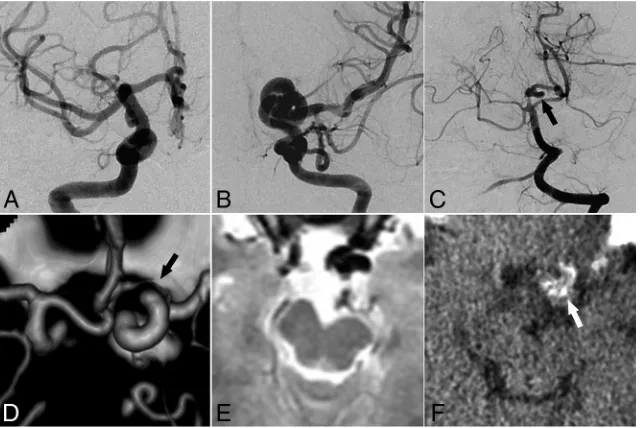
[image:3.594.56.376.47.261.2]FIG 2. Representative images of ICDE in a patient without PHACE syndrome (patient 6, a 28-year-old woman).AandB, Bilateral ICA angiography shows a tangled arterial mass of the left distal ICA and no observable left A1 segment.C, Left vertebral artery angiography shows a dolichoec-tasia of the left P1 and P2 segments (arrow).D, CT angiography shows hypoplasia of the left A1 segment (arrow).E, T2-weighted image of the brain shows hypoplasia of the left midbrain.F, CT image of vessel wall calcification in the left distal ICA (arrow).
Table 1: Arterial components and brain structures surrounding the distal ICA on the affected side versus the contralateral side
Affected Side (n= 20) Contralateral Side (n= 20) PValuea
A1 Dysplasia⫽15 (75.0%) Dysplasia⫽1 (5.0%) .001
M1 Dysplasia⫽2 (10.0%) Dysplasia⫽0 (0.0%) .487b
OA Ectopic⫽5 (25.0%) Ectopic⫽1 (5.0%) .219
AchoA Dysplasia⫽5 (25.0%) Dysplasia⫽0 (0.0%) .047b
PcomA Dysplasia⫽14 (70.0%) Dysplasia⫽3 (15.0%) .003
BA/PCA Dysplasia⫽14 (70.0%) Dysplasia⫽1 (5.0%) ⬍.001 Midbrain Hypoplasia⫽9 (45.0%) Hypoplasia⫽0 (0.0%) .001b
Note:—A1 indicates a segment of the anterior cerebral artery; M1, a segment of the MCA. BA/PCA indicates either BA or PCA, or both.
a
McNemar test.
b
[image:3.594.54.376.353.439.2]DISCUSSION
In our series of patients across a wide range of ages, we observed striking elongation and tortuosity of the distal ICA, regardless of the PHACE diagnostic status. We further observed that compared with the contralateral side, the ipsilateral arterial components and brain structures around the affected distal ICA more frequently showed dysplasia. Furthermore, the affected vessels had various manifestations, including stenosis, aneurysm, calcification, and vessel wall enhancement, and the disease evolved slowly with age according to follow-up radiologic imaging.
Embryologic Pathogenesis of ICDE of the Distal ICA
During the embryologic period of cere-bral artery formation, angiogenesis is mainly driven by hypoxia and related growth factors in the target tissue.11,12
We hypothesized that exposure of a spe-cific segment of the intracranial artery to a vasculogenetic trigger resulted in arte-rial lengthening. According to the ICA developmental anatomy proposed by Lasjaunias and Santoyo-Vazquez,13the ICA branches into a
cra-nial and caudal rami at stages I and II (3.5– 4 weeks); the former gives rise to the anterior cerebral artery and AchoA, whereas the latter gives rise to the PcomA, P1 segment, and upper BA. In this study, dolichoectasia at the anterior (A1 segment, AchoA, and CS segment) and posterior (PcomA, P1 segment, and upper BA) divi-sions of the ICA suggested that these regions were triggered during embryonic development. We observed no involvement of the supe-rior cerebellar artery in our cases (data not shown); because the su-perior cerebellar artery existed before ICA branching, the vasculoge-netic event may have occurred after the appearance of superior cerebellar artery.13Although the remaining PCA (segments P2–P4)
comprises the posterior choroidal branch of the caudal ramus at stage V, the involvement of the P2 segment in several cases suggests that the PCA precursor had also been affected.13
PHACE Syndrome
Cerebral arterial anomalies are observed in 91% of patients with PHACE syndrome,14and previous studies of PHACE syndrome
have reported a presentation of dolichoectasia of the internal ca-rotid arteries similar to that observed in our cases.2,4,15,16
There-fore, we searched for common features between our cases and PHACE syndrome cases.
First, we observed a strong female predominance in our pa-tient group in agreement with previous studies of PHACE syn-drome (up to 8:1).15,17,18Second, the timing of PHACE syndrome
was consistent with our speculated time course. Several studies of PHACE syndrome have reported that the teratogenic influence might occur from gestational weeks 3 to 5.5,8,14concurrent with
the regression of the embryonic capillary bed and active stemming of the craniocervical vasculature. Therefore, any influences on these 2 processes may result in a cutaneous hemangioma and trigeminal artery persistence.19
Third, several vascular anomalies have been reported in both patients with PHACE syndrome and in our patient group. In a previous study, A1 hypoplasia was reported as an intracranial anomaly affecting 8 of 12 patients with PHACE syndrome; this is similar to the findings of our study (15/20).20ICDE of the ICA was
accompanied by dolichoectasia of the posterior circulation in 6 of 7 patients with PHACE syndrome in a previous study.20An
aber-rant origin or course of the principal cerebral arteries, a major or minor PHACE syndrome criterion, was observed in 9 of 20 pa-tients (45%; 5 ectopic ophthalmic arteries and 4 other arteries).
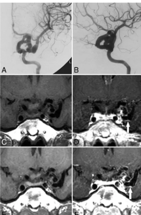
Fourth, 4 of the 20 patients in this study met the diagnostic criteria of PHACE syndrome, and an additional 7 patients were classified as possible cases of PHACE syndrome. In addition, some FIG 3. Evolution of vessel wall enhancement with aging in a patient
[image:4.594.51.379.64.140.2]with PHACE syndrome (patient 1, an 8-year-old girl).AandB, Left ICA angiography (anteroposterior and oblique views) reveals tortuosity in the left distal ICA with a small and long A1 segment.CandD, Noncon-trast and conNoncon-trast-enhanced vessel wall imaging at 8 years of age demonstrates vessel wall enhancement and slight vessel wall thicken-ing (arrowinD).EandF, Vessel wall imaging at 12 years of age shows a similar vessel wall enhancement pattern (arrowinF).
Table 2: Arterial components around the distal ICA on the affected side in patients with and without PHACE syndromea
PHACE (+) (n= 11) PHACE (−) (n= 9) PValueb
A1 Dysplasia⫽8 (72.7%) Dysplasia⫽7 (77.8%) 1.000
M1 Dysplasia⫽0 (0.0%) Dysplasia⫽2 (22.2%) .189
OA Ectopic⫽1 (9.1%) Ectopic⫽4 (44.4%) .127
AchoA Dysplasia⫽3 (27.3%) Dysplasia⫽2 (22.2%) 1.000
PcomA Dysplasia⫽7 (63.6%) Dysplasia⫽7 (77.8%) .642
Midbrain Dysplasia⫽5 (45.5%) Dysplasia⫽4 (44.4%) 1.000
a
PHACE includes cases of confirmed and possible PHACE syndrome; a small midbrain was not used as a criterion.
b
[image:4.594.54.284.166.516.2]patients exhibited ipsilateral midbrain hypoplasia. Because a lack of direct contact with the adjacent vessel does not support compres-sion-induced midbrain deformation (Fig 2E), we suspected that dolichoectasia of the arteries feeding the midbrain (BA, P1, or P2) mildly altered the blood supply and caused further hypoplasia. In 1 patient with PHACE (patient 10), hypoplasia of the ipsilateral cere-bellum and the ipsilateral midbrain was found to coexist, suggesting that these 2 structural anomalies shared a common origin. Therefore, if a smaller midbrain was defined as a posterior fossa anomaly asso-ciated with PHACE syndrome, an additional 4 patients in our study would meet the criteria for possible PHACE syndrome. Such lesions
might broaden the PHACE syndrome phenotype. According to a study by Heyer et al,20moderate effacement of the right
pons (Fig 2Cin the article by Heyer et al) and cerebral peduncle along with corresponding vascular anomalies was observed via MR imaging in a patient with PHACE syndrome. However, the author did not propose this finding as an anomaly.20
In our study, we found no significant differences in arterial component dys-plasia and brain structures between pa-tients positive and negative for PHACE (Table 2), suggesting that the 2 groups of patients share the same features and pathogenesis. Furthermore, although we did not detect an obvious cutaneous hemangioma in many of our patients, a previous study found that very small cu-taneous hemangiomas might be absent or regress spontaneously without prior recognition or reporting.4 The
above-mentioned aspects raise the intriguing possibility that a marked ICDE of the ICA might indicate an otherwise-unrecognized partial pheno-typic expression of PHACE syndrome. However, the spontaneous regression of cutaneous hemangiomas at an early age may cause the underestimation of the incidence of PHACE syndrome in this group of patients, which further induces underestimation of the relationship between the ICDE and PHACE syndrome.
Acquired Changes in the Involved Arterial Wall
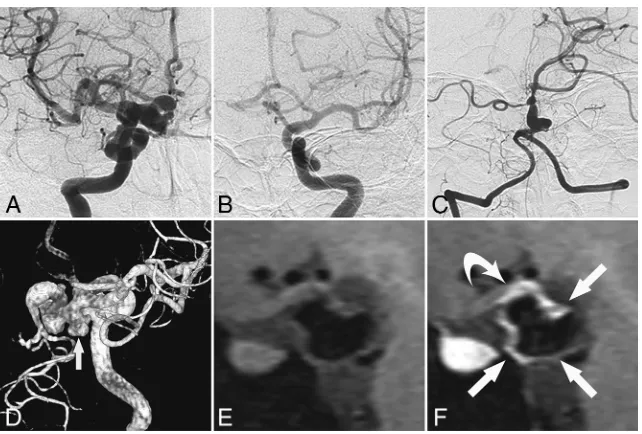
Normally, an abnormal mural angiogenesis likely causes an in-crease in the luminal caliber because the correct remodeling sig-nals induce apoptosis of the unnecessary vessel wall components. FIG 5. Vessel wall enhancement at the aneurysm wall and the parent artery wall in a patient
without PHACE syndrome (patient 11, a 45-year-old woman).A–C, Cerebral angiography shows dolichoectasia in the right distal ICA and basilar artery.D, 3D-DSA shows dolichoectasia in the right distal ICA and right PcomA as well as the formation of multiple aneurysms in the right PcomA (arrow).EandF, Non-contrast-enhanced and contrast-enhanced vessel wall imaging shows en-hancement of the aneurysm wall (arrowsinF) and the parent artery wall (curved arrowinF).
[image:5.594.53.532.48.254.2] [image:5.594.55.376.306.523.2]A lack of remodeling causes centripetal and longitudinal prolifer-ation and luminal reduction; however, this might also cause ecta-sias, elongated arteries, and aneurysms.13Although many cases
involving ICDE of the ICA, with or without PHACE syndrome, have been reported, little is known about the evolution of ves-sel wall lesions and relevant complications. Results from a fol-low-up study of vessel lesions may provide valuable prognostic information.
Progressive cerebral arterial stenosis and arterial occlusion and a Moyamoya-like vasculopathy leading to stroke have been described in infants with PHACE syndrome.2,20This progressive
cerebral vasculopathy corresponds with the proliferative phase of hemangioma growth, and as a result, the average age of experi-encing stroke among patients with PHACE syndrome is 8.8 months.21However, no ischemic stroke events were reported in
our present study, even among patients with a Moyamoya-like vasculopathy (patients 4, 8, 12). Notably, we observed vessel wall enhancement more frequently among young patients than older patients, indicating a regression in inflammation with aging. Ac-cordingly, we speculate that most arterial stenoses and occlusions formed within a short time during the prenatal or infant stage. Patients with mild lesions might pass through that period asymp-tomatically, and adult PHACE syndrome diagnoses may be inci-dental.1,22Due to the long-standing nature of the lesion, there is a
good chance of good collateral circulation formation secondary to arterial stenoses, which may present as nonsymptomatic steno-occlusive disease.
We further propose that ICDE of the distal ICA, with or with-out PHACE syndrome, might stabilize after a period of rapid pro-gression. Bracken et al15followed up several cases of PHACE in
neu-rodevelopmentally healthy patients for 1–12 years. McLaughlin et al5
reported a 24-year-old female patient with a pure arterial malfor-mation involving the distal ICA, PcomA, and PCA that was found on a CT scan obtained to determine the cause of a headache. When this patient was later followed up at 54 years of age, the abnormal vessels had not changed with time on MR images, and no symptoms relevant to the abnormal vessels were reported dur-ing the 31-year interval.5Similarly, our review of imaging data
collected during a long follow-up demonstrated a slow evolu-tion of vessel wall enhancement (patient 1) and slow progres-sion of both calcification and stenosis (patient 9). The vessel wall enhancement in patient 1 could be explained by the im-mature nature of the affected vessel wall, which may increase the permeability of the endothelium, with contrast leakage from the lumen into the arterial wall, and may be simultane-ously associated with an atherosclerotic-like process in the dys-plastic segments.
Treatment and Follow-Up
Many cases of ICDE with tortuous ICAs were identified inciden-tally, without relevant symptoms5,7,15; in these cases, the lesions
appeared stable on follow-up images and the patients did not receive medical treatment. However, several reports and our ob-servations suggested the need for regular imaging follow-up as well as medication in some cases. However, no specific treatment exists for dolichoectasia, and the surgical and medical therapies used to treat this condition have not been systematically
evalu-ated. Although anticoagulation and antiplatelet therapies might help in preventing an ischemic episode, some studies have indi-cated that aspirin and warfarin or both do not effectively reduce the stroke recurrence rates in patients with dolichoectasia and might increase the risk of hemorrhage in this population.23
How-ever, we note that these previous data were all with respect to the BAs.
Treatment for PHACE syndrome should address the afore-mentioned symptoms.2Corticosteroids and interferon have been
previously used to treat hemangiomas associated with PHACE syndrome; however, their efficacy in the treatment of acute-phase vessel wall inflammation remains unknown.16Occasionally, a pial
synangiosis procedure has been suggested for severe stenosis or occlusion of the distal ICA.20
We believe that attention should be paid to several cases in this study. One patient (patient 1) exhibited simultaneous vessel wall enhancement and calcification at 7 years of age, leading to our hypothesis that the affected vessel wall was prone to atherosclero-sis formation and secondary calcification. Questions also remain regarding the use of antiatherosclerosis therapies in young pa-tients. Another patient (patient 11,Fig 5) exhibited vessel wall enhancement in an aneurysm and its parent artery, which may be a risk factor for aneurysm rupture; accordingly, a pre-emptive aneurysm embolization was performed.24In another patient
(pa-tient 16), asymptomatic ICDE of the right ICA and hypoplasia of the right A1 segment were detected at 54 years of age, and a blood flow–related aneurysm of the left anterior communicating artery was observed at 69 years of age. This patient was later treated with coiling embolization. Therefore, we suggested a follow-up com-prising regular angiography studies (CTA or MRA) to demon-strate overall luminal changes and, if possible, vessel wall imaging to detect inflammation in the lesion.
Limitations
This study had several limitations. First, we found it difficult to objectively define “segmental dolichoectasia.” To overcome this problem, we included only cases with noncontroversial elonga-tion and unusual tortuosity relative to other segments or the con-tralateral ICA. Accordingly, we might have skipped many mild elongation cases and underestimated the number of relevant cases. Second, the definition of segmental arterial tortuosity is rather subjective. Lasjaunias et al9defined the ICA segments
ac-cording to embryogenic evolution. In this study, we considered ICDE of the distal ICA to be a congenital disease that may occur segmentally. Although we used this system to describe the ob-served lesions, we were unable to conclude that the lesions could be attributed to a similar congenital origin. Third, 2 patients in our study had bilateral ICA involvement, which has also been reported in patients with PHACE syndrome. However, a satisfac-tory interpretation of the bilateral pathogenesis could not be attained.20
CONCLUSIONS
vascular segments adjacent to the lesions, particularly in the ipsi-lateral proximal PCA. Imaging findings of affected patients dem-onstrated various mural abnormalities with a benign clinical course.
REFERENCES
1. Burch EA, Garzon MC, Parikh A, et al.A 65-year-old woman diag-nosed with PHACE syndrome.Pediatr Dermatol2013;30:e153–56
CrossRef Medline
2. Baccin CE, Krings T, Alvarez H, et al.A report of two cases with dolichosegmental intracranial arteries as a new feature of PHACES syndrome.Childs Nerv Syst2007;23:559 – 67CrossRef Medline
3. Metry DW, Dowd CF, Barkovich AJ, et al.The many faces of PHACE syndrome.J Pediatr2001;139:117–23CrossRef Medline
4. Rossi A, Bava GL, Biancheri R, et al.Posterior fossa and arterial abnormalities in patients with facial capillary haemangioma: pre-sumed incomplete phenotypic expression of PHACES syndrome. Neuroradiology2001;43:934 – 40CrossRef Medline
5. McLaughlin N, Raychev R, Duckwiler G, et al.Pure arterial malfor-mation of the posterior cerebral artery: importance of its recogni-tion.J Neurosurg2013;119:655– 60CrossRef Medline
6. Yuh SJ, Alkherayf F, Lesiuk H.Dolichoectasia of the vertebral basilar and internal carotid arteries: a case report and literature review. Surg Neurol Int2013;4:153CrossRef Medline
7. Nakahara I, Taki W, Tanaka M, et al.Dolichoectasia of the middle cerebral artery: case report. Neurol Med Chir (Tokyo)1995;35: 822–24CrossRef Medline
8. Metry D, Heyer G, Hess C, et al; PHACE Syndrome Research Confer-ence.Consensus statement on diagnostic criteria for PHACE syn-drome.Pediatrics2009;124:1447–56CrossRef Medline
9. Lasjaunias PL, Berenstein A, Ter Brugge KG.Surgical Neuroangio-graphy: Clinical Vascular Anatomy and Variations.2nd ed.Vol. 1. Berlin: Springer-Verlag; 2001
10. Samuels OB, Joseph GJ, Lynn MJ, et al.A standardized method for measuring intracranial arterial stenosis.AJNR Am J Neuroradiol
2000;21:643– 46Medline
11. Menshawi K, Mohr JP, Gutierrez J.A functional perspective on the embryology and anatomy of the cerebral blood supply.J Stroke
2015;17:144 –58CrossRef Medline
12. Plate KH.Mechanisms of angiogenesis in the brain.J Neuropathol Exp Neurol1999;58:313–20CrossRef Medline
13. Lasjaunias P, Santoyo-Vazquez A.Segmental agenesis of the internal carotid artery: angiographic aspects with embryological discus-sion.Anat Clin1984;6:133– 41CrossRef Medline
14. Haggstrom AN, Garzon MC, Baselga E, et al.Risk for PHACE syn-drome in infants with large facial hemangiomas.Pediatrics2010; 126:e418 –26CrossRef Medline
15. Bracken J, Robinson I, Snow A, et al.PHACE syndrome: MRI of intracerebral vascular anomalies and clinical findings in a series of 12 patients.Pediatr Radiol2011;41:1129 –38CrossRef Medline
16. Vermeer S, van Oostrom CG, Boetes C, et al.A unique case of PHACES syndrome confirming the assumption that PHACES syn-drome and the sternal malformation-vascular dysplasia associa-tion are part of the same spectrum of malformaassocia-tions.Clin Dysmor-phol2005;14:203– 06CrossRef Medline
17. Metry DW, Haggstrom AN, Drolet BA, et al.A prospective study of PHACE syndrome in infantile hemangiomas: demographic fea-tures, clinical findings, and complications.Am J Med Genet A2006; 140:975– 86Medline
18. Oza VS, Wang E, Berenstein A, et al.PHACES association: a neuro-radiologic review of 17 patients.AJNR Am J Neuroradiol2008;29: 807–13CrossRef Medline
19. Pascual-Castroviejo I, Vian˜o J, Moreno F, et al.Hemangiomas of the head, neck, and chest with associated vascular and brain anomalies: a complex neurocutaneous syndrome.AJNR Am J Neuroradiol1996; 17:461–71Medline
20. Heyer GL, Dowling MM, Licht DJ, et al.The cerebral vasculopathy of PHACES syndrome.Stroke2008;39:308 –16CrossRef Medline
21. Heyer GL, Millar WS, Ghatan S, et al.The neurologic aspects of PHACE: case report and review of the literature.Pediatr Neurol
2006;35:419 –24CrossRef Medline
22. Arora SS, Plato BM, Sattenberg RJ, et al.Adult presentation of PHACES syndrome.Interv Neuroradiol2011;17:137– 46CrossRef Medline
23. Passero SG, Calchetti B, Bartalini S.Intracranial bleeding in pati-ents with vertebrobasilar dolichoectasia.Stroke2005;36:1421–25
CrossRef Medline
24. Edjlali M, Gentric JC, Regent-Rodriguez C, et al.Does aneurysmal wall enhancement on vessel wall MRI help to distinguish stable from unstable intracranial aneurysms? Stroke 2014;45:3704 – 06