REVIEW ARTICLE
Challenges of the Anatomy and Diffusion Tensor
Tractography of the Meyer Loop
S.A. Mandelstam SUMMARY:This review addresses the complex and often controversial anatomy of the anterior bundle of the OR, also known as the Meyer loop. Before the advent of MR imaging, 2 main types of studies attempted to ascertain the “safe” distance for anterior temporal lobe resection to avoid postsurgical VFDs. There were those based first on postoperative VFD correlation and second on anatomic dissection studies. In the past decade, noninvasive diffusion MR imaging⫺based tractography tech-niques have been developed in an attempt to elucidate white matter connectivity. Although many of these techniques are still experimental, there are some clinical situations for which they may prove to be very helpful if properly performed and validated. The motivation for this review was to improve the outcome of patients with TLE undergoing temporal lobectomy: Would having anatomic information about the OR available to the neurosurgeon decrease the risk of postsurgical VFDs?
ABBREVIATIONS:ATL⫽anterior temporal lobectomy; CSD⫽constrained spherical deconvolution; DT-FT ⫽ diffusion tensor fiber tractography; DTI ⫽ diffusion tensor imaging; FA ⫽ fractional anisotropy; LGB⫽lateral geniculate body; LGN⫽lateral geniculate nucleus; OR⫽optic radiations; TLE⫽temporal lobe epilepsy; TP⫽temporal pole; VFD⫽visual field deficit
T
emporal lobectomy has become a common and successful form of epilepsy surgery, which can stop the occurrence of TLE in most appropriate patients who are refractory to med-ical treatment. A common complication of ATL is the occur-rence of a postoperative VFD due to disruption of the OR, specifically the anterior bundle, the Meyer loop. Unfortu-nately, the extent of a postoperative VFD cannot be accurately predicted by conventional MR imaging or from the extent of the resection performed. Risk of damage to the OR is substan-tial because the tracts are not separately visible under the op-erating microscope. As the Meyer loop transmits visual infor-mation from the contralateral superior field of both eyes, damage will cause homonymous superior quadrantanopia.1 The reported incidence of postoperative VFD varies from 68% to 100% of patients undergoing ATL. A study in Wales found that only 50% of 14 patients post⫺temporal lobectomy would be able to pass the visual examination required to drive.2InAustralia, a person with a quadrantanopia or hemianopia is ineligible for an unconditional driver’s license according to the “Medical Standards for Licensing and Clinical Manage-ment Guidelines” of theAustroadsdocument.3Ironically, a patient may be freed of driving constraints due to epilepsy yet be relegated to a lifetime prohibition of driving due to an ac-quired VFD.
The scope of this review is to revisit the anatomy of the OR with emphasis on the anterior fiber bundle, the Meyer loop, and its controversial relationship to the TP. The first part of the review will concentrate on the anatomic dissection and
postsurgical studies for determining the position of the ante-rior aspect of the loop. Subsequently, the focus will shift to the utility of DT-FT techniques in the identification of the bound-aries of the Meyer loop. Detailed technical MR imaging phys-ics is beyond the scope of this review and will not be addressed.
OR Anatomy
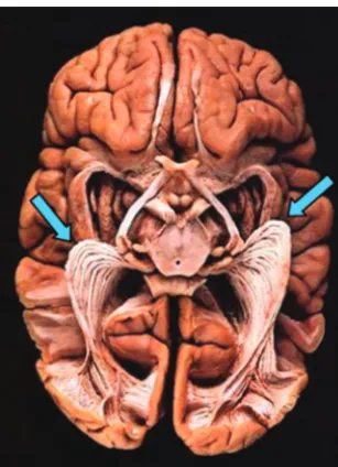
The OR consist of 4 white matter fiber bundles. The anterior bundle or the Meyer loop (Meyer 1907)4projects from the
LGB and runs anteriorly across the superior aspect of the an-terior tip of the ipsilateral temporal horn before making a sharp turn to pass posteriorly along the wall of the lateral ven-tricle to converge on the lower lip of the calcarine fissure.5,6
The central bundle leaves the LGB in a lateral direction and follows posteriorly along the lateral ventricular wall to the vi-sual cortex. The dorsal bundle extends directly posterior to meet the upper part of the calcarine cortex (Fig 1).
Surgical Studies
The boundary of the anterior fibers of the Meyer loop and its relationship to the TP has been controversial. Older studies used intraoperative estimates of resection size or brain dissec-tion (Table). There was no consistency among the reported locations, which varied from 30 to 45 mm posterior to the TP.7-9 In 1954, Penfield10 stated that resections extending
⬍6 cm posterior to the tip of the TP were “not likely” to result in VFD. These studies relied on the surgeon to subjectively estimate the resection size and the anterior extent of the Meyer loop in the absence of neuroimaging. The resection sizes were probably overestimated with underestimation of the Meyer loop because this would not be visualized surgically. This in-accuracy could explain why more recent studies by using dif-ferent methodologies show smaller measurements. In addi-tion, VFDs have not been assessed in a consistent manner. Older studies relied on the patient verbally reporting the def-icit, which is very unreliable. The incidence of VFDs seems to be increasing because of the increased sensitivity of automated perimetry for small defects compared with kinetic perimetry.2
From the Florey Neuroscience and Brain Research Institutes, Melbourne Brain Centre, Heidelberg, Victoria, Australia; and Department of Medical Imaging, Royal Children’s Hospital and University of Melbourne, Parkville, Victoria, Australia.
Please address correspondence to Simone A. Mandelstam, MD, Department of Medical Imaging, 2nd Floor, Royal Children’s Hospital, Flemington Rd, Parkville 3052, Victoria, Australia; e-mail: [email protected]
Anatomic Dissection Studies
The more recent group of anatomic studies uses the Klingler fiber dissection technique in cadaveric brains of previously healthy patients to look at the anatomy of the Meyer loop. This technique involves fixing a cadaveric brain in formalin, freez-ing it, and thawfreez-ing it before dissection. The freezfreez-ing and thaw-ing of the cadaveric brain allow ice crystals to form between myelinated nerve fibers, thereby separating them and facilitat-ing dissection (Fig 2).5,6,11Ebeling and Reulen (1988)11
dis-sected 25 brains and reported that the distance between the tip of the temporal lobe and the anterior edge of the Meyer loop is 27⫾3.5 mm with a range of 22–37 mm. They stated that the average location of the anterior edge of the Meyer loop is 5⫾ 3.9 mm anterior to the temporal horn tip. These studies also demonstrated differing distances from the OR to the TP (Fig 3). Choi et al5found an average of 31.4 mm, and Rubino et al6
found an average of 25 mm, which is similar to the measure-ments of Ebeling and Reulen. Kier et al12combined dissection
and MR imaging, a method they termed “anatomic dissection tractography,” and described the anterior extent of the Meyer loop to be at the level of the amygdala and not reaching the level of the temporal horn tip. However, Sincoff et al,13in a dissection
study, reported that the anterior bundle of the OR did cover the anterior tip of the lateral aspect of the temporal horn.
What becomes apparent looking at the different and con-flicting results of these studies is that there is great variability, which makes it difficult to give a generic recommendation to the neurosurgeon on the “safe” length of anterior temporal lobe that can be resected without causing a VFD. Potential sources of bias include relatively small sample sizes, interob-server and intersurgical variability, and different methodolo-gies. Dissection can only be used ex vivo and does not reliably
Fig 1.Schematic drawing of the human visual system.A, Right- and left-hemispheric fiber pathways.B, OR comprising the anterior bundle, the Meyer loop (orange), the central bundle (green), and the dorsal bundle (turquoise).
Distance between the anterior boundary of Meyer loop and the temporal pole tip as reported using differing techniques, 1954 –2010
Author/Year
Distance from Anterior Edge of the Meyer
Loop to Temporal Pole Tip (mm) Measurement Technique
Penfield 195410 60 Surgical resection, VFD
Bjork and Kugelberg 19577 30 Surgical resection, VFD, 96% partial Goldmann perimetry
Falconer and Wilson 19588 45 Surgical resection, VFD, 64% complete and 36%
incomplete Bjerrum
Marino and Rasmussen 19689 45 Surgical resection, VFD, 14% complete and 52%
incomplete Aimark and Tangent
Ebeling and Reulen 198811* 27 (22–37) Cadaver dissection (Klingler technique), 50 hemispheres
Krolak-Salmon et al 200021 20–31.3 MR imaging and automated static perimetry, 28% mild
28% moderate
Kier et al 200412 Anterior fibers do not reach temporal horn tip “Anatomic dissection tractography”
Rubino et al 20056 25 (22–30) Cadaver dissection (Klingler technique), 40 hemispheres
Barton et al 200525 24 (18–36) MR imaging and automated Goldmann perimetry, 100%
had VFD
Yamamoto et al 200518 37.3⫾2.5 DT-FT
Choi et al 20065 31.4 (28–34) Cadaver dissection (Klingler technique), 10 hemispheres
Nilsson et al 200714 44 (34–51) DT-FT
Taoka et al 200826 36.6 (30–43.2) DT-FT
Chen et al 200927 32.1 (20.9–51.5) DT-FT
Yogarajah et al 200932 Patients 24–43, controls 24–47 DT-FT
Wang et al 201028 36 (mean) DT-FT
* Criterion standard study.
REVIEW
[image:2.594.62.533.297.525.2]separate 1 fiber system from another. In fact, dissection of 1 white matter tract may lead to destruction of other tracts. For-malin shrinks brain tissue, which could influence measure-ments of anatomic distances, while the water freezing tech-nique has been found to cause fissuring of the specimen.14 Despite all these differences, there is likely an underlying fun-damental issue, which is the intersubject variability of the OR. There may even be significant hemispheric asymmetries within each individual, postulated to be due to expanded lan-guage areas in the left posterior temporal lobe displacing the OR on that side.15,16
Modern Imaging
The next part of this review will focus on the anatomy of the Meyer loop as demonstrated by modern imaging techniques. Unfortunately conventional MR imaging sequences cannot differentiate different white matter tracts. However, an MR imaging⫺based method for measuring the course of white matter tracts has been developed. This is known as DTI. Among the analyzing methods for DTI, DT-FT has been re-ported as robust for visualizing and evaluating white matter fiber direction and connectivity in the brain.17-19
Overview of DTI
DTI is an MR imaging technique that can be used to charac-terize the directional properties of the diffusion of water mol-ecules. It is based on the principle that diffusion is directed by anatomic microstructures (ie, white matter fibers).17-19
DT-FT offers the only noninvasive method for measuring the course of white matter tracts in vivo; however, DT-FT estimates have ongoing difficulty identifying the OR, espe-cially the Meyer loop.18
DTI indirectly evaluates the integrity of white matter by measuring water diffusion and directionality in 3D. From in-formation about direction of diffusion with a minimum of 6 different nonparallel directions, one can calculate the diffu-sion tensor and derive FA from this. FA values range from zero
(maximal isotropic diffusion) to 1 (maximal anisotropic dif-fusion). Major, medium, and minor eigenvalues specify rates of diffusivity along each of 3 orthogonal axes of a diffusion ellipsoid.17The direction of the largest eigenvector is the di-rection of greatest diffusivity and is assumed to align with the direction of fiber bundles. This is important in regions with densely packed axons such as white matter. Diffusion charac-teristics of a tissue provide information on its structural properties.
Data may be analyzed with a region-of-interest or voxel-based approach. For the voxel-voxel-based approach, all brains need to be normalized in common space. Although this is a statis-tically rigorous method, there may geometric distortions, eddy currents, magnetic susceptibility, patient motion, or im-age noise resulting in reduced sensitivity.
Region-of-interest approaches are also limited by user vari-ability because regions are manually outlined. To decrease user variability, the region of interest can be used as a seed point for tractography with a threshold for FA applied and the resulting voxels can be used for calculation.
DT-FT
DT-FT is the only available noninvasive technique that can delineate white matter tracts in vivo. Directional anisotropy information in each voxel provided by DTI is used to generate virtual maps of white matter tracts. Different algorithms de-termine how voxels should be connected according to their anisotropy and direction.17
There has been some confusion about the best technique, with 2 approaches described in the literature.
1. Deterministic: fiber assignment by continuous tracking. From a seed region of interest, tracking follows the largest tensor in each voxel and connects the voxels according to spe-cific thresholds for minimum FA and maximum change of direction between 2 voxels.
2. Probabilistic: fast-marching tractography and probabi-listic index of connectivity. These methods find the energeti-cally most favorable pathway between 2 voxels. The probabil-ity of connectivprobabil-ity between voxels from a seed point is calculated.20
The criterion standard for preoperative detection of the course of the OR is based on the gross dissection technique on frozen formalin-fixed tissue. The study by Ebeling and Re-ulen11has been used as the criterion standard for OR location measurements in 2 DT-FT methods,14,18and the data have
been confirmed by additional dissection5,6and clinical
stud-ies.21Krolak-Salmon et al21reported that 15 of 18 patients
presented with a postoperative visual field loss. They reported 2 cases in which the resection was limited to 20 mm from the TP but still produced a partial quadrantanopia.
The earlier reports of DT-FT of the OR were performed on very few patients.14,18,22,23Yamamoto et al18did basic deter-ministic fiber tracking on 5 volunteers and found difficulty identifying the OR and particularly the anterior extent of the Meyer loop. Powell et al22had similar issues performing fiber
tracking on 2 patients, of whom 1 had an intact Meyer loop and no postoperative VFD and the other had a disrupted Meyer loop with a postoperative VFD. Powell et al used 54 gradients with the assumption that this would improve the fiber tracking. Yamamoto et al in 200719experimented with
[image:3.594.92.246.42.254.2]using 6, 12, 40, and 81 gradients and found that there was no significant effect on visualization of the OR. They concluded that 6 directions are thus sufficient. In fact, this is true for the deterministic approach. No matter how many directions are used, the major eigenvector will be the same, and there is no true indication of the confidence that can be assigned to a particular tracking result. Therefore, the pathway may have no or little correlation with the underlying anatomy. The adop-tion of visualizaadop-tion strategies gives the impression that the viewer is looking at something “real,” and this could lead to serious errors.24This is far more likely to be the case in a region
where the fiber tracts bend and loop such as in the Meyer loop because these methods are inherently unable to take into ac-count crossing fibers.
The extent of brain shift related to surgery could also cause errors in the attempts to estimate the status of fiber tracts according to the resection extent alone.22,25Despite some
suc-cess, DT-FT estimates cause problems identifying the OR and particularly the Meyer loop section.18,22
Both the Nilsson and Yamamoto groups estimated the mean anterior position of the Meyer loop to be at least 1 cm posterior to estimates from dissection studies.14,18For
exam-ple, Nilsson et al14described the temporal horn as being 1.5 cm
anterior to the Meyer loop. They suggested that the discrep-ancy may be due to misidentification of the fibers during section or errors in the dissection estimates of absolute dis-tances. Distance errors are not an adequate explanation because the anatomic descriptions are detailed, with 1 study stating that the “anterior tip of the temporal horn was covered by the anterior optic radiation along its lateral half”13and
another confirming that in all of their specimens, “the anterior edge of the Meyer loop reached the tip of the temporal horn.”6 Taoka et al26looked retrospectively at the data of 14
pa-tients with hippocampal sclerosis who underwent temporal lobectomy. They were divided into 4 groups according to the size of the acquired VFD with group A having no deficit.
Trac-tography of the uncinate fasciculus was used to help recognize the most anterior point of the Meyer loop with the assumption that the loop is immediately behind the uncinate fasciculus. Although they claimed to have obtained tractography of the Meyer loop in “all the cases,” they also stated that there was no differentiating the loop from the uncinate fasciculus in 50% of cases.26Therefore, one would have to question the accuracy of
their Meyer loop reconstructions. Although they reportedly tried to evaluate postsurgical tractography of the OR, in most cases tractography could not be drawn. This problem was the-orized to be due to edema or gliosis within adjacent tissue, which would change the FA and thus lead to termination of the tracking. However, Chen et al27made the valid point that
there is a shift of the fiber tracts intraoperatively in vertical and horizontal planes at the resection site and that this would have to be factored into postoperative calculations.
Despite the limits of streamlined deterministic tracking, it is fairly simple computationally and seems easy to interpret if the viewer is unaware of the pitfalls. There are groups that continue to use this methodology. A study was recently pub-lished on a cohort from Southern China attempting to estab-lish an average local population value for the distance between the anterior tip of the Meyer loop and the TP. Interestingly, the scans were obtained on 16 patients with a variety of neurologic conditions ranging from brain tumors (not in the temporal lobe) to Parkinson disease. The 2 operators doing the analysis were a senior neurosurgeon and a junior radiologist using dif-ferent software programs. The conclusion was that the dis-tance from the Meyer loop to the TP is 36 mm in this popula-tion, and recommendations were given for surgical planning.28
This advice was based on multiple levels of accumulated errors (neurologically abnormal brains, operator and software differ-ences, unvalidated technique, and so forth), and utmost caution would be needed before accepting this as fact.
Hofer et al29used a new diffusion-weighted sequence with
a simple DT-FT algorithm, which is claimed to decrease
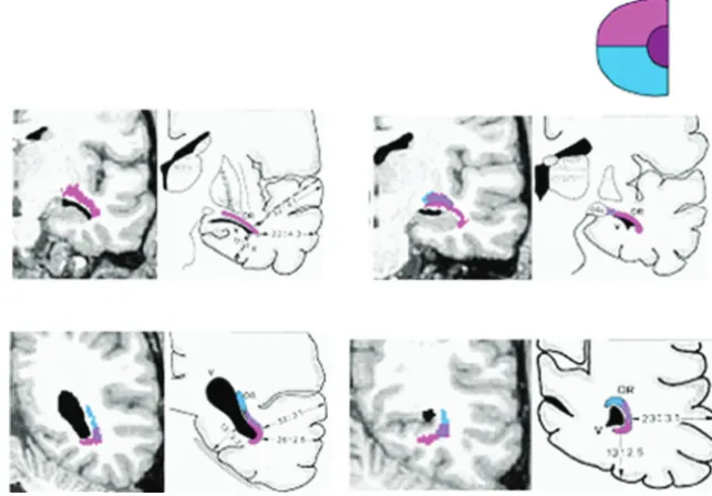
sus-Fig 3.Coronal sections showing the position of the direct, central, and the Meyer loop bundles within the OR. Each panel compares an MR image and estimated fibers (left) with illustrations from a dissection study (Ebeling and Reulen, 198810) (right). The positions of the OR bundles are shown by the color overlays, and the expected visual information carried by these bundles
[image:4.594.131.453.46.270.2]ceptibility artifacts and geometric distortions and improve spatial resolution. However, their resolution of 1.8 mm is still not adequate to avoid some recognized artifacts, including false-positive tracts. They then used a region of interest DT-FT method to generate tracking maps of the visual pathway. They reported that “most” fiber bundles could be fully or partially reconstructed in 5/6 subjects. In 1 subject, they were unable to reconstruct any bundles of the OR, and this was ascribed to the close vicinity and predominance of the inferior longitudinal fasciculus and the fronto-occipital fasciculus.29This result is
similar to the inability of Taoka et al26to separate the OR from
the uncinate fasciculus as discussed above. These studies continue to give an approximation of the pathways, but none can match the criterion standard of the dissection studies (Figs 4 and 5). Yet, the derived anatomy is thought to be increasingly valid with Ka-mada et al23showing, in 2 patients, that real-time perioperative
visual-evoked potentials confirmed the accuracy of tractography.
As the high false-negative rate of the streamlined determin-istic tracking approach with diffusion tensor data became rec-ognized, new algorithms by using probabilistic methods have been developed to attempt to locate valid pathways. Probabi-listic tracking algorithms generate multiple pathways from a given point in space. The end result is a set of multiple path-ways (streamlines) passing through the seed point. Counting how many times each voxel in the volume of interest is inter-sected by a streamline and expressed as a percentage then sum-marizes this information. The algorithm is run repeatedly to build up a pattern of possible paths through the data. High-angular-resolution diffusion imaging, with large numbers of gradient directions, increases this ability to estimate the prob-ability distributions of fiber directions and has been shown to increase the volume of tracts generated by tractography com-pared with methods using fewer gradient directions (Fig 6).30
Probabilistic tracking results give an indication of the pre-cision of the tracking result but say nothing about accuracy. However, inexperienced observers may still misinterpret these maps. There may appear to be a continuous pattern that is completely inaccurate with regard to true anatomy.24The
ac-curacy and precision for curved paths is lower than that for straight paths due to accumulated error, which is a major problem with tracking a structure that has the configuration of the Meyer loop. Sherbondy et al31used a probabilistic tracking
method on 8 volunteers to identify the “most likely” path
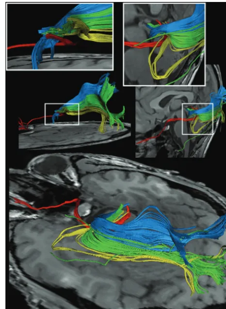
be-Fig 4.Top: Reconstructions of the optic nerves, tracts (red), and OR (yellow). Bottom: Dissection of the OR into the Meyer loop (yellow), the central bundle (green), and the dorsal bundle (blue). The use of 1.8-mm isotropic voxels reduces the voxel size of a more conventional 2.5-mm acquisition by a factor of 2.7. Nevertheless, this resolution is still not good enough to avoid “kissing” effects between the optic nerve and neighboring eye muscles. Reproduced with permission fromFrontiers in Neuroanatomyand Hofer et al.29
[image:5.594.300.533.43.361.2] [image:5.594.90.248.55.458.2]tween the lateral geniculate nucleus and the calcarine sulcus. Validity scores were independently assigned to every pathway connecting the LGN region of interest and the calcarine sulcus region of interest.31The findings were nearer to the criterion standard dissection results of Ebeling and Reulen,11with the
average distance of the most anterior position of Meyer loop measuring 28 mm from the TP.
Probabilistic multifiber tractography applied to diffusion MR imaging data acquired at 3T may be better able to cope with crossing and kissing fibers than deterministic models be-cause it allows many more possible local pathway orientations for each DTI sample point. This means that it is less likely to underestimate the anterior extent of the Meyer loop. Yogara-jah et al32combined retrospective probabilistic fiber tracking
in 20 controls and 21 postsurgical patients with postoperative assessment of VFDs. Nine of 21 patients had VFDs of up to 87% of the contralateral superior quadrant. The distances from the tip of the Meyer loop to the TP can be seen in the Table. Resection size was ascertained by using the Barton method, and Goldmann perimetry was performed by a con-sultant ophthalmologist blinded to the results of tractography and estimates of resection size. Using linear regression analy-sis, they demonstrated that the distance from the tip of the Meyer loop to the TP and the size of resection were significant predictors of the postoperative VFDs. There was a significant association between the anterior extent of the Meyer loop and whether a patient had a severe (⬎50%) quadrantic VFD (P⫽
.01). If the anterior extent of the loop was⬍35 mm from the TP, patients were more likely to have severe VFDs irrespective of the size of the resection.32This finding is in keeping with the
observations of Chen et al27that the change in the size of the
Meyer loop postresection for each subject allowed calculation of the individual preserve ratio for the prediction of the post-operative VFDs. Despite the limitations of the study, which included lack of blinding of the investigators involved in im-age processing and processing in the coronal plane only, these
results are similar to those of the dissection studies and the VFD-based study of Barton et al.25Operator-dependent
fac-tors such as variability in seed voxel placement and the choice of connectivity threshold to select between tract and nontract voxels are potential causes of significant variability. This leads to the tracts being “virtual” representations of underlying white matter.
One of the biggest problems with probabilistic tractogra-phy methods is lack of validation against a histologic standard, causing difficulty in evaluating individual methods and quan-titatively comparing methods.24,30 Clatworthy et al30 have
started to address this by describing a method of OR tractog-raphy using an existing well-validated probabilistic algorithm with the ability to accommodate multiple fiber directions within each voxel. A method of quantitatively assessing the accuracy of tract images is described on the basis of receiver operating characteristic analysis. This compares generated tracts with probabilistic histologic data of the OR in MR im-aging space and provides an estimate of the frequencies of both false-positive and false-negative voxels. Seed voxel placement is automated, removing differences between operators as a source of tract image variability. Threshold selection is objec-tive, based on a quantitative comparison of tract images and histologic reference data in stereotaxic space. Their results are described as showing a good match between the resulting binary tract images and the reference data by using a second group to validate the parameters generated in the first group of subjects. Although once again the sample size is small, this study is a good first step toward addressing the issue of validation.
In general, there are limitations common to all existing tractography methods. False-positive tracts can be generated that are not part of the tract of interest. This is relevant when the anterior projections from the OR may actually reflect the inferior occipitofrontal fibers.20Peripheral voxels to the
cen-tral tract may be represented as having lower probability and may be removed at the thresholding process with inclusion of regions around the central tract, which makes the central tract appear more bulky than it actually is. Voxel values, which rep-resent the probability of tracing a pathway through the diffu-sion, do not relate in a straightforward way to the anatomic strength of the underlying pathways.24Voxel size used in DTI
can be between 1 and 15 mm3, but an axon measures
approx-imately 0.01 mm. Therefore, the signal from 1 voxel represents thousands of axons that may have different directions. Algo-rithms can have difficulty tracking fibers crossing in a voxel.16
Although the probabilities will reflect anatomically meaning-ful features such as axonal packing density and myelination, they will also be influenced by factors such as the length of the pathway (because probabilities will decay with distance) and tract geometry.30
The Future
At this point in time, to my knowledge, there is no single val-idated recommended method of DT-FT in the literature. Early attempts at tracking the OR have demonstrated mixed success, though it seems that newer probabilistic methods may be more representative of the OR than deterministic methods. Further work is needed to improve and validate fiber tracking algorithms and to develop multimodal techniques for assess-ment of brain connectivity.24The anatomic measurements of
[image:6.594.92.246.47.274.2]the anterior border of the Meyer loop have been controversial for half a century, and fiber tracking, with its inherent limita-tions, is not simplifying this issue. The general consensus seems to be that there is great individual variation in this an-atomically complex region.
It is always important to bring the patient back into focus. Tractography studies are showing great promise in the delin-eation of the Meyer loop, and effort should be made to use this information to direct the neurosurgical approach to the indi-vidual patient to avoid a VFD or to appropriately counsel the patient regarding a VFD if deemed surgically unavoidable (Fig 7). New tractography algorithms and methods are being de-vised to deal with the issues of crossing fibers to improve the reliability of the resulting estimates of the fiber orientation density within each voxel.33
Prospective clinical studies by using these methods are needed in which the preoperative tractography results are used by the neurosurgeons in modifying the surgical approach. The major questions are the following:
● How accurate are DT-FT-based studies of the OR?
● Can a criterion standard validated tractography algorithm be developed for standardized assessment of the OR?
● Will tractography of the OR significantly decrease the inci-dence and severity of postoperative VFDs in patients with TLE who undergo ATL?
Acknowledgments
I acknowledge Shawna Farquharson for the processing of Fig-ure 7.
References
1. Jacobson DM. The localizing value of a quadrantanopia. Arch Neurol
1997;54:401– 04
2. Pathak-Ray V, Ray A, Walters R, et al.Detection of visual field defects in pa-tients after anterior temporal lobectomy for mesial temporal sclerosis: estab-lishing eligibility to drive.Eye2002;16:744 – 48
3.Medical standards for licensing and clinical management guidelines: section 23 vision and eye disorders—assessing fitness to drive.Austroads Inc2006; 23.2.3 and 23.3.1:96 –100
4. Meyer A.The connections of the occipital lobes and the present status of the cerebral visual affections.Trans Assoc Am Physicians1907;22:7–15
5. Choi C, Rubino PA, Fernandez-Miranda JC, et al.Meyer’s loop and the optic radiations in the transsylvian approach to the mediobasal temporal lobe.
Neurosurgery2006;59(4 suppl):228 –36
6. Rubino PA, Rhoton AL Jr, Tong X, et al.Three-dimensional relationships of the optic radiation.Neurosurgery2005;57(4 suppl):219 –27
7. Bjork A, Kugelberg E.Visual field defects after temporal lobectomy.Acta Oph-thalmol (Copenh)1957;35:210 –16
8. Falconer MA, Wilson JL.Visual field changes following anterior temporal lobectomy: their significance in relation to Meyer’s loop of the optic radia-tion.Brain1958;81:1–14
9. Marino R Jr, Rasmussen T.Visual field changes after temporal lobectomy in man.Neurology1968;18:825–35
10. Penfield W.Temporal lobe epilepsy.Br J Surg1954;41:337– 43
11. Ebeling U, Reulen H-J.Neurosurgical topography of the optic radiation in the temporal lobe.Acta Neurochir1988;92:29 –36
12. Kier EL, Staib LH, Davis LM, et al.MR imaging of the temporal stem: anatomic dissection tractography of the uncinate fasciculus, inferior occipitofrontal fasciculus, and Meyer’s loop of the optic radiation.AJNR Am J Neuroradiol
2004;25:677–91
13. Sincoff EH, Tan Y, Abdulrauf SI.White matter fiber dissection of the optic radiations of the temporal lobe and implications for surgical approaches to the temporal horn.J Neurosurg2004;101:739 – 46
14. Nilsson D, Starck G, Ljungberg M, et al.Intersubject variability in the anterior extent of the optic radiation assessed by tractography. Epilepsy Res
2007;77:11–16
15. Wang F, Sun T, Li XL, et al.Diffusion tensor tractography of the temporal stem on the inferior limiting sulcus.J Neurosurg2008;108:775– 81
16. Jeelani Nu O, Jindahra P, Tamber MS, et al.‘Hemispherical asymmetry in Meyer’s Loop: a prospective study of visual-field deficits in 105 cases under-going anterior temporal lobe resection for epilepsy.J Neurol Neurosurg Psychi-atry2010;81:985–91. Epub 2010 Jun 27
17. Mori S, van Zijl PC.Fiber tracking: principles and strategies—a technical review.NMR Biomed2002;15:468 – 80
18. Yamamoto T, Yamada K, Nishimura T, et al.Tractography to depict three layers of visual field trajectories to the calcarine gyri.Am J Ophthalmol
2005;140:781– 85
19. Yamamoto A, Miki Y, Urayama S, et al.Diffusion tensor fiber tractography of the optic radiation: analysis with 6-, 12-, 40-, and 81-directional motion-prob-ing gradients—a preliminary study.AJNR Am J Neuroradiol2007;28:92–96 20. Ciccarelli O, Toosy AT, Parker GJ, et al.Diffusion tractography based group
mapping of major white-matter pathways in the human brain.Neuroimage
2003;19:1545–55
21. Krolak-Salmon P, Guenot M, Tiliket C, et al.Anatomy of optic nerve radiations as assessed by static perimetry and MRI after tailored temporal lobectomy.
Br J Ophthalmol2000;84:884 – 89
22. Powell HW, Parker GJ, Alexander DC, et al.MR tractography predicts visual field defects following temporal lobe resection.Neurology2005;65:596 –99 23. Kamada K, Todo T, Morita A, et al.Functional monitoring for visual pathway
using real-time visual evoked potentials and optic-radiation tractography.
Neurosurgery2005;57(1 suppl):121–27
24. Jones DK.Challenges and limitations of quantifying brain connectivity in vivo with diffusion MRI.Imaging in Medicine2010;2:341–55
25. Barton JJ, Hefter R, Chang B, et al.The field defects of anterior temporal lobectomy: a quantitative reassessment of Meyer’s loop. Brain 2005; 128:2123–33
26. Taoka T, Sakamoto M, Nakagawa H, et al.Diffusion tensor tractography of the Meyer loop in cases of temporal lobe resection for temporal lobe epilepsy: correlation between postsurgical visual field defect and anterior limit of Mey-er’s loop on tractography.AJNR Am J Neuroradiol2008;29:1329 –34 27. Chen X, Weigel D, Ganslandt O, et al.Prediction of visual field deficits by
diffusion tensor imaging in temporal lobe epilepsy surgery.Neuroimage
2009;45:286 –97
28. Wang YX, Zhu XL, Deng M, et al.The use of diffusion tensor tractography to measure the distance between the anterior tip of Meyer’s loop and the tempo-ral pole in a cohort from Southern China.J Neurosurg2010;113:1144 –51. Epub 2010 Aug 20
29. Hofer S, Karaus A, Frahm J.Reconstruction and dissection of the entire human visual pathway using diffusion tensor MRI.Front Neuroanat2010;4:15 30. Clatworthy PL, Williams GB, Acosta-Cabronero J, et al.Probabilistic
tractog-raphy of the optic radiations: an automated method and anatomical valida-tion.Neuroimage2010;49:2001–12
31. Sherbondy AJ, Dougherty RF, Napel S, et al.Identifying the human optic radi-ation using diffusion imaging and fiber tractography.J Vis2008;8:1–11 32. Yogarajah M, Focke NK, Bonelli S, et al.Defining Meyer’s loop-temporal lobe
resections, visual field deficits and diffusion tensor tractography.Brain
2009;132:1656 – 68
33. Tournier J-D, Calamante F, Connelly A.Robust determination of the fiber orientation distribution in diffusion MRI: non-negativity constrained super-resolved spherical deconvolution.Neuroimage2007;35:1459 –72
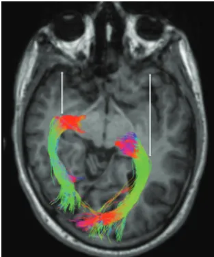
Fig 7.Reconstructed OR by using the probabilistic method of CSD32in a 17-year-old male
[image:7.594.91.248.44.232.2]