• No results found

Motion Compensation Techniques in Neonatal and Fetal MR Imaging

N/A
N/A
Protected

Academic year: 2020

Share "Motion Compensation Techniques in Neonatal and Fetal MR Imaging"

Copied!
13
0
0

Loading.... (view fulltext now)

Full text

Figure

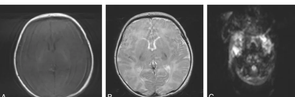
FIG 1. T1-weighted (A), T2-weighted (B), and diffusion-weighted (C) axial MR brain images of a 5-day-old full-term neonate without motionartifacts acquired at 1.5T.
FIG 3. T1-weighted gradient-echo axial brain images at 1.5T (TR, 142 ms; TE, 6 ms; section thickness, 4 mm; scanning time, 16 seconds) of aBreathing artifacts appear in the form of high-signal ghosts in the operator-selected phase-encoding direction and severely degrade image28-week-old fetus without motion artifacts after a successful breath-hold (A) and with motion artifacts after an incomplete breath-hold (B).quality.
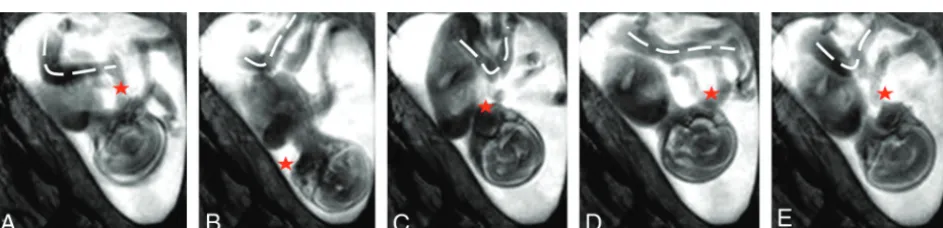
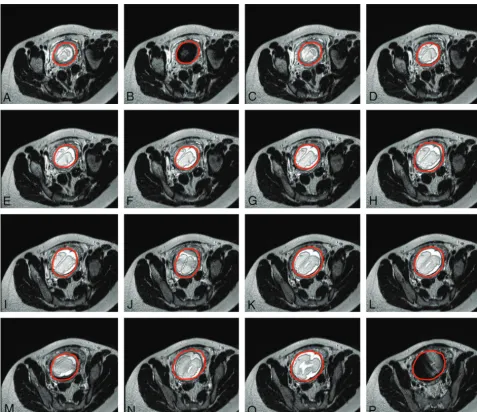
FIG 5. Successive axially planned sections of a single-shot fast-spin-echo acquisition at 1.5T (TR, 1000 ms; TE, 127 ms; section thickness, 4 mm;(plane, fetal movement resulted in plane transposition in the produced images (scanning duration, 26 seconds) o
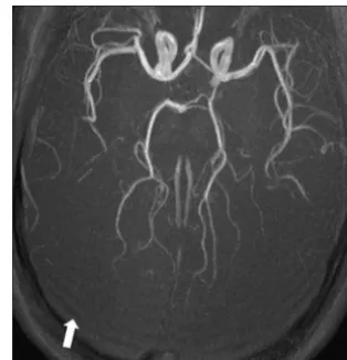
FIG 6. Ringing artifacts (ing artifacts occur due to data undersampling, and should not bearrow) at the back of the brain of a 4-week-old full-term neonate on an axial maximum intensity projection of anoptimized neonatal MR angiography protocol79 acquired
+5

References

Related documents