ORIGINAL RESEARCH
Treatment of Intracranial Aneurysms by
Functional Reconstruction of the Parent Artery:
The Budapest Experience with the Pipeline
Embolization Device
I. Szikora Z. Berentei Z. Kulcsar M. Marosfoi Z.S. Vajda W. Lee A. Berez P.K. Nelson
BACKGROUND AND PURPOSE:Aneurysm treatment by intrasaccular packing has been associated with a relatively high rate of recurrence. The use of mesh tubes has recently gained traction as an alternative therapy. This article summarizes the midterm results of using an endoluminal sleeve, the PED, in the treatment of aneurysms.
MATERIALS AND METHODS: A total of 19 wide-neck aneurysms were treated in 18 patients: 10 by implantation of PEDs alone and 9 by a combination of PED and coils. Angiographic and clinical results were recorded immediately and at 6 months following treatment.
RESULTS: Immediate angiographic occlusion was achieved in 4 and flow reduction, in another 15 aneurysms. Angiography at 6 months demonstrated complete occlusion in 17 and partial filling in 1 of 18 patients. There was no difference between coil-packed and unpacked aneurysms. Of 28 side branches covered byⱖ1 device, the ophthalmic artery was absent immediately in 1 and at 6 months in another 2 cases. One patient experienced abrupt in-stent thrombosis resulting in a transient neurologic deficit, and 1 patient died due to rupture of a coexisting aneurysm. All giant aneurysms treated with PED alone were demonstrated by follow-up cross-sectional imaging to have involuted by 6 months.
CONCLUSIONS:Treatment of large, wide-neck, or otherwise untreatable aneurysms with functional reconstruction of the parent artery may be achieved with relative safety using dedicated flow-modifying devices with or without adjunctive use of intrasaccular coil packing.
ABBREVIATIONS:AICA⫽anterior inferior cerebellar artery; BA⫽basilar artery; Immed⫽ Imme-diate; DSA⫽digital subtraction angiography; ICA⫽internal carotid artery; ID⫽internal diameter; MRA⫽MR angiography; N/A⫽not applicable; PcomA⫽posterior communicating artery; PED⫽ Pipeline Embolization Device; PITA⫽Pipeline Embolization Device for Intracranial Treatment of Aneurysms; Recan⫽recanalization; SAH⫽subarachnoid hemorrhage; SCA⫽superior cerebellar artery; Sup⫽superior
T
he application of mesh tube implants for the treatment of intracranial aneurysms was first proposed in the early 1990s.1-3Early experimental work suggested 3 potential mech-anisms of action: improved anatomic reconstruction of the parent artery, functional modification of flow across the ori-fice of the aneurysm, and biologic repair of the aneurysm neck by neointimal overgrowth.1Flow conditions within the parent artery and the aneurysm itself have long been thought to play an important role in aneurysm growth and rupture.4-11Yet,the early application of stent-assisted aneurysm therapy was primarily focused on geometric reconstruction of the parent artery through improved coil packing of the aneurysm. A few clinical case reports, however, suggested that improved out-comes associated with the use of stents may be attributable to stent-induced modification of flow within and around the an-eurysm,12,13providing the impetus for development of higher coverage endoluminal sleeves for the treatment of cerebral an-eurysms.14The purpose of this study was to analyze the early clinical experience with the first dedicated flow-modifying de-vice, the PED (Chestnut Medical, Menlo Park, California).
Materials and Methods
Indications
Between February 2007 and July 2008, a total of 19 aneurysms in 18 patients were treated at the National Institute of Neurosurgery, Budapest. Nine patients were treated in a multicenter international safety trial (the PITA study) enrolling a total of 31 patients. Eight patients were treated in a single-center trial (the PITA II Budapest study), enrolling 8 patients total. These later patients were not in-cluded in the PITA trial. Both studies were conducted under an ethics Received September 25, 2009; accepted after revision November 16.
From the Department of Neuroradiology (I.S., Z.B., Z.K., M.M., Z.S.V., W.L.), National Institute of Neurosurgery, Budapest, Hungary; Department of Neuroradiology (P.K.N.), New York University Medical Center, New York, New York; and Chestnut Medical Technologies Inc (A.B.), Menlo Park, California.
This work was partially supported by a grant from the Hungarian National Scientific and Research Fund (OTKA 73773).
Please address correspondence to Istvan Szikora, MD, PhD, National Institute of Neuro-surgery, Neuroradiology, Amerikai ut 57, Budapest, 1145, Hungary; e-mail: [email protected]
Indicates open access to non-subscribers at www.ajnr.org
Indicates article with supplemental on-line table.
DOI 10.3174/ajnr.A2023
INTERVENTIONAL
ORIGINAL
committee⫺approved protocol. One additional patient was treated following European conformity mark approval of the device.
Two aneurysms were treated as incidental findings; 10 because of headaches; 6 due to mass effect; and 1 due to recanalization following previous endovascular treatment. Regarding aneurysm morphology, sidewall and fusiform aneurysms were treated; we considered these to have a high likelihood of failure and/or recurrence with conventional endovascular techniques either because of a large or giant aneurysm or an unfavorable neck-to-sack ratio.
Periprocedural Angiographic Evaluation of the Aneurysms
Morphologic characteristics of the aneurysms and parent arteries, such as diameter and volume, were measured on 3D reconstructed images derived from rotational angiography before the procedure. All measurements were performed on an Advantage Windows Worksta-tion by using ADW 4.2 software (GE Healthcare, Milwaukee, Wiscon-sin). Flow modification after PED implantation was classified as ei-ther complete stasis (if no contrast material entered the aneurysm following deployment of PED), significant flow reduction (if contrast stagnation was seen within the aneurysm at the late venous phase of the angiographic series), or slow flow (if the contrast circulation within the aneurysm became slower but without contrast stagnation into late venous phase images). Aneurysm occlusion was rated as either complete (if no contrast material entered the aneurysm by completion of the procedure) or incomplete (if any contrast filling of the aneurysm was seen on the last angiogram).
Operative Techniques
The Device.The PED is a woven wire mesh tube made of 25% platinum and 75% cobalt-nickel alloy. It is designed to provide 30%– 35% metal coverage of the inner surface of the target vessel with a pore size of 0.02– 0.05 mm2at a nominal diameter. The PED is attached to a pusher wire, compatible with a 3F (0.027-inch ID) microcatheter. The pusher wire has a platinum coil tip, which extends 15 mm beyond the distal edge of the PED. The device is delivered via a 0.027-inch ID microcatheter, requiring a 6F guide catheter support. Once posi-tioned across the aneurysmal segment, the delivery wire is held while the distal one-third to one-half of the PED is carefully unsheathed. Once the unsheathed segment begins to expand, its distal end is re-leased from the delivery wire by clockwise rotation of the wire. The proximal segment of the PED is then deployed by gentle advancement of the wire. Once the PED is fully deployed, it may be recrossed by advancing the microcatheter over the indwelling delivery wire, which can then be exchanged for a second PED. This provides a safe possi-bility of deploying multiple PEDs through the same microcatheter if necessary. At any point up to final deployment, the PED may be captured and removed from the body.
Procedural Protocol.In each case, a 3F neurovascular microcath-eter (Renegade High Flow, Boston Scientific, Natick, Massachusetts; or Mass Transit, Cordis [Johnson and Johnson], Miami Lakes, Flor-ida) was placed across the neck of the aneurysm either directly or over an exchange wire that was previously advanced into a distal branch with the help of a smaller microcatheter. The number of PEDs to be deployed was decided on the basis of flow reduction, but multiple coverage of clinically significant side branches (such as the anterior choroidal artery or the SCA and the AICA in the basilar territory) was carefully avoided. Clinically forgiving branches, such as the ophthal-mic artery, were liberally covered by as many PEDs as thought neces-sary to achieve satisfactory flow reduction. Earlier in the series, several aneurysms were treated by PED-supported coil packing of the
aneu-rysm. For those, a dual-marker microcatheter (Excelsior SL 10, Bos-ton Scientific) was placed within the aneurysm before PED implan-tation to secure the possibility of additional coil placement into the sac in case flow modification was considered unsatisfactory. With growing experience, we abandoned secondary coiling except in cases in which early aneurysm occlusion of an intradural aneurysm was desirable.
Periprocedural Medication.Each patient was given 75 mg of clo-pidogrel and 100 mg of aspirin per day for at least 48 hours before and 6 weeks following the procedure. Aspirin, 100 mg/day, is still contin-ued. Patients were anticoagulated by an intravenous bolus of sodium-heparin to achieve a double the normal activated clotting time value. Heparin anticoagulation was discontinued by the end of procedure.
Follow-Up
Patients were clinically followed at 30 days, 6 months, and 1.5–2 years after the procedure. Angiographic follow-up was performed routinely at 6 months. Clinical follow-up examinations were performed by the authors during office visits. Ophthalmologic symptoms were evalu-ated by an independent ophthalmologist. Angiographic results were evaluated by using both hard copy films and DICOM files by an in-dependent core lab for patients treated in the clinical studies and by the authors for the first patient treated outside the studies (patient 18, On-line Table and Tables 1 and 2). Cross-sectional imaging follow-up by either CT or MR imaging was performed at 6 months for aneu-rysms presenting with mass effect. Eleven patients (12 aneuaneu-rysms) had late imaging follow-up by MRA between 1.5 and 2 years postembolization.
Results
Aneurysm Location and Morphologic Data
Eighteen aneurysms were located on the intracranial ICA and 1, on the basilar trunk (BA). Four aneurysms were giant (ⱖ25 mm), 10 were large (10 –25 mm), and 5 were small (⬍10 mm) according to the Yasargil classification with a mean largest diameter of 16 mm. The mean aneurysm volume was 1.26 cm3, ranging from 0.047 to 3.969 cm3. All aneurysms had wide necks, ranging from 4 to 18 mm, with a mean neck size of 7.4 mm (On-line Table and Tables 1 and 2).
Procedural Techniques
Angiographic Results
Flow Modification and Aneurysm Occlusion.After
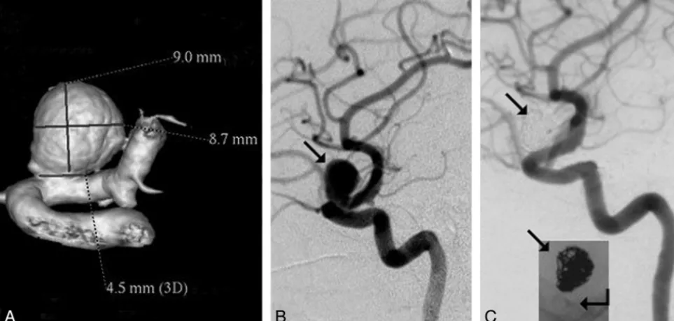
place-ment of the PED construct, angiographic evaluation demon-strated complete stasis in 3 aneurysms, significant flow reduc-tion in 13 (Fig 1A⫺C), and slow flow in 3. Complete aneurysm occlusion was found in 3 cases of PED-supported coil packing and in 1 case following flow modification alone.
Periprocedural Complications
Thirty-nine of 41 PEDs were successfully deployed in the de-sired position. Two PEDs were introduced into the microcath-eter; however, they could not be deployed due to friction in a highly tortuous ICA. In addition, a gentle balloon dilation (Hyperform Balloon; ev3, Irvine, California) was needed to
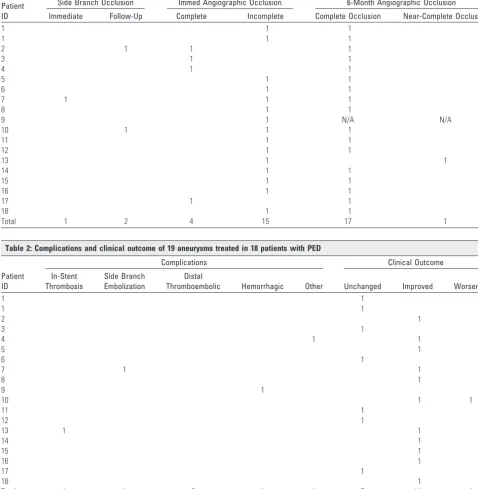
[image:3.594.53.532.63.552.2]fully open the distal section of 1 device. Clinical complications occurred in 4 cases. One patient had a mild postprocedural hemiparesis lasting for 2 days. The patient fully recovered, and no ischemic changes were seen on CT. This event was primar-ily considered the result of contrast overload due to difficulties of catheter navigation and a lengthy procedure, but a CT-negative thromboembolic complication could not be ex-cluded. One patient had an embolic occlusion of a retinal ar-tery branch, resulting in a small visual field deficit following PED-supported coil packing of her complex paraophthalmic aneurysm. One patient who we subsequently learned had not taken her antiplatelet medication experienced acute intrapro-cedural in-stent thrombosis within the ICA. It was initially recanalized with tirofiban (Aggrastat) but re-occluded 2 days Table 1: Immediate and 6 months angiographic results of 19 aneurysms treated with PED
Patient ID
Side Branch Occlusion Immed Angiographic Occlusion 6-Month Angiographic Occlusion
Immediate Follow-Up Complete Incomplete Complete Occlusion Near-Complete Occlusion
1 1 1
1 1 1
2 1 1 1
3 1 1
4 1 1
5 1 1
6 1 1
7 1 1 1
8 1 1
9 1 N/A N/A
10 1 1 1
11 1 1
12 1 1
13 1 1
14 1 1
15 1 1
16 1 1
17 1 1
18 1 1
[image:3.594.51.530.307.553.2]Total 1 2 4 15 17 1
Table 2: Complications and clinical outcome of 19 aneurysms treated in 18 patients with PED
Patient ID
Complications Clinical Outcome
In-Stent Thrombosis
Side Branch Embolization
Distal
Thromboembolic Hemorrhagic Other Unchanged Improved Worsened
1 1
1 1
2 1
3 1
4 1 1
5 1
6 1
7 1 1
8 1
9 1
10 1 1
11 1
12 1
13 1 1
14 1
15 1
16 1
17 1
18 1
later, despite being treated with double antiplatelets. The pa-tient had a transient hemiparesis. Having good collateral cir-culation, she fully recovered and the ICA later recanalized (Fig 2).
One patient died secondary to a diffuse SAH within 5 hours following treatment. Repeat angiography failed to demon-strate the source of bleeding; however, autopsy suggested rup-ture of a small coexisting ICA bifurcation aneurysm distal to the target lesion. Given the patient’s tortuous anatomy and difficult catheter navigation, this could have resulted from a guidewire perforation during either the exchange manipula-tion or PED deployment. Small angiographically occult leak-age might have led to a significant collection of blood within the subarachnoid space until it became symptomatic. Alto-gether, permanent nonrestricting morbidity occurred in 1, and mortality, in another patient.
Normal Branches Arising from the Parent Artery in Which the PED Would Be Deployed
A total of 28 visible side branches were covered, having an estimated diameter of 0.5–1 mm, including the ophthalmic artery in 17, the PcomA in 5, the anterior choroidal artery in 4, and the AICA and the posterior inferior cerebellar artery in 1 patient each. The ophthalmic artery was covered by 2 PEDs in 6, and by 3 and 4 in 1 case each. The PcomA was covered by 2 devices in 1 patient. The remaining branches had single
cov-erage only. One ophthalmic artery was immediately nonvisu-alized on posttreatment angiography and resulted in a retinal branch occlusion. Two ophthalmic arteries, each covered by 3 or 4 devices, were found occluded at the 6-month follow-up. These delayed occlusions were clinically silent. No additional side branch occlusions were detected, including branches of the basilar trunk (Fig 3).
Imaging Follow-Up
Eighteen of the 19 aneurysms were studied by conventional angiography at 6 months following treatment (1 patient died). All but 1 aneurysm was completely occluded, without evi-dence of neck remnant, and exhibited smooth remodeling of the parent artery (Fig 1A⫺D). In the patient with in-stent occlusion of the ICA, delayed recanalization of the parent ar-tery was associated with partial residual filling of a bilobed giant aneurysm (Fig 2). Additional coil packing did not seem to make a difference in late aneurysm occlusion (9/9 loosely coiled and 8/9 uncoiled aneurysms thrombosed completely, despite the generally larger sizes of aneurysms in the uncoiled cohort; 1.6 cm3versus 0.9 cm3by volume) (On-line Table and Tables 1 and 2, Figs 4 and 5).
Large and giant aneurysms treated exclusively with PEDs were demonstrated by cross-sectional imaging (CT or MR im-aging) to have involuted, with resolution of mass effect in all Fig 1.Giant cavernous carotid aneurysm, with the patient presenting with third and sixth nerve palsy due to mass effect (patient 16, On-line Table and Tables 1 and 2), treated with flow modification.
[image:4.594.53.532.40.412.2]patients at 6-month follow-up, except the patient with resid-ual filling of her giant ICA aneurysm (Figs 1 and 3).
Clinical Follow-Up
Seven patients remained clinically unchanged. Six patients with compression syndromes have either completely or sig-nificantly improved. Five of 10 patients presenting with head-aches reported relief of their symptoms. One patient died
(On-line Table and Tables 1 and 2). There was no relationship between the immediate angiographic findings and clinical outcome (On-line Table and Tables 1 and 2).
Late Imaging Follow-Up
[image:5.594.55.534.42.551.2]occlusion. All parent vessels were patent proximal and distal to the aneurysm, and no aneurysm recurrence was seen by MRA. The treated segment of the parent artery could not be visual-ized due to the limitations of the technique.
Discussion
The potential of intravascular stent technology in the treat-ment of aneurysms has been proposed, and its technical feasi-bility, assessed in animal experiments in the early 1990s.1-3It was hypothesized that stent implantation may impact the re-sults of endovascular treatment by facilitating attenuated
coil-packing, modifying local flow dynamics, and providing a framework for the proliferating intima to cover the aneurysm.1
Further laboratory experiments demonstrated that de-pending on their mesh attenuation, stents alter and reduce the velocities of vortical flow within the aneurysm, increasing the intra-aneurysmal circulation time and affecting momentum exchange between the sac and the parent vessel.15It was also shown that in addition to the porosity, the geometry of the stent has an impact on aneurysm thrombosis, depending on the local flow patterns.16,17
Fig 3.Giant dissecting aneurysm of the basilar trunk in a 16-year-old boy presenting with mass effect and sudden onset of hemiparesis (On-line Table and Tables 1 and 2, patient 18.).A, T1-weighted sagittal MR image demonstrates a basilar trunk aneurysm with mass effect (arrow).
[image:6.594.54.536.42.593.2]For clinical practice, following the application of modified balloon-expandable coronary stents,18open cell self-expand-able intracranial stents became availself-expand-able for the morphologic reconstruction of the parent vessel across the orifice of the aneurysm.12,13,18-24 More recently, closed cell-design stents were added to the therapeutic arsenal, presumably providing better coverage of the aneurysm neck.25-27Even though most clinical reports published to date have mainly focused on the utility of stents in facilitating coil packing of complex neck aneurysms, several observations suggested that stents not only promote improved packing but may independently contrib-ute to the long-term stability of the aneurysm even with lower packing attenuation.13,18This assertion is supported by re-ports of aneurysms found to thrombose spontaneously after stent placement alone12,13or following double-stent coverage of the orifice without coil packing,18further highlighting the
significance of stent design as proposed by Lieber et al15,16and Barath et al17in earlier experimental studies.
The above results allowed a conceptual change in the ap-plication of stent technology in aneurysm treatment. Instead of using stents as an adjunctive device, one can apply the in-traluminar sleeves primarily for functional anatomic recon-struction of the parent artery by altering flow dynamics and inducing intimal proliferation. Early clinical experiences with 2 such devices, the Silk stent (Balt Extrusion, Montmorency, France)28and the PED,29-31have recently been reported. Nine of the 18 cases reported here are part of the first clinical trial designed to study the safety and efficacy of a flow-diverter device, the PED (the PITA trial).
Because our intention was to find an effective treatment technique for aneurysms otherwise difficult to treat, large, gi-ant, fusiform, or wide-neck (as defined by a neck⬎4 mm or a Fig 4.Results of aneurysm treatment by a combination of flow modification and additional coil packing.AandB, Nine-millimeter-diameter paraophthalmic aneurysm (arrow,B) treated with flow modification and coil packing (patient 3, On-line Table and Tables 1 and 2).C, Six-month follow-up angiogram demonstrates complete occlusion of the aneurysm with reconstruction of the parent artery. Arrows demonstrate subtraction artifacts and nonsubtracted view of the coil mass (23.3% volumetric packing attenuation); broken arrow demonstrates a single PED within the ICA.
Fig 5.Results of aneurysm treatment with flow modification only.AandB, Aneurysm with size and location similar to the one on Fig 4 (10-mm diameter paraophthalmic ICA aneurysm;
[image:7.594.54.536.44.274.2] [image:7.594.57.535.329.499.2]neck-to-sac ratio⬍1:2) lesions were treated exclusively. Over-all, the aneurysms were larger compared with those in most endovascular series, having a mean diameter of 16 mm and a mean volume of 1.26 cm3. The PED is a single layer of woven wire mesh design with a pore size of 0.020 – 0.052 mm2.. Its flexibility and conformability to the implanted parent arteries were satisfactory. All devices were successfully navigated into position with moderate effort by using standard microcath-eters. Two of 41 PEDs could not be deployed due to friction and were easily removed together with the microcatheter. In-troduction and gentle inflation of a microballoon was neces-sary to release and fully open the distal tip of 1 PED that failed to detach from its delivery wire. This, too, was related to fric-tion due to proximal tortuosity. Thirty-nine devices were ac-ceptably deployed across the target lesion and apposed to the parent artery wall.
The exact degree of flow reduction sufficient for aneurysm thrombosis and its importance in achieving ultimate aneu-rysm occlusion are unclear and are likely to remain subjective in each case until flow and thrombosis simulations become incorporated into routine clinical practice. The aim is to pro-long circulation time (contrast residence) within the aneu-rysm. Intra-aneurysmal circulation (vorticity) has been shown to be directly related to the surface area of the orifice32-34and can be reduced by decreasing the free surface available for fluid exchange. PED provides approximately 30%–35% surface coverage at a nominal opening as a single layer, but its design allows the overlapping of multiple devices, which can be used to strategically increase the degree of aneurysm coverage, pro-viding 2 options for significantly reducing intra-aneurysmal circulation: the primary use of multiple coaxial PEDs or (par-ticularly where perforator branches restrict the number of PEDs that can safely be used) reducing intra-aneurysmal flow with adjunctive coiling of the aneurysm.10In this series, we used both approaches.
In very large and giant aneurysms, in which it is difficult to achieve a significant effect on flow with coils, we elected to use multiple PED coverage. Aneurysms covered byⱖ3 devices were practically twice as large as those treated with single or double PED coverage (On-line Table and Tables 1 and 2). To avoid occlusion of important side branches, we first used a single long bridging PED to create a stable scaffold, followed by shorter devices placed strategically over the aneurysm neck to enhance coverage of the orifice but minimize that of the parent artery proximal and distal to the aneurysm.
For smaller aneurysms, at the beginning, additional coiling was performed if complete stasis to the subjective satisfaction of the operator was not achieved. Technically, coil packing following PED coverage of the neck was very easy. With grow-ing experience, we learned that complete stasis is not required for ultimate aneurysm occlusion; if the flow is visibly reduced by angiography, the aneurysm is likely to thrombose, and an-eurysm packing is discontinued. Applying both techniques provided an opportunity for retrospective comparison of an-eurysms that had additional coiling with those that were treated with PED coverage alone. Analysis of the 6-month fol-low-up results demonstrated that, in general, the coil-packing density was very low. Further, even though unpacked aneu-rysms had significantly larger volume than those that were adjunctively coiled (On-line Table and Tables 1 and 2),
aneu-rysms of similar size and location occluded equally, regardless of coil packing (On-line Table and Tables 1 and 2, Figs 4 and 5). On the other hand, all but 1 coiled aneurysm was treated with a single PED, while unpacked aneurysms were covered by multiple overlapping devices. We concluded that for many aneurysms, PED coverage itself is an effective treatment, and for the future, we restricted additional coil packing to cases in which there is an increased risk of aneurysm rupture (such as multiple aneurysms) or the need to restrict PED coverage to a single device due to the vicinity of significant side branches.
Patency of side branches is a significant concern for the application of multilayer high-mesh-attenuation stents. In our study, 1 acute occlusion was attributed to severe catheter-induced ICA vasospasm rather than stent coverage. This was supported by the reopening of the ophthalmic artery at 6 months. Both branches that had delayed occlusion were cov-ered by either 2 or 4 overlapping devices. A single-layer PED did not cause any side branch thrombosis. Having this positive experience in the relatively forgiving area of the supraclinoid ICA encouraged us to use the technique of targeted multilayer PED coverage in a large fusiform basilar trunk aneurysm, which was treated by 5 coaxial PEDs. In this case, coverage of the SCA and AICA segments was limited to single anchoring devices, while 3 additional short PEDs were overlapped across the aneurysm neck. No visible branches of the BA occluded, and no clinical signs of perforator-related ischemia were de-tected within 8 months of follow-up (Fig 3).
Another significant concern regarding high-metal-surface-area stents is their potential thrombogenicity, particularly if multiple devices are used. In-stent thrombosis occurred in 1 case in a patient retrospectively found not to have been suit-ably covered with antiplatelet agents. The occluded ICA re-opened later, while the patient was on proper medication (Fig 2). This suggests that the PED has an acceptable thrombotic profile, but rigorous antiplatelet periprocedural medication is necessary.
Similar to the application of other intracranial stents, the necessity of aggressive antiaggregation raises further concerns about potential bleeding complications. In our study, a patient died due to diffuse SAH. This patient had a very small aneu-rysm on the ipsilateral ICA bifurcation distal to her giant mul-tilobulated target aneurysm on the proximal ICA. Control an-giography failed to demonstrate a source of bleeding; however, an autopsy suggested rupture of the small distal aneurysm, likely related to wire perforation. This suggests that patients having an additional aneurysm distal to the target lesion carry higher risk because perforation of the distal aneurysm may occur during the exchange maneuver or PED placement. While such perforation might otherwise remain clinically si-lent, it may become catastrophic under double antiaggrega-tion. Treatment of the distal aneurysm before the target lesion should be considered.
6 months in a single case without any flow restriction. The patient is being followed clinically and by MR imaging and MRA.
Effective treatment of mass effect is difficult with intrasac-cular aneurysm packing. Treatment with flow diversion re-sulted not only in angiographic aneurysm occlusion but also in reduction of mass effect. This result is proved by the elimi-nation or improvement of cranial nerve neuropathies and headaches as well as by involution of the aneurysmal mass and resolution of the mass effect at 6 months by CT or MR imaging (Figs 1 and 3).
The long-term results are yet to be studied. We do not have angiographic results for more than 6 months; however, time-of-flight MRA in 11 patients (12 aneurysms at 18 –24 months) indicated persistent occlusion of the aneurysms and good flow within the parent arteries proximal and distal to the treated segments. Patients have been clinically followed forⱕ12–18 months so far. No further clinical events have been recorded. This article summarizes short-term results of only a small cohort of patients. On the basis of these results, application of this technology seems to be beneficial for patients with large and giant proximal aneurysms, particularly those having mass effect. Further studies are warranted to collect longer term follow-up data and to investigate the potential of the tech-nique for treatment of more distally located aneurysms.
Conclusions
Treatment of large, wide-neck, or otherwise untreatable aneu-rysms with functional reconstruction of the parent artery may be achieved with relative safety by using dedicated flow-mod-ifying devices. Currently available technology using the PED provides high efficacy with lasting effects in proximal intracra-nial sidewall aneurysms. Additional coil packing is not re-quired to achieve aneurysm thrombosis, but it is technically feasible and can be applied if early aneurysm occlusion is de-sired (ie, acute SAH) or when treating aneurysms in which eloquent branch vessels limit the number of PEDs. Dual-agent antiaggregation seems to be essential in avoiding thromboem-bolic complications and needs to be seriously considered in cases of multiple aneurysms. Longer follow-up and larger studies are warranted.
References
1. Szikora I, Guterman LR, Wells KM, et al.Combined use of stents and coils to treat experimental wide-necked carotid aneurysms: preliminary results.
AJNR Am J Neuroradiol1994;15:1091–102
2. Turjman F, Massoud TF, Ji C, et al.Combined stent implantation and endosac-cular coil placement for treatment of experimental wide-necked aneurysms: a feasibility study in swine.AJNR Am J Neuroradiol1994;15:1087–90 3. Massoud TF, Turjman F, Ji C, et al.Endovascular treatment of fusiform
aneu-rysms with stents and coils: technical feasibility in a swine model.AJNR Am J Neuroradiol1995;16:1953– 63
4. Burleson AC, Strother CM, Turitto VT.Computer modeling of intracranial saccular and lateral aneurysms for the study of their hemodynamics. Neuro-surgery1995;37:774 – 82, discussion 782– 84
5. Cebral JR, Castro MA, Burgess JE, et al.Characterization of cerebral aneu-rysms for assessing risk of rupture by using patient-specific computational hemodynamics models.AJNR Am J Neuroradiol2005;26:2550 –59
6. Foutrakis GN, Yonas H and Sclabassi RJ.Finite element methods in the simu-lation and analysis of intracranial blood flow.Neurol Res1997;19:174 – 86 7. Foutrakis GN, Yonas H, Sclabassi RJ.Saccular aneurysm formation in curved
and bifurcating arteries.AJNR Am J Neuroradiol1999;20:1309 –17 8. Graves VB, Strother CM, Partington CR, et al.Flow dynamics of lateral carotid
artery aneurysms and their effects on coils and balloons: an experimental study in dogs.AJNR Am J Neuroradiol1992;13:189 –96
9. Jou LD, Quick CM, Young WL, et al.Computational approach to quantifying hemodynamic forces in giant cerebral aneurysms.AJNR Am J Neuroradiol
2003;24:1804 –10
10. Kerber CW, Heilman CB.Flow in experimental berry aneurysms: method and model.AJNR Am J Neuroradiol1983;4:374 –77
11. Strother CM.In vitro study of haemodynamics in a giant saccular aneurysm model: influence of flow dynamics in the parent vessel and effects of coil em-bolisation.Neuroradiology1995;37:159 – 61
12. Lylyk P, Ferrario A, Pasbo´n B, et al.Buenos Aires experience with the Neuro-form self-expanding stent for the treatment of intracranial aneurysms.J Neu-rosurg2005;102:235– 41
13. Fiorella D, Albuquerque FC, Deshmukh VR, et al.Usefulness of the Neuroform stent for the treatment of cerebral aneurysms: results at initial (3– 6-mo) fol-low-up.Neurosurgery2005;56:1191–201, discussion 1201– 02
14. Nelson PK, Sahlein D, Shapiro M, et al.Recent steps toward a reconstructive endovascular solution for the orphaned, complex-neck aneurysm. Neurosur-gery2006;59(5 suppl 3):S77–92, discussion S3–13
15. Lieber BB, Stancampiano AP, Wakhloo AK.Alteration of hemodynamics in aneurysm models by stenting: influence of stent porosity.Ann Biomed Eng
1997;25:460 – 69
16. Lieber BB, Gounis MJ.The physics of endoluminal stenting in the treatment of cerebrovascular aneurysms.Neurol Res2002;24(suppl 1):S33– 42
17. Bara´th K, Cassot F, Ru¨fenacht DA, et al.Anatomically shaped internal carotid artery aneurysm in vitro model for flow analysis to evaluate stent effect.AJNR Am J Neuroradiol2004;25:1750 –59
18. Szikora I, Berentei Z, Kulcsar Z, et al.Endovascular treatment of intracranial aneurysms with parent vessel reconstruction using balloon and self-expand-able stents.Acta Neurochir (Wien)2006;148:711–23, discussion 723. Epub 2006 May 17
19. Benitez RP, Silva MT, Klem J, et al.Endovascular occlusion of wide-necked aneurysms with a new intracranial microstent (Neuroform) and detachable coils.Neurosurgery2004;54:1359 – 67, discussion 1368
20. Fiorella D, Albuquerque FC, Han P, et al.Preliminary experience using the Neuroform stent for the treatment of cerebral aneurysms.Neurosurgery2004; 54:6 –16, discussion 16 –17
21. Howington JU, Hanel RA, Harrigan MR, et al.The Neuroform stent, the first microcatheter-delivered stent for use in the intracranial circulation. Neuro-surgery2004;54:2–5
22. Li YX, Li XF, Wu SC, et al.Application of Neuroform stent in the treatment of intracranial aneurysm [in Chinese].Zhongguo YiXue Ke Xue Yuan Xue Bao
2004;26:647–50
23. Cross DT 3rd, Moran CJ, Derdeyn CP, et al.Neuroform stent deployment for treatment of a basilar tip aneurysm via a posterior communicating artery route.AJNR Am J Neuroradiol2005;26:2578 – 81
24. Sani S, Lopes DK.Treatment of a middle cerebral artery bifurcation aneurysm using a double Neuroform stent “Y” configuration and coil embolization: technical case report.Neurosurgery2005;57(1 suppl):E209, discussion E209 25. Weber W, Bendszus M, Kis B, et al.A new self-expanding nitinol stent
(Enter-prise) for the treatment of wide-necked intracranial aneurysms: initial clini-cal and angiographic results in 31 aneurysms.Neuroradiology2007;49:555– 61. Epub 2007 May 3
26. Peluso JP, van Rooij WJ, Sluzewski M, et al.A new self-expandable nitinol stent for the treatment of wide-neck aneurysms: initial clinical experience.AJNR Am J Neuroradiol2008;29:1405– 08. Epub 2008 Apr 24
27. Lubicz B, Franc¸ois O, Levivier M, et al.Preliminary experience with the Enter-prise stent for endovascular treatment of complex intracranial aneurysms: potential advantages and limiting characteristics.Neurosurgery2008;62:1063– 69, discussion 1069 –70
28. Brouwer PA.Placement of the new Silk stent.Paper presented at: Fourth Intra-cranial Stent Symposium, May 5–9, 2008; Ankara, Turkey
29. Fiorella D, Kelly ME, Albuquerque FC, et al.Curative reconstruction of a giant midbasilar trunk aneurysm with the pipeline embolization device. Neurosur-gery2009;64:212–17, discussion 217
30. Fiorella D, Woo HH, Albuquerque FC, et al.Definitive reconstruction of cir-cumferential, fusiform intracranial aneurysms with the Pipeline emboliza-tion device.Neurosurgery2008;62:1115–20, discussion 1120 –21
31. Lylyk P, Miranda C, Ceratto R, et al.Curative endovascular reconstruction of cerebral aneurysms with the Pipeline embolization device: the Buenos Aires experience.Neurosurgery2009;64:632– 42, discussion 642– 43, quiz N6 32. Paal G, Ugron A, Szikora I.Flow in simplified and real models of intracranial
aneurysms.Int J Heat Fluid2007;28:653– 64
33. Ujiie H, Tachibana H, Hiramatsu O, et al.Effects of size and shape (aspect ratio) on the hemodynamics of saccular aneurysms: a possible index for sur-gical treatment of intracranial aneurysms.Neurosurgery1999;45:119 –29, dis-cussion 129 –30