addenda and errata
e22
#2007 International Union of Crystallography doi:10.1107/S1600536807011130 Acta Cryst.(2007). E63, e22–e23 Acta Crystallographica Section EStructure Reports
Online
ISSN 1600-5368
On the polymorphism of thiamine dichloride
monohydrate (Vitamin B1)
Frank H. Herbsteina* and Shengzhi Hub
a
Schulich Faculty of Chemistry, Technion–Israel Institute of Technology, Haifa, Israel 32000, and bDepartment of Chemistry, Xiamen University, Xiamen, People’s Republic of China
Correspondence e-mail: [email protected]
Received 21 October 2006 Accepted 9 March 2007
#2007 International Union of Crystallography All rights reserved
To date the structures of only two polymorphs of thiamine dichloride monohydrate have been reported in the literature.
Comment
The crystal structure of a new polymorph of Vitamin B1 has recently been described (Balasubramanian et al., 2006). According to these authors, this is thethirdpolymorphic form of this important biochemical to be identified and they note that ‘two different forms [were] reported previously’ by Kraut & Reed (1962) and Suhet al.(1982); similar statements appear in theirAbstractand elsewhere in their text. These statements about thenumberof polymorphs require careful examination. The reportedcell dimensions summarized in Table 1 clearly fall into two groups – firstly Cambridge Structural Database (CSD, Version 1.8; Allen et al., 2002) refcodes THIAMC, THIAMC01 and THIAMC12, and then separately polymorph III (THIAMC13). In the first group, the values ofa,b(unique) and unit-cell volume are very similar but the values ofandc
differ, as do the assigned space groups. One immediately suspects that revised choices ofandcwould give essentially the same unit cells and the same space group for all three members of the first group. This has been confirmed by transforming THIAMC12 to space group P21/c, as shown in
Table 1. An alternative but equivalent method of demon-strating the equivalence of the group I structures is viathe reduced cells, not reproduced here but given in the CSD. Suh
et al.(1982, see p. 116) recognized that they and Kraut & Reed studied the same polymorph. For convenience, we designate the group I structure as the P21/n polymorph and the
THIAMC13 structure as the P21/c polymorph; standard
designations require knowledge of the thermodynamic rela-tions between the polymorphs.
[image:1.610.43.567.630.729.2]We note that the differences in cell dimensions for the various independent determinations are far larger than their
Table 1
Cell dimensions reported for Vitamin B1 (A˚ ,, A˚3 ).
Measurements at nominal 300 K unless stated otherwise. Standard uncertainties as in publications; those of III were measured ‘from 25 reflections’.
Refcode
Polymorph
designation a b/ c
Unit cell volume Z
Reported space
group Reference
Group I results
THIAMC I 6.99 (1) 20.59 (2) 114.0 (1) 12.73 (2) 1673.8 4 P21/c Kraut & Reed (1962)
THIAMC01† II 6.975 20.555 98.78 11.727 1661.16 4 P21/n Suhet al.(1982)
THIAMC12 296K Not given 6.9928 (2) 20.6631 (10) 98.699 (2) 11.7695 (5) 1681.0 (2) 4 P21/n Teet al.(2003)
THIAMC12 reoriented toP21/c 6.9928 20.6631 114.369 12.775 1681.0 4 P21/c
Group II results
THIAMC13 173 K III 9.1437 (2) 7.3438 (2) 92.112 (1) 24.7447 (6) 1660.47 (7) 4 P21/c Balsubramanianet al.(2006)
reported standard uncertainties, suggesting unspecified systematic differences; dehydration (Teet al., 2003) does not appear to provide an explanation. Comparison of torsion angles (Table 2) provides some more information; it is not clear whether the differences in torsion angles for the three examples of group I are due to real structural differences. Teet al. (2003) describe the P21/n polymorph as ‘a
nonstoichio-metric solvate, a class of solvates where the water molecules occupy voids in a stable network that does not collapse after dehydration.’
References
Allen, F. H. (2002).Acta Cryst.B58, 380–388.
Balasubramanian, T., Jebas, S. R., Thamotharan, S., Rheinwald, G. & Lang, A. G. (2006).Acta Cryst. E62, o290–o292.
Kraut, J. & Reed, H. J. (1962).Acta Cryst.15, 747–757.
Suh, I.-H. & Kim, Y.-I. (1982).Rep. R. I. Chem. Spect. Chungnam,3, 36– 45.
Suh, I.-H., Kim, Y.-I., Yoon, M. J., Ku, Y. & Ahn, S. T. (1982).J. Korean Phys. Soc.15, 114–121.
Te, R. L., Griesser, U. J., Morris, K. B., Byrn, S. R. & Stowell, J. G. (2003).Cryst. Growth Design,3, 997–1004.
addenda and errata
Acta Cryst.(2007). E63, e22–e23 Herbstein and Hu C
[image:2.610.46.566.128.182.2]12H18N4OS2+2Cl H2O
e23
Table 2Some torsion angles () calculated from the published atomic coordinates.
The nomenclature follows that of Balsubramanianet al.(2006). Standard uncertainties are about 0.1
. As the molecules are chiral (although the crystals are racemic) it is necessary to specify the enantiomer when making comparisons; all our values refer to the enantiomer withD79. There are some differences of
sign between our values and those of Balsubramanianet al.(2006), presumably due to different choices of enantiomer.
Refcode T(C2–N1–C7–C8) D(N1–C7–C8–C9) S (S1–C1–C4–C5) S (C1–C4–C5–O1) (C7–C8–C9–N2)
THIAMC 170.8 76.1 103.4 53.8 176.8
THIAMC01 170.6 74.8 100.8 50.6 177.2
THIAMC12 170.9 75.7 103.1 53.6 3.5
organic papers
o290
Balasubramanianet al. C12H18N4OS2+2ClH2O doi:10.1107/S1600536805041619 Acta Cryst.(2006). E62, o290–o292
Acta Crystallographica Section E Structure Reports
Online
ISSN 1600-5368
Thiamine dichloride monohydrate: vitamin
B1 (form III)
T. Balasubramanian,a Samuel Robinson Jebas,a
S. Thamotharan,aG. Rheinwaldb and A. G. Langb*
aDepartment of Physics, National Institute of
Technology, Tiruchirappalli 620 015, India, and
bInstitute of Chemistry, Chemnitz University of
Technology, Germany
Correspondence e-mail: [email protected]
Key indicators
Single-crystal X-ray study
T= 173 K
Mean(C–C) = 0.002 A˚
Rfactor = 0.033
wRfactor = 0.081
Data-to-parameter ratio = 17.5
For details of how these key indicators were automatically derived from the article, see http://journals.iucr.org/e.
#2006 International Union of Crystallography Printed in Great Britain – all rights reserved
Two different forms of the title compound, C12H18N4OS2+ -2ClH2O, have already been reported [Kraut & Reed (1962).
Acta Cryst.15, 747–757; Suhet al.(1982).J. Korean Phys. Soc.
15, 114–121]. In this third form, in which the H atoms were located, a different conformation is observed. The planes of the pyrimidine and thiazolium rings are at a dihedral angle of 79 (15). The structure contains two weak O—H Cl hydrogen bonds.
Comment
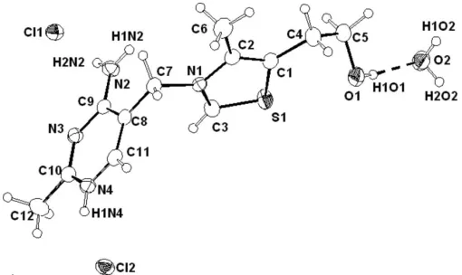
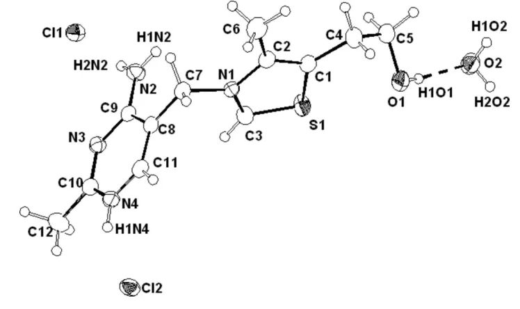
Thiamine, as its pyrophosphate ester, is a coenzyme in a number of important metabolic processes, such as the decar-boxylation of-keto acids and the transfer of aldehyde or acyl groups (Krampitz, 1969). In the present work, the H atoms were located in a difference map, leading to a better under-standing of the hydrogen-bonding pattern in vitamin B1. A molecular diagram of the title compound, (I), is given in Fig. 1. Bond angles within the pyrimidine ring are all close to 120.
The small difference between the angle of 120.93 (12)at N4
and 118.76 (12) at N3 is somewhat surprising in view of the
fact that N4 is protonated whereas N3 is not. It has already been pointed out that one would expect the angle at the ring N atom in pyrimidines to approach 125 for the protonated or
otherwise substituted case or 106 for the unsubstituted case
(Pauling & Corey, 1956).
The bond lengths and angles in this structure are similar to those of the two different forms reported previously [forms I (Kraut & Reed, 1962) and II (Suhet al., 1982)]. Both of the aromatic rings in the cation are essentially planar. The conformation of the cation is described by two torsion angles,
’T(C2—N1—C7—C8) and’P(N1—C7—C8—C9), centring the methylene atom C7 to the respective rings (Pletcher & Sax, 1972). Three conformations have been observed for thiamine, namely F, S and V conformations. The values of’T and’Pdetermine whether the conformation is F, S or V. The torsion angle of ’Tis 0 for F,100 for S and 90 for V (Shinet al., 1977). The value of’Pis90for F,150for S and 90 for V (Shin et al., 1977). The F conformation has
been the most frequently found in thiamine derivatives. The
present structure, in which the torsion angles’T= 179.18 (11) and’P=79.27 (15), also belongs to the F conformation.
The C5-hydroxyethyl side chain is folded back towards the thiazolium ring from the opposite side to the pyrimidine ring. The thiazolium ring is described by the two torsion angles’5
(S1—C1—C4—C5) and ’5 (C1—C4—C5—O1). In the
present work, the values of ’5 and ’5 are 24.2 (20) and 63.35 (18), respectively. However, these angles differ widely from those of the two forms reported earlier. We have calculated these angles’5and’5as 103.4 (6) and 53.76 (7),
respectively, for form I, and100.8 (3) and 50.6 (8),
respec-tively, for form II. The values of’5and’5for most thiamine
derivatives have been observed to lie in the range60 to90
(Shinet al., 1977) and within 10from60(Shinet al., 1977).
The essential difference occurs in the angle’5of the present
structure. This difference appears due to the packing requirements for the hydroxy group participating in hydrogen bonding. The geometries of the hydrogen bonds are given in Table 2. This structure contains two weak O—H Cl and N— H Cl hydrogen bonds.
Experimental
Thiamine hydrochloride (660 mg, Sigma) was dissolved in 75 ml of water. The mixture was heated for about 4 h and allowed to cool. After a week, colourless block-shaped crystals appeared.
Crystal data
C12H18N4OS2+2Cl
H2O Mr= 355.28
Monoclinic,P21=c a= 9.1437 (2) A˚
b= 7.3438 (2) A˚
c= 24.7447 (6) A˚
= 92.112 (1) V= 1660.47 (7) A˚3
Z= 4
Dx= 1.421 Mg m3 MoKradiation Cell parameters from 25
reflections
= 2–30.8 = 0.53 mm1
T= 173 (2) K Block, colourless 0.50.320.24 mm
Data collection
Bruker SMART CCD diffractometer
!scans
Absorption correction: multi-scan (SADABS; Bruker, 1998)
Tmin= 0.817,Tmax= 0.881
11933 measured reflections
4716 independent reflections 3913 reflections withI> 2(I)
Rint= 0.026
max= 30.8
h=12!11
k=10!10
l=35!29
Refinement
Refinement onF2 R[F2> 2(F2)] = 0.033 wR(F2) = 0.081
S= 1.05 4716 reflections 270 parameters
All H-atom parameters refined
w= 1/[2
(Fo2) + (0.0341P)2 + 0.5004P]
whereP= (Fo2+ 2Fc2)/3 (/)max< 0.001
[image:4.610.44.299.70.222.2]max= 0.27 e A˚3 min=0.27 e A˚3
Table 1
Selected geometric parameters (A˚ ,).
S1—C3 1.6753 (15) S1—C1 1.7229 (14) O1—C5 1.419 (2) N1—C3 1.3146 (17) N1—C2 1.3897 (17) N1—C7 1.4883 (17)
N2—C9 1.3161 (17) N3—C10 1.3087 (17) N3—C9 1.3570 (17) N4—C11 1.3477 (19) N4—C10 1.3497 (19)
C3—S1—C1 91.68 (7) C3—N1—C2 113.93 (11) C3—N1—C7 123.82 (12) C2—N1—C7 122.25 (11) C10—N3—C9 118.76 (12) C11—N4—C10 120.93 (12) C2—C1—S1 109.86 (10) C4—C1—S1 122.80 (11) C1—C2—N1 112.36 (11) N1—C2—C6 120.17 (13)
N1—C3—S1 112.17 (11) O1—C5—C4 108.20 (13) N1—C7—C8 112.35 (11) N2—C9—N3 116.55 (12) N2—C9—C8 122.23 (13) N3—C9—C8 121.21 (12) N3—C10—N4 122.08 (13) N3—C10—C12 119.72 (14) N4—C10—C12 118.21 (13) N4—C11—C8 120.59 (13)
C1—C4—C5—O1 63.35 (18) N1—C7—C8—C11 103.29 (14)
[image:4.610.313.566.205.388.2]N1—C7—C8—C9 79.27 (15) C7—C8—C9—N2 3.1 (2)
Table 2
Hydrogen-bond geometry (A˚ ,).
D—H A D—H H A D A D—H A
O2—H1O2 Cl2i
0.82 (2) 2.40 (3) 3.2120 (14) 171 (2) O2—H2O2 Cl1ii
0.84 (3) 2.32 (3) 3.1636 (13) 175 (2) N2—H1N2 Cl2iii
0.85 (2) 2.36 (2) 3.1934 (14) 166 (2) O1—H1O1 O2 0.83 (2) 1.95 (2) 2.7555 (17) 166 (2) N2—H2N2 Cl1 0.83 (2) 2.36 (2) 3.1744 (13) 168 (2) N4—H1N4 Cl2 0.85 (2) 2.28 (2) 3.0659 (12) 153 (2)
Symmetry codes: (i)xþ1;y1 2;zþ
1
2; (ii)x;yþ 1 2;zþ
1
2; (iii)xþ1;y;z.
All H atoms were found in a difference Fourier map and refined isotropically [C—H = 0.89 (3)–1.003 (19) A˚ ].
Data collection:SMART(Bruker, 1998); cell refinement:SAINT (Bruker, 1998); data reduction:SAINT; program(s) used to solve structure:SIR97(Altomare et al., 1999); program(s) used to refine structure: SHELXL97 (Sheldrick, 1997); molecular graphics: ZORTEP (Zsolnai & Pritzkow, 1995); software used to prepare material for publication:SHELXL97.
References
Altomare, A., Burla, M. C., Camalli, M., Cascarano, G. L., Giacovazzo, C., Guaghardi, A., Moliterni, A. G., Polidori, G. & Spagna, R. (1999).J. Appl. Cryst.32, 115–119.
Bruker (1998).SMART,SAINTandSADABS. Bruker AXS Inc., Madison, Wisconsin, USA.
Krampitz, L. O. (1969).Annu. Rev. Biochem.38, 213–240. Kraut, J. & Reed, H. J. (1962).Acta Cryst.15, 747–757.
Pauling, L. & Corey, R. B. (1956).Arch. Biochem. Biophys.65, 164–181.
organic papers
Acta Cryst.(2006). E62, o290–o292 Balasubramanianet al. C
12H18N4OS2+2ClH2O
o291
Figure 1 [image:4.610.313.565.451.523.2]Pletcher, J. & Sax, M. (1972).J. Am. Chem. Soc.94, 3998–4005. Sheldrick, G. M. (1997).SHELXL97. University of Go¨ttingen, Germany. Shin, W., Pletcher, J., Blank, G. & Sax, M. (1977).J. Am. Chem. Soc.99, 3491–
3499.
Suh, I.-H., Kim, Y.-J., Yoon, M. J., Ku, Y. & Ahn, S. T. (1982).J. Korean Phys. Soc.15, 114–121.
Zsolnai, L. & Pritzkow, H. (1995). ZORTEP. University of Heidelberg, Germany.
organic papers
o292
Balasubramanianet al. Csupporting information
sup-1
Acta Cryst. (2006). E62, o290–o292supporting information
Acta Cryst. (2006). E62, o290–o292 [doi:10.1107/S1600536805041619]
Thiamine dichloride monohydrate: vitamin B1 (form III)
T. Balasubramanian, Samuel Robinson Jebas, S. Thamotharan, G. Rheinwald and A. G. Lang
S1. Comment
Thiamine, as its pyrophosphate ester, is a coenzyme in a number of important metabolic processes such as the
de-carboxylation of α-keto acids and the transfer of aldehyde or acyl groups (Krampitz, 1969). In the present work, the H
atoms being located by difference map leads to the better understanding of the hydrogen-bonding pattern in vitamin B1.
The molecular diagram of the title compound, (I), is given in Fig. 1. Bond angles within the pyrimidine ring are all close
to 120°. The small difference between the angle of 120.93 (12)° at N4 and 118.76 (12)° at N3 is some what surprising in
view of the fact that N4 is externally bonded to an H atom whereas N3 is not. It has already been pointed out that one
would expect the angle at the ring N atom in pyrimidines to approach 125° for the externally bonded case or 106° for the
non-bonded case (Pauling & Corey, 1956).
The bond lengths and bond angles in this structure are similar to those of the two different forms reported previously
[forms I (Kraut & Reed, 1962) and II (Suh et al., 1982) {please check assignment of references}]. Both of the aromatic
rings in this molecule are virtually planar with small deviations from strict planarity. The conformation of the thiamine
molecule is described by two torsion angles, φT and φP, centering the methylene atom C7 to the respective rings φT [C2—
N1—C7—C8] and φP [N1—C7—C8—C9] (Pletcher & Sax, 1972). Basically three kinds of conformers have been
observed in the thiamine molecule, namely F, S and V conformations. The value of φT and φP decides the conformation
whether belongs to F, S and V conformation. The torsion angle of φT is 0° for F, ±100° for S and ±90° for V (Shin et al.,
1977). The value of φP is ±90° for F, ±150° for S and ±90° for V (Shin et al., 1977). Among them, the F conformation has
been most frequently found in thiamine derivatives. The present structure in which the torsion angle of φT = 179.18 (11)°,
and φP = −79.27 (15)° also belongs to the F conformation. Thus the F conformation is the predominant one of the free
thi-amine molecules.
The C5-hydroxyethyl side chain is folded back towards the thiazolium ring from the opposite side to the pyrimidine
moiety. The thiazolium ring is described by the two torsion angles φ5a [S1—C1—C4—C5] and φ5 b [C1—C4—C5—O1].
In the present work, the values of φ5a and φ5 b are 24.2 (20) and −63.35 (18)°, respectively. However, these angles differ
widely from those of the two forms reported earlier. We have calculated these angles φ5a and φ5 b as 103.4 (6) and
53.76 (7)°, respectively, for form I, and −100.8 (3) and 50.6 (8)°, respectively, for form II. The values of φ5a and φ5 b for
most of the thiamine derivatives have been observed to lie in the range ±60 to ±90° (Shin et al., 1977) and within 10°
from ±60° (Shin et al., 1977). The essential difference occurs in the angle φ5a of the present structure. This difference
appears due to the packing specificity required for the hydroxy group participating in hydrogen bonding. The geometries
of the hydrogen bonds are given in Table 2. This structure contains two weak but distinct O—H···Cl and N—H···Cl
hydrogen bonds. The interatomic distances H1O2···Cl2 and H2O2···Cl1 are 2.40 (3) and 2.32 (3) Å, respectively, which
are shorter than the normal van der Waals distance (3.0) Å. These hydrogen bonds must be weaker than N—H···Cl bonds,
for which the hydrogen to chloride distances are 2.359 (19), 2.36 (2) and 2.28 (2) Å in this structure. The hydrogen-bond
supporting information
sup-2
Acta Cryst. (2006). E62, o290–o2923.163 (13) and 3.212 (14) Å, which is in the range of normal van der Waals distance (3.2 Å).
S2. Experimental
Thiamine hydrochloride (660 mg, Sigma) was dissolved in 75 ml of water. The mixture was heated for about 4 h and
allowed to cooling. After a week, colorless block-shaped crystals appeared.
S3. Refinement
All H atoms were found in a difference Fourier map and refined isotropically [C—H = 0.89 (3)–1.003 (19) Å]. O2···H101
[1.95 (2) Å] hydrogen bond is caught up using SHELXL program. The unusual hydrogen bonding is novel and found to
[image:7.610.115.489.214.436.2]be very weak.
Figure 1
View of (I), with 50% probability displacement ellipsoids.
Thiamine dichloride monohydrate
Crystal data
C12H18N4OS2+·2Cl−·H2O
Mr = 355.28
Monoclinic, P21/c
Hall symbol: -P 2y bc
a = 9.1437 (2) Å
b = 7.3438 (2) Å
c = 24.7447 (6) Å
β = 92.112 (1)°
V = 1660.47 (7) Å3
Z = 4
F(000) = 744
Dx = 1.421 Mg m−3
Mo Kα radiation, λ = 0.71073 Å Cell parameters from 25 reflections
θ = 2–30.8°
µ = 0.53 mm−1
T = 173 K Block, colorless 0.5 × 0.32 × 0.24 mm
Data collection
Bruker SMART CCD diffractometer
ω scans
Absorption correction: empirical (using intensity measurements)
(SADABS; Bruker, 1998)
supporting information
sup-3
Acta Cryst. (2006). E62, o290–o2924716 independent reflections 3913 reflections with I > 2σ(I)
Rint = 0.026
θmax = 30.8°, θmin = 1.7°
h = −12→11
k = −10→10
l = −35→29
Refinement
Refinement on F2 Least-squares matrix: full
R[F2 > 2σ(F2)] = 0.033
wR(F2) = 0.081
S = 1.05 4716 reflections 270 parameters
0 restraints
All H-atom parameters refined
w = 1/[σ2(F
o2) + (0.0341P)2 + 0.5004P] where P = (Fo2 + 2Fc2)/3
(Δ/σ)max < 0.001 Δρmax = 0.27 e Å−3 Δρmin = −0.27 e Å−3
Special details
Geometry. All e.s.d.'s (except the e.s.d. in the dihedral angle between two l.s. planes) are estimated using the full covariance matrix. The cell e.s.d.'s are taken into account individually in the estimation of e.s.d.'s in distances, angles and torsion angles; correlations between e.s.d.'s in cell parameters are only used when they are defined by crystal symmetry. An approximate (isotropic) treatment of cell e.s.d.'s is used for estimating e.s.d.'s involving l.s. planes.
Fractional atomic coordinates and isotropic or equivalent isotropic displacement parameters (Å2)
x y z Uiso*/Ueq
Cl1 0.73143 (4) 0.61805 (5) −0.093135 (14) 0.02631 (8)
Cl2 −0.13005 (4) 0.87135 (5) 0.087101 (15) 0.02970 (9)
S1 0.60833 (4) 0.53995 (5) 0.197213 (15) 0.03026 (10)
O1 0.75506 (13) 0.42930 (16) 0.29537 (4) 0.0330 (2)
H1O1 0.796 (2) 0.332 (3) 0.3025 (9) 0.055 (7)*
O2 0.91842 (15) 0.13742 (17) 0.33306 (5) 0.0350 (3)
H1O2 0.978 (3) 0.186 (3) 0.3542 (9) 0.056 (7)*
H2O2 0.873 (3) 0.066 (3) 0.3532 (10) 0.064 (7)*
N1 0.55130 (12) 0.82985 (15) 0.14875 (4) 0.0196 (2)
N2 0.58901 (13) 0.75687 (18) 0.01444 (5) 0.0257 (2)
H1N2 0.6531 (19) 0.791 (2) 0.0383 (7) 0.029 (4)*
H2N2 0.618 (2) 0.706 (3) −0.0133 (8) 0.037 (5)*
N3 0.35590 (12) 0.71385 (16) −0.01825 (5) 0.0237 (2)
N4 0.15847 (13) 0.81725 (17) 0.02986 (5) 0.0263 (2)
H1N4 0.067 (2) 0.825 (3) 0.0347 (8) 0.043 (5)*
C1 0.68837 (15) 0.73632 (18) 0.22234 (5) 0.0230 (3)
C2 0.64574 (15) 0.88011 (18) 0.19145 (5) 0.0217 (3)
C3 0.52270 (16) 0.65450 (19) 0.14677 (6) 0.0258 (3)
H3 0.4614 (19) 0.603 (2) 0.1201 (7) 0.030 (4)*
C4 0.78891 (19) 0.7385 (2) 0.27160 (7) 0.0337 (3)
H4A 0.733 (2) 0.775 (3) 0.3017 (8) 0.044 (5)*
H4B 0.856 (2) 0.832 (3) 0.2658 (8) 0.045 (5)*
C5 0.86387 (18) 0.5601 (3) 0.28388 (7) 0.0357 (4)
H5A 0.937 (2) 0.578 (3) 0.3136 (8) 0.039 (5)*
H5B 0.919 (2) 0.523 (3) 0.2514 (8) 0.040 (5)*
C6 0.6878 (2) 1.0741 (2) 0.19913 (7) 0.0327 (3)
supporting information
sup-4
Acta Cryst. (2006). E62, o290–o292H6B 0.603 (2) 1.152 (3) 0.2053 (8) 0.052 (6)*
H6C 0.749 (2) 1.089 (3) 0.2314 (9) 0.052 (6)*
C7 0.49062 (15) 0.96432 (18) 0.10890 (6) 0.0224 (3)
H7A 0.5733 (17) 1.025 (2) 0.0934 (6) 0.020 (4)*
H7B 0.4346 (18) 1.049 (2) 0.1290 (7) 0.029 (4)*
C8 0.39391 (14) 0.87760 (17) 0.06647 (5) 0.0209 (2)
C9 0.44822 (14) 0.78210 (17) 0.02089 (5) 0.0203 (2)
C10 0.21472 (15) 0.72945 (19) −0.01256 (6) 0.0249 (3)
C11 0.24659 (15) 0.89251 (19) 0.06859 (6) 0.0241 (3)
H11 0.1983 (18) 0.956 (2) 0.0970 (7) 0.025 (4)*
C12 0.11179 (19) 0.6489 (3) −0.05357 (8) 0.0366 (4)
H12B 0.017 (4) 0.663 (4) −0.0458 (12) 0.097 (10)*
H12A 0.126 (3) 0.529 (4) −0.0564 (10) 0.078 (8)*
H12C 0.116 (3) 0.709 (4) −0.0897 (13) 0.110 (11)*
Atomic displacement parameters (Å2)
U11 U22 U33 U12 U13 U23
Cl1 0.02476 (16) 0.02817 (17) 0.02614 (16) −0.00201 (12) 0.00298 (12) −0.00412 (13)
Cl2 0.01952 (15) 0.0377 (2) 0.03200 (18) 0.00201 (13) 0.00257 (12) −0.00273 (15)
S1 0.0424 (2) 0.01704 (16) 0.03040 (19) −0.00494 (14) −0.01116 (15) 0.00375 (13)
O1 0.0365 (6) 0.0313 (6) 0.0311 (6) −0.0021 (5) −0.0024 (4) 0.0069 (5)
O2 0.0421 (7) 0.0326 (6) 0.0303 (6) −0.0064 (5) −0.0001 (5) 0.0036 (5)
N1 0.0210 (5) 0.0164 (5) 0.0216 (5) 0.0003 (4) 0.0027 (4) 0.0004 (4)
N2 0.0197 (5) 0.0321 (6) 0.0255 (6) 0.0023 (5) 0.0029 (5) −0.0030 (5)
N3 0.0222 (5) 0.0247 (6) 0.0243 (6) 0.0001 (4) 0.0017 (4) 0.0017 (5)
N4 0.0177 (5) 0.0317 (6) 0.0295 (6) 0.0029 (5) 0.0027 (4) 0.0073 (5)
C1 0.0272 (6) 0.0206 (6) 0.0212 (6) −0.0036 (5) 0.0019 (5) −0.0012 (5)
C2 0.0257 (6) 0.0190 (6) 0.0206 (6) −0.0014 (5) 0.0037 (5) −0.0025 (5)
C3 0.0311 (7) 0.0186 (6) 0.0271 (7) −0.0039 (5) −0.0058 (5) 0.0012 (5)
C4 0.0422 (9) 0.0320 (8) 0.0261 (7) −0.0115 (7) −0.0082 (6) 0.0029 (6)
C5 0.0304 (8) 0.0462 (9) 0.0301 (8) −0.0042 (7) −0.0038 (6) 0.0126 (7)
C6 0.0467 (9) 0.0191 (7) 0.0322 (8) −0.0067 (6) −0.0001 (7) −0.0041 (6)
C7 0.0244 (6) 0.0177 (6) 0.0251 (6) 0.0019 (5) 0.0022 (5) 0.0026 (5)
C8 0.0216 (6) 0.0197 (6) 0.0214 (6) 0.0028 (5) 0.0022 (5) 0.0047 (5)
C9 0.0204 (6) 0.0186 (6) 0.0221 (6) 0.0013 (5) 0.0030 (5) 0.0049 (5)
C10 0.0236 (6) 0.0244 (7) 0.0267 (7) −0.0010 (5) −0.0004 (5) 0.0063 (5)
C11 0.0238 (6) 0.0245 (6) 0.0242 (6) 0.0039 (5) 0.0052 (5) 0.0054 (5)
C12 0.0290 (8) 0.0403 (9) 0.0400 (9) −0.0058 (7) −0.0073 (7) 0.0006 (8)
Geometric parameters (Å, º)
S1—C3 1.6753 (15) C2—C6 1.4863 (19)
S1—C1 1.7229 (14) C3—H3 0.931 (18)
O1—C5 1.419 (2) C4—C5 1.504 (2)
O1—H1O1 0.83 (2) C4—H4A 0.95 (2)
O2—H1O1 1.95 (2) C4—H4B 0.93 (2)
supporting information
sup-5
Acta Cryst. (2006). E62, o290–o292O2—H2O2 0.84 (3) C5—H5B 1.003 (19)
N1—C3 1.3146 (17) C6—H6A 0.94 (2)
N1—C2 1.3897 (17) C6—H6B 0.98 (2)
N1—C7 1.4883 (17) C6—H6C 0.96 (2)
N2—C9 1.3161 (17) C7—C8 1.4904 (19)
N2—H1N2 0.854 (19) C7—H7A 0.971 (16)
N2—H2N2 0.83 (2) C7—H7B 0.958 (17)
N3—C10 1.3087 (17) C8—C11 1.3544 (18)
N3—C9 1.3570 (17) C8—C9 1.4325 (18)
N4—C11 1.3477 (19) C10—C12 1.481 (2)
N4—C10 1.3497 (19) C11—H11 0.963 (17)
N4—H1N4 0.85 (2) C12—H12B 0.90 (3)
C1—C2 1.3526 (19) C12—H12A 0.89 (3)
C1—C4 1.499 (2) C12—H12C 1.00 (3)
C3—S1—C1 91.68 (7) O1—C5—H5B 110.5 (11)
C5—O1—H1O1 108.2 (16) C4—C5—H5B 108.4 (11)
H1O1—O2—H1O2 106.8 (18) H5A—C5—H5B 106.9 (15)
H1O1—O2—H2O2 113.6 (17) C2—C6—H6A 111.0 (13)
H1O2—O2—H2O2 103 (2) C2—C6—H6B 112.0 (12)
C3—N1—C2 113.93 (11) H6A—C6—H6B 108.8 (18)
C3—N1—C7 123.82 (12) C2—C6—H6C 110.7 (13)
C2—N1—C7 122.25 (11) H6A—C6—H6C 110.0 (18)
C9—N2—H1N2 121.6 (12) H6B—C6—H6C 104.1 (17)
C9—N2—H2N2 120.3 (13) N1—C7—C8 112.35 (11)
H1N2—N2—H2N2 118.0 (17) N1—C7—H7A 107.0 (9)
C10—N3—C9 118.76 (12) C8—C7—H7A 111.7 (9)
C11—N4—C10 120.93 (12) N1—C7—H7B 106.3 (10)
C11—N4—H1N4 116.0 (13) C8—C7—H7B 109.2 (10)
C10—N4—H1N4 123.0 (13) H7A—C7—H7B 110.1 (14)
C2—C1—C4 127.34 (13) C11—C8—C9 116.36 (13)
C2—C1—S1 109.86 (10) C11—C8—C7 120.23 (12)
C4—C1—S1 122.80 (11) C9—C8—C7 123.36 (12)
C1—C2—N1 112.36 (11) N2—C9—N3 116.55 (12)
C1—C2—C6 127.47 (13) N2—C9—C8 122.23 (13)
N1—C2—C6 120.17 (13) N3—C9—C8 121.21 (12)
N1—C3—S1 112.17 (11) N3—C10—N4 122.08 (13)
N1—C3—H3 122.6 (11) N3—C10—C12 119.72 (14)
S1—C3—H3 125.2 (11) N4—C10—C12 118.21 (13)
C1—C4—C5 114.60 (14) N4—C11—C8 120.59 (13)
C1—C4—H4A 107.9 (12) N4—C11—H11 116.0 (10)
C5—C4—H4A 109.7 (12) C8—C11—H11 123.4 (10)
C1—C4—H4B 105.5 (13) C10—C12—H12B 113.7 (19)
C5—C4—H4B 112.1 (13) C10—C12—H12A 110.9 (17)
H4A—C4—H4B 106.7 (17) H12B—C12—H12A 106 (2)
O1—C5—C4 108.20 (13) C10—C12—H12C 113.1 (18)
O1—C5—H5A 113.6 (11) H12B—C12—H12C 102 (2)
supporting information
sup-6
Acta Cryst. (2006). E62, o290–o292C3—S1—C1—C2 0.35 (11) C2—N1—C7—C8 179.18 (11)
C3—S1—C1—C4 −179.97 (13) N1—C7—C8—C11 103.29 (14)
C4—C1—C2—N1 −179.84 (13) N1—C7—C8—C9 −79.27 (15)
S1—C1—C2—N1 −0.18 (15) C10—N3—C9—N2 177.41 (13)
C4—C1—C2—C6 −0.3 (2) C10—N3—C9—C8 −3.04 (19)
S1—C1—C2—C6 179.37 (13) C11—C8—C9—N2 −179.32 (13)
C3—N1—C2—C1 −0.15 (17) C7—C8—C9—N2 3.1 (2)
C7—N1—C2—C1 −179.47 (12) C11—C8—C9—N3 1.15 (18)
C3—N1—C2—C6 −179.74 (14) C7—C8—C9—N3 −176.39 (12)
C7—N1—C2—C6 0.95 (19) C9—N3—C10—N4 2.6 (2)
C2—N1—C3—S1 0.42 (16) C9—N3—C10—C12 −177.33 (13)
C7—N1—C3—S1 179.73 (10) C11—N4—C10—N3 −0.3 (2)
C1—S1—C3—N1 −0.44 (12) C11—N4—C10—C12 179.67 (14)
C2—C1—C4—C5 −156.16 (15) C10—N4—C11—C8 −1.7 (2)
S1—C1—C4—C5 24.2 (2) C9—C8—C11—N4 1.19 (19)
C1—C4—C5—O1 −63.35 (18) C7—C8—C11—N4 178.81 (12)
C3—N1—C7—C8 −0.07 (18)
Hydrogen-bond geometry (Å, º)
D—H···A D—H H···A D···A D—H···A
O2—H1O2···Cl2i 0.82 (2) 2.40 (3) 3.2120 (14) 171 (2)
O2—H2O2···Cl1ii 0.84 (3) 2.32 (3) 3.1636 (13) 175 (2)
N2—H1N2···Cl2iii 0.854 (19) 2.359 (19) 3.1934 (14) 165.9 (15)
O1—H1O1···O2 0.83 (2) 1.95 (2) 2.7555 (17) 166 (2)
N2—H2N2···Cl1 0.83 (2) 2.36 (2) 3.1744 (13) 167.8 (18)
N4—H1N4···Cl2 0.85 (2) 2.28 (2) 3.0659 (12) 152.7 (17)