inorganic papers
i246
Smolentsev and Naumov Ca(ClO2)2 doi:10.1107/S160053680503415X Acta Cryst.(2005). E61, i246–i248 Acta Crystallographica Section E
Structure Reports Online
ISSN 1600-5368
Calcium chlorite, Ca(ClO
2)
2, from X-ray powder
diffraction data
Anton I. Smolentsev* and Dmitry Yu. Naumov
Nikolaev Institute of Inorganic Chemistry, SB Russian Academy of Sciences, Akad. Lavrentiev prosp. 3, Novosibirsk 90, 630090, Russian Federation
Correspondence e-mail: [email protected]
Key indicators
Powder X-ray study
T= 293 K
Mean(Cl–O) = 0.003 A˚
Rfactor = 0.127
wRfactor = 0.145
Data-to-parameter ratio = 6.77
For details of how these key indicators were automatically derived from the article, see http://journals.iucr.org/e.
#2005 International Union of Crystallography Printed in Great Britain – all rights reserved
The structure of calcium chlorite, Ca(ClO2)2, has been refined
from X-ray powder diffraction data using the Rietveld method. The compound crystallizes in the orthorhombic space group Ccca (Ccce), with Z = 4. The structure is based on separate layers parallel to theacplane, consisting of calcium cations that are coordinated by chlorite anions; the O atoms form almost ideal square antiprisms. Within the layers, each anion bridges four metal cations. The Ca atoms are located on special positions of 222 symmetry, the Cl atoms lie on twofold axes and the O atoms are in general positions. The compound is isostructural with strontium chlorite, Sr(ClO2)2, and lead
chlorite, Pb(ClO2)2.
Comment
Recently, we carried out the crystal structure determination of barium chlorite hydrate, Ba(ClO2)23.5H2O (Smolentsev &
Naumov, 2005a). To continue our systematic structural char-acterization of alkaline earth metal chlorites, the present investigation has been performed. Furthermore, obtaining crystal data for calcium chlorite seemed appropriate because it was investigated previously (together with the strontium salt) by Riganti & Garrini (1960), who could not index the X-ray powder pattern satisfactorily and only proposed a pseudo-cubic cell with a lattice parameter of 5.80 A˚ (5.97 A˚ for the strontium salt). The results of our study are reported here.
Ca(ClO2)2 is isostructural with Sr(ClO2)2 (Smolentsev &
Naumov, 2005d) and Pb(ClO2)2 (Okuda et al., 1990). The
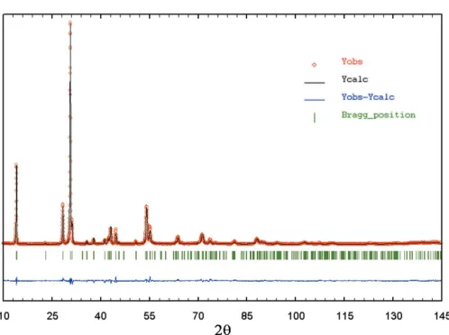
experimental and calculated powder profiles, as well as the difference plot, are shown in Fig. 1. The structure of the title
[image:1.610.208.457.532.718.2]Received 7 October 2005 Accepted 21 October 2005 Online 27 October 2005
Figure 1
Experimental and calculated powder profiles of Ca(ClO2)2, with
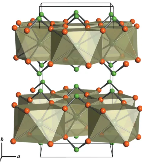
compound is layered. Separate layers are parallel to the ac
plane and consist of calcium cations coordinated by chlorite anions. The full coordination sphere of the cation includes eight chlorite O atoms forming an almost ideal square anti-prism. Within a layer these square antiprisms share four edges. Each ClO2
anion serves as a tetradentate bridging ligand bonding four metal cations (Fig. 2). The shortest Cl Cl distance between adjacent layers is 3.541 (1) A˚ . Satisfactory geometric parameters of the ClO2
anion (i.e.corresponding to those in other chlorites) were obtained through a weak restraint of the Cl—O distance to 1.57 (2) A˚ , which is the average value of the most accurate distances reported by Tarimci & Schempp (1975), Tarimci et al. (1976), Pakkanen (1979), Castellani Bisi (1984), Okuda et al. (1990), Marsh (1991), and Smolentsev & Naumov (2005a,b,c). At the same time, the O—Cl—O angle of 115.3 (3) is comparable to the
average of 110 (2)found in the literature.
In the previously studied Ba(ClO2)23.5H2O, a layered
structure is also realized, but in this compound, layers are formed with the participation of water molecules. Never-theless, general details of the layer construction are similar to those in the title chlorite. Unfortunately, comparison with anhydrous Ba(ClO2)2cannot be made because of difficulties
with indexing and structure solution (caused either by a non-monophase pattern or high texture), but analysis of the diffractogram showed that Ba(ClO2)2most probably belongs
to a different crystal system than the calcium member. Among the compounds with comparable anions of other acids, the most similarity is observed for some hypophosphites, viz. Pb(H2PO2)2, Sr(H2PO2)2 and Ba(H2PO2)2 (Kuratieva et al.,
2005), which have almost identical positional relationships of structure building units – metal cationsM2+(M= Sr, Ba and Pb) and anions with similar shape. Barium hypophosphite is closely related to the title compound. The two compounds have the same space group and similar lattice and positional parameters. This fact confirms the assumption that the ClO2
and H2PO2 anions may perform similar structural roles in
many cases, which is the result of their geometry and elec-tronic configuration.
Experimental
Polycrystalline Ca(ClO2)2was synthesized at room temperature by
reacting an aqueous suspension of calcium peroxide with an excess of chlorine dioxide, followed by precipitation from solution by adding a 3:1 mixture of ethanol and diethyl ether. A white crystalline powder resulted.
Crystal data
Ca(ClO2)2
Mr= 174.98
Orthorhombic,CccaðCcceÞ a= 5.7434 (7) A˚
b= 12.6002 (9) A˚
c= 5.7405 (7) A˚
V= 415.43 (8) A˚3
Z= 4
Dx= 2.798 Mg m
3
CuKradiation
Wavelength of incident radiation: 1.54178 A˚
T= 293 (2) K
Specimen shape: flat sheet 10100.1 mm
Particle morphology: plate-like, white
Data collection
Philips PW1700 powder diffractometer
Specimen mounting: drifted powder on standard quartz sample holder
Specimen mounted in reflection mode
Scan method: step 2min= 5.0, 2max= 145.0
Increment in 2= 0.02
Refinement
Rp= 0.127
Rwp= 0.145
Rexp= 0.080
RB= 0.043
S= 1.81
Profile function: split pseudo-Voigt 210 reflections
31 parameters
w= 1/[2(Y
i)]
(/)max= 0.01
Preferred orientation correction:
Icorr=Iobs[G2+(1G2)
exp(G12)], whereis the acute
angle between the scattering vector and the normal to the crystallites,G1=2.69 (2),G2=
0.102 (1), axis [010] (Rietveld, 1969)
Table 1
Selected geometric parameters (A˚ ,).
Ca—O 2.481 (3)
Ca—Oi
2.499 (3)
Cl—O 1.585 (3)
O—Cl—Oii 115.3 (3)
Symmetry codes: (i)xþ1 2;y;zþ
1
2; (ii)xþ1;y;zþ 1 2.
The list of peak positions was obtained from the X-ray raw data using the program WINPLOTR (Roisnel & Rodriguez-Carvajal, 2005) and used for indexing by theCRYSFIREsuite (Shirley, 2000). An orthorhombic cell was selected as that best corresponding to the observed diffractogram. The calculated cell parameters,aandc, are in agreement with the pseudo-cubic parameter reported by Riganti & Garrini (1960). In fact, the cell metric is extremely close to tetragonal. Probably this was the reason why previous authors had difficulty in indexing the powder patterns. Systematic absences,hkl withh +k
inorganic papers
Acta Cryst.(2005). E61, i246–i248 Smolentsev and Naumov Ca(ClO
[image:2.610.49.294.68.354.2]2)2
i247
Figure 2odd,hk0 withhorkodd,h0lwithhorlodd, and 0klwithkorlodd, showed that the space group is Ccca (Ccce). Proceeding from this information, the Pb(ClO2)2 structure [Ccca (Ccce), Z = 4, a =
6.004 (1) A˚ ,b= 12.504 (2) A˚ ,c= 6.010 (1) A˚ ; Okudaet al., 1990] was chosen as a trial model for the full-profile Rietveld refinement. The Rietveld refinement was performed in two general steps. First, the validity of cell parameters and space group was confirmed using the profile-matching method (split pseudo-Voigt peak-shape function). At this stage, only the zero shift, background polynomial coefficients, cell parameters, peak-shape and FWHM variables were varied. In subsequent refinements, the scale factor, and positional and isotropic displacement parameters for all atoms were refined. The Cl—O distances were weakly restrained to 1.57 (2) A˚ . This also improved the O—Cl—O angles. No other restraints were used. Since the structure is layered in the [010] direction, refinement of preferred orientation coefficients was also applied.
Data collection: APD1700 Software(Philips, 1989); cell refine-ment:FULLPROF2k (Rodriguez-Carvajal, 2004); program(s) used to refine structure:FULLPROF2k; molecular graphics:WINPLOTR (Roisnel & Rodriguez-Carvajal, 2005) and BS (Ozawa & Kang, 2004); software used to prepare material for publication: WINPLOTR.
The authors thank N. V. Kuratieva for useful comments during the preparation of this paper.
References
Castellani Bisi, C. (1984).Acta Cryst.C40, 1120–1121.
Kuratieva, N. V., Naumova, M. I., Podberezskaya, N. V. & Naumov D. Y. (2005).Acta Cryst.C61, i14–i16.
Marsh, R. E. (1991).Acta Cryst.C47, 1775.
Okuda, M., Ishihara, M., Yamanaka, M., Ohba, S. & Saito, Y. (1990).Acta Cryst.C46, 1755–1759.
Ozawa, T. C. & Kang, S. J. (2004).Balls & Sticks(BS). Version 1.51. http:// www.softbug.com/toycrate/bs.
Pakkanen, T. (1979).Acta Cryst.B35, 2670–2672.
Philips (1989). APD1700 Software. Version 4.0. Philips Electronics Inc., Natick, MA, USA.
Rietveld, H. M. (1969).J. Appl. Cryst.2, 65–71.
Riganti, V. & Garrini, E. (1960).Gazz. Chim. Ital.90, 321–327.
Rodriguez-Carvajal, J. (2004).FULLPROF2k. Version of November 2004. Laboratoire Leon Brillouin (CEA/CNRS), CEA-Saclay, 91191 Gif-sur-Yvette Cedex, France.
Roisnel, T. & Rodriguez-Carvajal, J. (2005).WINPLOTR. Version of February 2005. Laboratoire Leon Brillouin (CEA/CNRS), CEA-Saclay, 91191 Gif-sur-Yvette Cedex, France.
Shirley, R. (2000). CRYSFIRE User’s Manual. Guildford, England: The Lattice Press.
Smolentsev, A. I. & Naumov, D. Y. (2005a).Acta Cryst.C61, i49–i50. Smolentsev, A. I. & Naumov, D. Y. (2005b).Acta Cryst.C61, i17–i19. Smolentsev, A. I. & Naumov, D. Y. (2005c).Acta Cryst.E61, i38–i40. Smolentsev, A. I. & Naumov, D. Y. (2005d).Acta Cryst.E61, i249–i250. Tarimci, C., Rosenstein, R. D. & Schempp, E. (1976).Acta Cryst.B32, 610–
612.
Tarimci, C. & Schempp, E. (1975).Acta Cryst.B31, 2146–2149.
inorganic papers
i248
Smolentsev and Naumov Ca(ClOsupporting information
sup-1 Acta Cryst. (2005). E61, i246–i248
supporting information
Acta Cryst. (2005). E61, i246–i248 [https://doi.org/10.1107/S160053680503415X]
Calcium chlorite, Ca(ClO
2)
2, from X-ray powder diffraction data
Anton I. Smolentsev and Dmitry Yu. Naumov
calcium chlorate(III)
Crystal data Ca(ClO2)2 Mr = 174.98
Orthorhombic, Ccca(Ccce) a = 5.7434 (7) Å
b = 12.6002 (9) Å c = 5.7405 (7) Å V = 415.43 (8) Å3
Z = 4
Dx = 2.798 Mg m−3
Cu Kα radiation, λ = 1.54178 Å T = 293 K
Particle morphology: plate-like white
flat_sheet, 10 × 10 mm
Data collection
Philips PW1700 powder diffractometer
Radiation source: sealed X-ray tube Graphite monochromator
Specimen mounting: drifted powder on standard quartz sample holder
Data collection mode: reflection Scan method: step
2θmin = 5.041°, 2θmax = 145.041°, 2θstep = 0.020°
Refinement
Least-squares matrix: full Rp = 0.127
Rwp = 0.145 Rexp = 0.080 RBragg = 0.043 7001 data points
Profile function: split pseudo-Voigt 31 parameters
1 restraint
Weighting scheme based on measured s.u.'s w = 1/[σ2(Y
i)] (Δ/σ)max = 0.01
Background function: six-coefficients polinomial function
Preferred orientation correction: Icorr = Iobs[G2+(1 – G2)exp(G1α2)], where α is the acute angle between the scattering vector and the normal to the crystallites, G1 = -2.69(2), G2 = 0.102(1), axis [010], (Rietveld, 1969)
Special details
Experimental. The experimental data were collected with Cu K? radiation (λ1=1.54060 Å, λ2=1.54439 Å) on a Philips PW1700 automatic diffractometer with Si as external standard.
Fractional atomic coordinates and isotropic or equivalent isotropic displacement parameters (Å2)
x y z Uiso*/Ueq
Ca 0.00000 0.25000 0.25000 0.0054 (6)*
Cl 0.50000 0.08227 (11) 0.25000 0.0172 (4)*
supporting information
sup-2 Acta Cryst. (2005). E61, i246–i248
Atomic displacement parameters (Å2)
U11 U22 U33 U12 U13 U23
? ? ? ? ? ? ?
Geometric parameters (Å, º)
Ca—O 2.481 (3) Cl—O 1.585 (3)
Ca—Oi 2.499 (3)
O—Cl—Oii 115.3 (3)