metal-organic papers
Acta Cryst.(2005). E61, m1373–m1375 doi:10.1107/S1600536805018660 Schilleret al. [Re(C
7H12NO2S3)(C18H15P)]C3H6O
m1373
Acta Crystallographica Section EStructure Reports
Online
ISSN 1600-5368
[2-Carboxy-2,2
000,2
000000-nitrilotris(ethanethiolato)-j
4N
,
S
,
S
000,
S
000000](triphenylphosphine-j
P
)rhenium(III)
acetone solvate
Eik Schiller,aWerner Kraus,b* Gu¨nter Reck,b Hartmut Spiesa and Hans-Ju¨rgen Pietzscha
a
Forschungszentrum Rossendorf, Institut fu¨r Bioanorganische und Radiopharmazeutische Chemie, Postfach 510119, 01314 Dresden, Germany, andb
Bundesanstalt fu¨r Material-forschung und -pru¨fung, Richard-Willsta¨tter-Strasse 11, D-12489 Berlin, Germany
Correspondence e-mail: [email protected]
Key indicators
Single-crystal X-ray study
T= 273 K
Mean(C–C) = 0.018 A˚ Disorder in main residue
Rfactor = 0.074
wRfactor = 0.214
Data-to-parameter ratio = 16.8
For details of how these key indicators were automatically derived from the article, see http://journals.iucr.org/e.
#2005 International Union of Crystallography Printed in Great Britain – all rights reserved
The title compound, [Re(C7H12NO2S3)(C18H15P)]C3H6O, crystallizes from a solution in chloroform–acetone–cyclo-hexane with enantiomers disordered equally over each molecular site. Hydrogen bonds between the carboxyl groups form dimers in the crystal structure.
Comment
The application of radionuclides for the internal radiotherapy of cancer has been intensely investigated over the last decade (Larson & Krenning, 2005). Among the multitude of potential radionuclides suitable for therapeutic application in nuclear medicine, rhenium-188 is one of the most attractive (Knapp, 1998). This is due to its nuclide properties and its daily availability from a 188W/188Re generator. The major draw-backs in internal radionuclide therapy have been low target uptake and accumulation of radioactivity in non-target organs, such as kidney and liver. To overcome these problems, several strategies have been suggested. Increasing hydrophilicity of the ligands and coupling of hydrophilic molecules are possible approaches.
Rhenium complexes with tetradentate/monodentate NS3/P (‘4+1’) coordination are promising candidates for the devel-opment of rhenium-188 complexes of highin vivostability. 4+1 complexes containing the tris-(2-mercaptoethyl)amine chelator (NS3) are of high lipophilicity, giving rise to accu-mulation of these compounds in the liver (Pietzsch et al., 2001). These unfavourable biodistribution characteristics were diminished by the introduction of a carboxyl group into the NS3 framework, resulting in increased hydrophilicity of the corresponding rhenium complexes (Seifert et al., 2004). The new ligand can easily be modified by coupling different amines
viaan amide bond, thus increasing the variability of the 4+1 approach. The crystal structure of the first rhenium 4+1 complex with a carboxyl group containing a tripodal tetra-dentate NS3ligand, the title complex, (I), is reported here.
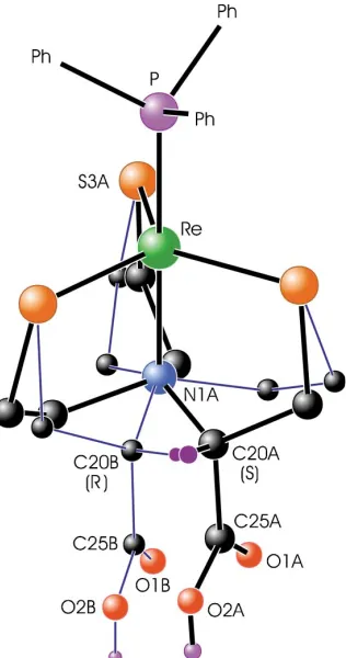
Compound (I) crystallizes as a disordered racemic struc-ture. For clarity, only the structure of one component molecule
is depicted in Fig. 1. With respect to the triphenylphosphine group, there are two different arrangements of the NS3 chelator, which occupy the same site in the disordered crystal structure, as shown in Fig. 2. The two arrangements are approximately related by a partial mirror plane through atoms Re, S3A, P and N1A. This mirror plane converts the 20(R) configuration of the molecule into a 20(S) configuration.
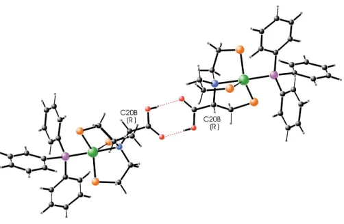
Fig. 3 shows the linkage of two molecules via hydrogen bonds between the carboxyl groups, forming a dimer in the crystal structure. There are four possibilities for this dimer formation, namely linkage of two 20(R) molecules, 20(S) pairs, and two different combinations of 20(R) and 20(S) molecules, disordered over the centre of symmetry of the space group. The corresponding symmetry-independent hydrogen bonds are given in Table 2. From the geometry of the hydrogen bonds, it can be concluded that the formation of 20(R)–20(R) and 20(S)–20(S) dimers is energetically favoured. Never-theless, the refinement of (I) in the space group P1 as an ordered twinned structure with two symmetry-independent molecules gave no satisfactory results.
Experimental
The title compound was prepared according to the method published by Spieset al.(1995). Slow solvent evaporation of a solution of (I) in chloroform–acetone–cyclohexane (8:1:1) gave green crystals suitable for X-ray analysis. Spectroscopic analysis: 1H NMR (400 MHz, methanol-d4/CDCl3,, p.p.m.): 2.44 (td,J= 12.54 and 5.31 Hz, 1H), 2.73 (m, 2H), 2.86 (td,J= 13.05 and 4.30 Hz, 1H), 3.08 (m, 2H), 3.20 (m, 1H), 3.37 (m, 1H), 3.44 (m, 1H), 3.65 (m, 2H), 7.27 (m, 9 Harom), 7.43 (m, J = 8.70 and 8.70 Hz, 6 Harom);
13
C NMR (101 MHz, methanol-d4/CDCl3,, p.p.m.): 46.76 (d,J= 4.6 Hz, 1C, SCH2), 49.14 (d,J= 4.6 Hz, 1C, SCH2), 50.70 (d,J= 4.6 Hz, 1C, SCH2), 55.81 (s, 1C, NCH2), 57.11 (s, 1C, NCH2), 70.78 (s, 1C, NCH), 127.85 (d,J= 9.1 Hz, 6 Carom), 129.02 (s, 3 Carom), 134.34 (d,J= 11.4 Hz, 6 Carom), 144.04 (d,
J = 46.2 Hz, 3 Carom), 172.00 [s, 1C, C(O)]; 31
P NMR (162 MHz, methanol-d4/CDCl3,, p.p.m.): 31.80 (s); IR (KBr, cm
1
): 1435 (P— Carom), 1714 (C O); ESI
+
MS (m/z): 687M+.
Crystal data
[Re(C7H12NO2S3)(C18H15P)]
-C3H6O
Mr= 744.90
Triclinic,P1 a= 9.624 (6) A˚ b= 12.811 (7) A˚ c= 13.392 (9) A˚ = 112.231 (5)
= 96.527 (6)
= 90.078 (6)
V= 1516.8 (17) A˚3
Z= 2
Dx= 1.631 Mg m 3
MoKradiation Cell parameters from 928
reflections = 2.9–28.2
= 4.29 mm1
T= 273 (2) K Plate, green
0.170.150.02 mm
Data collection
Bruker SMART CCD area-detector diffractometer
!scans
Absorption correction: multi-scan (SADABS; Sheldrick, 1996) Tmin= 0.452,Tmax= 0.914
7570 measured reflections
5244 independent reflections 4168 reflections withI> 2(I) Rint= 0.063
max= 25.0
h=11!11 k=14!15 l=13!15
Refinement
Refinement onF2 R[F2> 2(F2)] = 0.074
wR(F2) = 0.214 S= 0.99 5244 reflections 312 parameters
H-atom parameters constrained w= 1/[2(F
o2) + (0.1647P)2]
whereP= (Fo2+ 2Fc2)/3
(/)max= 0.001 max= 2.46 e A˚
3
min=2.02 e A˚ 3
metal-organic papers
m1374
Schilleret al. [Re(C [image:2.610.97.255.314.614.2]7H12NO2S3)(C18H15P)]C3H6O Acta Cryst.(2005). E61, m1373–m1375
Figure 1
The structure of one of the two disordered complexes in (I), showing 30% probability displacement ellipsoids.
Figure 2
Table 1
Selected geometric parameters (A˚ ,).
Re—S1A 2.219 (11) Re—S3A 2.216 (12) Re—N1A 2.228 (15) Re—S2A 2.243 (12) Re—P 2.315 (3) P—C7 1.822 (11) P—C1 1.829 (12) P—C13 1.835 (12) S1A—C19A 1.860 (17)
S2A—C21A 1.862 (18) S3A—C23A 1.862 (18) O1A—C25A 1.220 (9) O2A—C25A 1.297 (9) N1A—C20A 1.511 (17) N1A—C22A 1.526 (17) N1A—C24A 1.529 (17) C20A—C25A 1.53 (3)
S1A—Re—S3A 124 (3) S1A—Re—N1A 86.0 (8) S3A—Re—N1A 86.6 (10) S1A—Re—S2A 115 (3) S3A—Re—S2A 119 (3)
N1A—Re—S2A 83.8 (7) C7—P—Re 117.2 (3) C1—P—Re 117.3 (3) C13—P—Re 114.2 (3)
Table 2
Hydrogen-bond geometry (A˚ ,).
D—H A D—H H A D A D—H A
O2A—H16 O1Bi
0.82 1.83 2.64 (3) 176 O2B—H28 O1Ai
0.82 1.82 2.64 (2) 179 O2A—H16 O1Ai
0.82 2.18 2.88 (3) 144 O2B—H28 O1Bi
0.82 2.24 2.93 (2) 142
Symmetry code: (i)xþ1;yþ1;zþ2.
The atoms of the disordered ligand were refined isotropically and with restrained bond lengths. All H atoms were positioned geome-trically and refined as riding, with C—H = 0.93–0.97 A˚ and O—H = 0.82 A˚ , and withUiso(H) = 1.2Ueq(parent atom) (1.3Ueqfor OH and methyl groups). There is one acetone molecule in the asymmetric unit. This solvent molecule is disordered over two positions, assumed to have equal site occupancy, and was refined with isotropic displa-cement parameters. The highest peak and the deepest hole in the final difference Fourier map are located 0.59 A˚ from C28A(in the disor-dered solvent molecule) and 0.88 A˚ from Re, respectively.
Data collection:SMART(Bruker, 1997); cell refinement:SAINT
(Bruker, 1997); data reduction: SAINT; program(s) used to solve
structure:SHELXS97(Sheldrick, 1997); program(s) used to refine structure: SHELXL97 (Sheldrick, 1997); molecular graphics:
SHELXTL (Bruker, 1997); software used to prepare material for publication:SHELXTL.
References
Bruker (1997).SMART,SAINTandSHELXTL. Bruker AXS Inc., Madison, Wisconsin, USA.
Knapp, F. F. (1998).Cancer Biother. Radiopharm.13, 337–349. Larson, S. M. & Krenning, E. P. (2005).J. Nucl. Med.46, 1S–204S.
Pietzsch, H.-J., Gupta, A., Syhre, R., Leibnitz, P. & Spies, H. (2001).Bioconj. Chem.12, 544–583.
Seifert, S., Ku¨nstler, J.-U., Schiller, E., Pietzsch, H.-J., Pawelke, B., Bergmann, R. & Spies H. (2004).Bioconj. Chem.15, 856–863.
Sheldrick, G. M. (1996).SADABS. University of Go¨ttingen, Germany. Sheldrick, G. M. (1997). SHELXS97 and SHELXL97. University of
Go¨ttingen,.
Spies, H., Glaser, M., Pietzsch, H.-J., Hahn, F. E. & Lu¨gger, T. (1995).Inorg. Chim. Acta,240, 465–478.
metal-organic papers
Acta Cryst.(2005). E61, m1373–m1375 Schilleret al. [Re(C
7H12NO2S3)(C18H15P)]C3H6O
m1375
Figure 3
supporting information
sup-1 Acta Cryst. (2005). E61, m1373–m1375
supporting information
Acta Cryst. (2005). E61, m1373–m1375 [https://doi.org/10.1107/S1600536805018660]
[2-Carboxy-2,2
′
,2
′′
-nitrilotris(ethanethiolato)-
κ
4N
,
S
,
S
′
,
S
′′
](triphenylphosphine-κ
P
)rhenium(III) acetone solvate
Eik Schiller, Werner Kraus, G
ü
nter Reck, Hartmut Spies and Hans-J
ü
rgen Pietzsch
[2-Carboxy-2,2′,2′′-nitrilotris(ethanethiolato)- κ4N,S,S′,S′′](triphenylphosphine-κP)rhenium(III) acetone solvate
Crystal data
[Re(C7H12NO2S3)(C18H15P)]·C3H6O
Mr = 744.90
Triclinic, P1
a = 9.624 (6) Å
b = 12.811 (7) Å
c = 13.392 (9) Å
α = 112.231 (5)°
β = 96.527 (6)°
γ = 90.078 (6)°
V = 1516.8 (17) Å3
Z = 2
F(000) = 740
Dx = 1.631 Mg m−3
Mo Kα radiation, λ = 0.71073 Å
Cell parameters from 928 reflections
θ = 2.9–28.2°
µ = 4.29 mm−1
T = 273 K
Plate, green
0.17 × 0.15 × 0.02 mm
Data collection
Bruker SMART CCD area-detector diffractometer
Radiation source: fine-focus sealed tube Graphite monochromator
ω scans
Absorption correction: multi-scan (SADABS; Sheldrick, 1996)
Tmin = 0.452, Tmax = 0.914
7570 measured reflections 5244 independent reflections 4168 reflections with I > 2σ(I)
Rint = 0.063
θmax = 25.0°, θmin = 1.7°
h = −11→11
k = −14→15
l = −13→15
Refinement
Refinement on F2
Least-squares matrix: full
R[F2 > 2σ(F2)] = 0.074
wR(F2) = 0.214
S = 0.99
5244 reflections 312 parameters 155 restraints
Primary atom site location: structure-invariant direct methods
Secondary atom site location: difference Fourier map
Hydrogen site location: inferred from neighbouring sites
H-atom parameters constrained
w = 1/[σ2(F
o2) + (0.1647P)2] where P = (Fo2 + 2Fc2)/3 (Δ/σ)max = 0.001
supporting information
sup-2 Acta Cryst. (2005). E61, m1373–m1375
Special details
Geometry. All e.s.d.'s (except the e.s.d. in the dihedral angle between two l.s. planes) are estimated using the full covariance matrix. The cell e.s.d.'s are taken into account individually in the estimation of e.s.d.'s in distances, angles and torsion angles; correlations between e.s.d.'s in cell parameters are only used when they are defined by crystal symmetry. An approximate (isotropic) treatment of cell e.s.d.'s is used for estimating e.s.d.'s involving l.s. planes.
Refinement. Refinement of F2 against ALL reflections. The weighted R-factor wR and goodness of fit S are based on F2, conventional R-factors R are based on F, with F set to zero for negative F2. The threshold expression of F2 > σ(F2) is used
only for calculating R-factors(gt) etc. and is not relevant to the choice of reflections for refinement. R-factors based on F2
are statistically about twice as large as those based on F, and R- factors based on ALL data will be even larger.
Fractional atomic coordinates and isotropic or equivalent isotropic displacement parameters (Å2)
x y z Uiso*/Ueq Occ. (<1)
Re 0.69315 (5) 0.73931 (4) 0.70161 (4) 0.0440 (2)
P 0.8194 (3) 0.8396 (3) 0.6273 (3) 0.0421 (7)
C1 1.0050 (12) 0.8725 (9) 0.6792 (8) 0.048 (3)
C2 1.0466 (13) 0.9086 (10) 0.7897 (10) 0.071 (4)
H1 0.9809 0.9110 0.8362 0.086*
C3 1.1824 (12) 0.9407 (11) 0.8311 (13) 0.084 (5)
H2 1.2089 0.9648 0.9056 0.100*
C4 1.2805 (14) 0.9377 (11) 0.7637 (12) 0.079 (5)
H3 1.3732 0.9598 0.7928 0.095*
C5 1.2436 (11) 0.9027 (11) 0.6542 (13) 0.081 (5)
H4 1.3101 0.9009 0.6084 0.097*
C6 1.1064 (11) 0.8701 (10) 0.6131 (11) 0.066 (4)
H5 1.0807 0.8457 0.5384 0.080*
C7 0.7635 (9) 0.9804 (9) 0.6435 (8) 0.041 (2)
C8 0.8499 (12) 1.0703 (8) 0.6508 (9) 0.058 (3)
H6 0.9458 1.0609 0.6521 0.069*
C9 0.8008 (13) 1.1724 (9) 0.6560 (10) 0.073 (4)
H7 0.8624 1.2306 0.6608 0.087*
C10 0.6609 (12) 1.1877 (11) 0.6541 (11) 0.083 (5)
H8 0.6253 1.2562 0.6575 0.099*
C11 0.5737 (15) 1.1004 (9) 0.6470 (11) 0.085 (5)
H9 0.4780 1.1106 0.6459 0.102*
C12 0.6222 (11) 0.9989 (10) 0.6417 (10) 0.067 (4)
H10 0.5595 0.9414 0.6367 0.080*
C13 0.8227 (10) 0.7693 (8) 0.4801 (9) 0.048 (3)
C14 0.8559 (11) 0.6573 (9) 0.4364 (12) 0.060 (3)
H11 0.8754 0.6189 0.4826 0.072*
C15 0.8610 (12) 0.6002 (13) 0.3258 (13) 0.072 (4)
H12 0.8834 0.5248 0.2983 0.086*
C16 0.8327 (12) 0.6567 (12) 0.2582 (12) 0.077 (5)
H13 0.8365 0.6194 0.1841 0.093*
C17 0.7988 (12) 0.7674 (12) 0.2976 (10) 0.078 (5)
H14 0.7791 0.8053 0.2509 0.094*
C18 0.7946 (11) 0.8215 (12) 0.4070 (9) 0.063 (4)
supporting information
sup-3 Acta Cryst. (2005). E61, m1373–m1375
S1A 0.858 (3) 0.620 (3) 0.707 (3) 0.062 (6) 0.50
S2A 0.687 (8) 0.8885 (19) 0.858 (3) 0.057 (6) 0.50
S3A 0.512 (5) 0.699 (8) 0.573 (5) 0.051 (6) 0.50
O1A 0.530 (2) 0.4485 (17) 0.8505 (17) 0.082 (6)* 0.50
O2A 0.644 (2) 0.5808 (18) 0.9992 (12) 0.076 (6)* 0.50
H16 0.6086 0.5443 1.0296 0.098* 0.50
N1A 0.5683 (18) 0.6520 (16) 0.7800 (15) 0.053 (3)* 0.50
C19A 0.770 (3) 0.530 (2) 0.765 (2) 0.056 (5)* 0.50
H17 0.7190 0.4665 0.7073 0.067* 0.50
H18 0.8397 0.5009 0.8052 0.067* 0.50
C20A 0.673 (2) 0.601 (2) 0.8390 (18) 0.060 (7)* 0.50
H19 0.7297 0.6635 0.8957 0.072* 0.50
C21A 0.564 (3) 0.842 (2) 0.932 (2) 0.055 (5)* 0.50
H20 0.6171 0.8289 0.9921 0.066* 0.50
H21 0.5008 0.9012 0.9618 0.066* 0.50
C22A 0.482 (3) 0.736 (2) 0.860 (2) 0.069 (8)* 0.50
H22 0.4035 0.7559 0.8192 0.082* 0.50
H23 0.4451 0.7010 0.9043 0.082* 0.50
C23A 0.399 (4) 0.603 (3) 0.608 (2) 0.051 (4)* 0.50
H24 0.3137 0.6410 0.6291 0.061* 0.50
H25 0.3734 0.5366 0.5425 0.061* 0.50
C24A 0.461 (3) 0.566 (2) 0.696 (2) 0.064 (7)* 0.50
H26 0.5058 0.4956 0.6632 0.076* 0.50
H27 0.3861 0.5523 0.7327 0.076* 0.50
C25A 0.6064 (16) 0.5335 (14) 0.8952 (14) 0.068 (8)* 0.50
S1B 0.665 (8) 0.8858 (19) 0.855 (3) 0.057 (6) 0.50
S2B 0.852 (3) 0.610 (3) 0.678 (3) 0.062 (6) 0.50
S3B 0.516 (5) 0.685 (8) 0.569 (5) 0.051 (6) 0.50
O1B 0.455 (2) 0.5415 (18) 0.9015 (17) 0.085 (7)* 0.50
O2B 0.5944 (13) 0.6568 (11) 1.0462 (11) 0.092 (7)* 0.50
H28 0.5548 0.6234 1.0774 0.119* 0.50
N1B 0.5755 (13) 0.6327 (11) 0.7644 (11) 0.053 (3)* 0.50
C19B 0.5989 (13) 0.8090 (11) 0.9347 (11) 0.055 (5)* 0.50
H29 0.6492 0.8391 1.0075 0.066* 0.50
H30 0.5008 0.8238 0.9409 0.066* 0.50
C20B 0.613 (3) 0.6841 (15) 0.8876 (15) 0.061 (7)* 0.50
H31 0.7138 0.6738 0.8998 0.074* 0.50
C21B 0.778 (2) 0.501 (3) 0.721 (2) 0.056 (5)* 0.50
H32 0.7989 0.4261 0.6733 0.067* 0.50
H33 0.8177 0.5127 0.7948 0.067* 0.50
C22B 0.622 (3) 0.5122 (19) 0.716 (3) 0.076 (9)* 0.50
H34 0.5847 0.4696 0.7548 0.091* 0.50
H35 0.5807 0.4784 0.6410 0.091* 0.50
C23B 0.378 (4) 0.624 (3) 0.620 (2) 0.051 (4)* 0.50
H36 0.2904 0.6595 0.6128 0.061* 0.50
H37 0.3632 0.5436 0.5756 0.061* 0.50
C24B 0.418 (2) 0.640 (3) 0.7360 (19) 0.057 (7)* 0.50
supporting information
sup-4 Acta Cryst. (2005). E61, m1373–m1375
H39 0.3889 0.7130 0.7822 0.068* 0.50
C25B 0.5442 (17) 0.6184 (14) 0.9444 (13) 0.065 (7)* 0.50
O3A 0.221 (3) 0.811 (2) 1.040 (2) 0.100 (4)* 0.50
C26A 0.108 (3) 0.757 (2) 1.0252 (16) 0.100 (4)* 0.50
C27A −0.032 (3) 0.814 (3) 1.037 (3) 0.100 (4)* 0.50
H40 −0.0199 0.8887 1.0366 0.130* 0.50
H41 −0.0633 0.8198 1.1041 0.130* 0.50
H42 −0.0998 0.7706 0.9773 0.130* 0.50
C28A 0.092 (4) 0.629 (2) 0.994 (3) 0.100 (4)* 0.50
H43 0.1818 0.5965 0.9815 0.130* 0.50
H44 0.0278 0.5964 0.9287 0.130* 0.50
H45 0.0577 0.6137 1.0516 0.130* 0.50
O3B 0.245 (3) 0.822 (2) 1.073 (2) 0.100 (4)* 0.50
C26B 0.162 (3) 0.742 (2) 1.0578 (16) 0.100 (4)* 0.50
C27B 0.003 (3) 0.754 (4) 1.056 (3) 0.100 (4)* 0.50
H46 −0.0168 0.8328 1.0816 0.130* 0.50
H47 −0.0317 0.7213 1.1027 0.130* 0.50
H48 −0.0424 0.7164 0.9833 0.130* 0.50
C28B 0.222 (4) 0.625 (2) 1.040 (3) 0.100 (4)* 0.50
H49 0.3209 0.6295 1.0384 0.130* 0.50
H50 0.1782 0.5706 0.9721 0.130* 0.50
H51 0.2030 0.6030 1.0982 0.130* 0.50
Atomic displacement parameters (Å2)
U11 U22 U33 U12 U13 U23
Re 0.0415 (3) 0.0445 (3) 0.0534 (3) −0.00623 (19) 0.0069 (2) 0.0268 (2)
P 0.0368 (15) 0.0433 (16) 0.0512 (17) −0.0024 (12) 0.0068 (13) 0.0232 (14)
C1 0.050 (7) 0.044 (7) 0.060 (8) −0.006 (5) 0.010 (6) 0.030 (6)
C2 0.062 (9) 0.084 (11) 0.070 (10) −0.024 (8) 0.001 (7) 0.034 (8)
C3 0.067 (10) 0.105 (14) 0.082 (11) −0.029 (10) −0.013 (9) 0.045 (10)
C4 0.039 (8) 0.093 (12) 0.097 (13) −0.007 (7) −0.015 (8) 0.032 (10)
C5 0.029 (7) 0.088 (12) 0.129 (16) −0.001 (7) 0.006 (8) 0.046 (11)
C6 0.047 (8) 0.085 (11) 0.082 (10) 0.006 (7) 0.017 (7) 0.046 (9)
C7 0.043 (6) 0.044 (6) 0.041 (6) −0.005 (5) 0.008 (5) 0.019 (5)
C8 0.046 (7) 0.045 (7) 0.088 (10) −0.005 (5) 0.013 (7) 0.031 (7)
C9 0.068 (10) 0.041 (8) 0.115 (13) −0.004 (7) 0.029 (9) 0.032 (8)
C10 0.077 (11) 0.055 (9) 0.113 (14) 0.025 (8) −0.002 (10) 0.032 (9)
C11 0.058 (9) 0.060 (10) 0.155 (18) 0.005 (7) 0.008 (10) 0.063 (11)
C12 0.052 (8) 0.052 (8) 0.094 (11) −0.009 (6) 0.011 (7) 0.026 (8)
C13 0.043 (6) 0.057 (8) 0.052 (7) 0.001 (5) 0.007 (5) 0.028 (6)
C14 0.066 (9) 0.040 (7) 0.072 (9) 0.000 (6) 0.013 (7) 0.016 (7)
C15 0.065 (9) 0.058 (9) 0.078 (11) 0.004 (7) 0.007 (8) 0.010 (8)
C16 0.066 (10) 0.105 (14) 0.048 (8) −0.004 (9) 0.015 (7) 0.012 (9)
C17 0.075 (11) 0.099 (14) 0.062 (10) 0.015 (10) 0.006 (8) 0.033 (10)
C18 0.055 (8) 0.077 (10) 0.055 (8) 0.007 (7) −0.002 (6) 0.025 (7)
S1A 0.063 (3) 0.071 (5) 0.073 (18) 0.007 (3) 0.013 (7) 0.051 (9)
supporting information
sup-5 Acta Cryst. (2005). E61, m1373–m1375
S3A 0.053 (3) 0.044 (17) 0.057 (3) −0.011 (6) 0.004 (3) 0.022 (7)
S1B 0.048 (19) 0.063 (2) 0.058 (2) −0.016 (2) 0.012 (6) 0.0208 (17)
S2B 0.063 (3) 0.071 (5) 0.073 (18) 0.007 (3) 0.013 (7) 0.051 (9)
S3B 0.053 (3) 0.044 (17) 0.057 (3) −0.011 (6) 0.004 (3) 0.022 (7)
Geometric parameters (Å, º)
Re—S1A 2.219 (11) C19A—H18 0.970
Re—S2B 2.215 (11) C20A—C25A 1.53 (3)
Re—S3A 2.216 (12) C20A—H19 0.980
Re—S3B 2.216 (12) C21A—C22A 1.490 (18)
Re—N1B 2.221 (12) C21A—H20 0.970
Re—N1A 2.228 (15) C21A—H21 0.970
Re—S2A 2.243 (12) C22A—H22 0.970
Re—S1B 2.242 (11) C22A—H23 0.970
Re—P 2.315 (3) C23A—C24A 1.497 (18)
P—C7 1.822 (11) C23A—H24 0.970
P—C1 1.829 (12) C23A—H25 0.970
P—C13 1.835 (12) C24A—H26 0.970
C1—C2 1.382 (9) C24A—H27 0.970
C1—C6 1.384 (9) S1B—C19B 1.860 (18)
C2—C3 1.359 (8) S2B—C21B 1.862 (17)
C2—H1 0.930 S3B—C23B 1.862 (18)
C3—C4 1.369 (8) O1B—C25B 1.220 (9)
C3—H2 0.930 O2B—C25B 1.295 (9)
C4—C5 1.363 (8) O2B—H28 0.820
C4—H3 0.930 N1B—C22B 1.520 (17)
C5—C6 1.371 (8) N1B—C20B 1.527 (16)
C5—H4 0.930 N1B—C24B 1.532 (16)
C6—H5 0.930 C19B—C20B 1.495 (17)
C7—C12 1.380 (9) C19B—H29 0.970
C7—C8 1.386 (9) C19B—H30 0.970
C8—C9 1.371 (8) C20B—C25B 1.53 (3)
C8—H6 0.930 C20B—H31 0.980
C9—C10 1.359 (8) C21B—C22B 1.503 (19)
C9—H7 0.930 C21B—H32 0.970
C10—C11 1.365 (8) C21B—H33 0.970
C10—H8 0.930 C22B—H34 0.970
C11—C12 1.361 (8) C22B—H35 0.970
C11—H9 0.930 C23B—C24B 1.496 (18)
C12—H10 0.930 C23B—H36 0.970
C13—C18 1.383 (9) C23B—H37 0.970
C13—C14 1.383 (9) C24B—H38 0.970
C14—C15 1.39 (2) C24B—H39 0.970
C14—H11 0.930 O3A—C26A 1.241 (10)
C15—C16 1.363 (8) C26A—C28A 1.534 (10)
C15—H12 0.930 C26A—C27A 1.532 (10)
supporting information
sup-6 Acta Cryst. (2005). E61, m1373–m1375
C16—H13 0.930 C27A—H41 0.960
C17—C18 1.369 (8) C27A—H42 0.960
C17—H14 0.930 C28A—H43 0.960
C18—H15 0.930 C28A—H44 0.960
S1A—C19A 1.860 (17) C28A—H45 0.960
S2A—C21A 1.862 (18) O3B—C26B 1.247 (10)
S3A—C23A 1.862 (18) C26B—C27B 1.540 (10)
O1A—C25A 1.220 (9) C26B—C28B 1.540 (10)
O2A—C25A 1.297 (9) C27B—H46 0.960
O2A—H16 0.820 C27B—H47 0.960
N1A—C20A 1.511 (17) C27B—H48 0.960
N1A—C22A 1.526 (17) C28B—H49 0.960
N1A—C24A 1.529 (17) C28B—H50 0.960
C19A—C20A 1.488 (18) C28B—H51 0.960
C19A—H17 0.970
S1A—Re—S2B 9 (2) C19A—C20A—N1A 111.3 (19)
S1A—Re—S3A 124 (3) C19A—C20A—C25A 110.0 (17)
S2B—Re—S3A 116 (3) N1A—C20A—C25A 113.8 (14)
S1A—Re—S3B 120 (3) C19A—C20A—H19 107.1
S2B—Re—S3B 112 (3) N1A—C20A—H19 107.1
S3A—Re—S3B 5 (5) C25A—C20A—H19 107.1
S1A—Re—N1B 81.3 (8) C22A—C21A—S2A 112 (2)
S2B—Re—N1B 84.6 (8) C22A—C21A—H20 109.3
S3A—Re—N1B 85.0 (10) S2A—C21A—H20 109.3
S3B—Re—N1B 82.8 (10) C22A—C21A—H21 109.3
S1A—Re—N1A 86.0 (8) S2A—C21A—H21 109.3
S2B—Re—N1A 89.9 (8) H20—C21A—H21 107.9
S3A—Re—N1A 86.6 (10) C21A—C22A—N1A 113 (2)
S3B—Re—N1A 84.9 (11) C21A—C22A—H22 109.0
N1B—Re—N1A 7.0 (7) N1A—C22A—H22 109.0
S1A—Re—S2A 115 (3) C21A—C22A—H23 109.0
S2B—Re—S2A 123 (3) N1A—C22A—H23 109.0
S3A—Re—S2A 119 (3) H22—C22A—H23 107.8
S3B—Re—S2A 123 (4) C24A—C23A—S3A 116 (3)
N1B—Re—S2A 90.5 (6) C24A—C23A—H24 108.2
N1A—Re—S2A 83.8 (7) S3A—C23A—H24 108.2
S1A—Re—S1B 119 (2) C24A—C23A—H25 108.2
S2B—Re—S1B 128 (3) S3A—C23A—H25 108.2
S3A—Re—S1B 114 (4) H24—C23A—H25 107.4
S3B—Re—S1B 118 (3) C23A—C24A—N1A 114 (2)
N1B—Re—S1B 87.4 (6) C23A—C24A—H26 108.8
N1A—Re—S1B 80.5 (7) N1A—C24A—H26 108.8
S2A—Re—S1B 6 (4) C23A—C24A—H27 108.8
S1A—Re—P 96.3 (7) N1A—C24A—H27 108.8
S2B—Re—P 92.6 (7) H26—C24A—H27 107.7
S3A—Re—P 94.0 (9) O1A—C25A—O2A 124.8 (19)
supporting information
sup-7 Acta Cryst. (2005). E61, m1373–m1375
N1B—Re—P 176.3 (4) O2A—C25A—C20A 109.2 (14)
N1A—Re—P 176.8 (5) C19B—S1B—Re 99.8 (9)
S2A—Re—P 93.1 (5) C21B—S2B—Re 105.8 (14)
S1B—Re—P 96.3 (5) C23B—S3B—Re 105.7 (18)
C7—P—C1 99.7 (4) C25B—O2B—H28 109.5
C7—P—C13 103.3 (4) C22B—N1B—C20B 111.4 (16)
C1—P—C13 102.8 (5) C22B—N1B—C24B 111.8 (17)
C7—P—Re 117.2 (3) C20B—N1B—C24B 109.5 (15)
C1—P—Re 117.3 (3) C22B—N1B—Re 109.0 (14)
C13—P—Re 114.2 (3) C20B—N1B—Re 105.4 (9)
C2—C1—C6 117.6 (12) C24B—N1B—Re 109.6 (12)
C2—C1—P 119.6 (8) C20B—C19B—S1B 114.7 (13)
C6—C1—P 122.7 (8) C20B—C19B—H29 108.6
C3—C2—C1 120.7 (13) S1B—C19B—H29 108.6
C3—C2—H1 119.6 C20B—C19B—H30 108.6
C1—C2—H1 119.6 S1B—C19B—H30 108.6
C2—C3—C4 120.4 (14) H29—C19B—H30 107.6
C2—C3—H2 119.8 C19B—C20B—N1B 112.9 (12)
C4—C3—H2 119.8 C19B—C20B—C25B 115.3 (13)
C5—C4—C3 120.6 (14) N1B—C20B—C25B 112.5 (13)
C5—C4—H3 119.7 C19B—C20B—H31 105.0
C3—C4—H3 119.7 N1B—C20B—H31 105.0
C4—C5—C6 118.7 (14) C25B—C20B—H31 105.0
C4—C5—H4 120.7 C22B—C21B—S2B 108 (2)
C6—C5—H4 120.7 C22B—C21B—H32 110.1
C5—C6—C1 122.0 (13) S2B—C21B—H32 110.1
C5—C6—H5 119.0 C22B—C21B—H33 110.1
C1—C6—H5 119.0 S2B—C21B—H33 110.1
C12—C7—C8 116.0 (11) H32—C21B—H33 108.5
C12—C7—P 118.1 (8) C21B—C22B—N1B 115 (2)
C8—C7—P 125.7 (8) C21B—C22B—H34 108.6
C9—C8—C7 123.2 (11) N1B—C22B—H34 108.6
C9—C8—H6 118.4 C21B—C22B—H35 108.6
C7—C8—H6 118.4 N1B—C22B—H35 108.6
C10—C9—C8 119.2 (12) H34—C22B—H35 107.5
C10—C9—H7 120.4 C24B—C23B—S3B 111 (3)
C8—C9—H7 120.4 C24B—C23B—H36 109.4
C9—C10—C11 118.7 (13) S3B—C23B—H36 109.4
C9—C10—H8 120.7 C24B—C23B—H37 109.4
C11—C10—H8 120.7 S3B—C23B—H37 109.4
C12—C11—C10 122.2 (13) H36—C23B—H37 108.0
C12—C11—H9 118.9 C23B—C24B—N1B 113 (2)
C10—C11—H9 118.9 C23B—C24B—H38 109.0
C11—C12—C7 120.7 (12) N1B—C24B—H38 109.0
C11—C12—H10 119.6 C23B—C24B—H39 109.0
C7—C12—H10 119.6 N1B—C24B—H39 109.0
C18—C13—C14 116.0 (12) H38—C24B—H39 107.8
supporting information
sup-8 Acta Cryst. (2005). E61, m1373–m1375
C14—C13—P 119.8 (9) O1B—C25B—C20B 125.8 (14)
C13—C14—C15 122.2 (14) O2B—C25B—C20B 109.4 (14)
C13—C14—H11 118.9 O3A—C26A—C28A 125.0 (17)
C15—C14—H11 118.9 O3A—C26A—C27A 122.1 (16)
C16—C15—C14 118.8 (14) C28A—C26A—C27A 113 (3)
C16—C15—H12 120.6 C26A—C27A—H40 109.5
C14—C15—H12 120.6 C26A—C27A—H41 109.5
C15—C16—C17 121.1 (14) H40—C27A—H41 109.5
C15—C16—H13 119.4 C26A—C27A—H42 109.5
C17—C16—H13 119.4 H40—C27A—H42 109.5
C16—C17—C18 118.8 (14) H41—C27A—H42 109.5
C16—C17—H14 120.6 C26A—C28A—H43 109.5
C18—C17—H14 120.6 C26A—C28A—H44 109.5
C17—C18—C13 123.1 (13) H43—C28A—H44 109.5
C17—C18—H15 118.4 C26A—C28A—H45 109.5
C13—C18—H15 118.4 H43—C28A—H45 109.5
C19A—S1A—Re 101.7 (13) H44—C28A—H45 109.5
C21A—S2A—Re 104.5 (13) O3B—C26B—C27B 122.3 (16)
C23A—S3A—Re 102.3 (18) O3B—C26B—C28B 118.6 (16)
C25A—O2A—H16 109.5 C27B—C26B—C28B 119 (3)
C20A—N1A—C22A 110.5 (19) C26B—C27B—H46 109.5
C20A—N1A—C24A 114.0 (18) C26B—C27B—H47 109.5
C22A—N1A—C24A 105.2 (19) H46—C27B—H47 109.5
C20A—N1A—Re 106.1 (12) C26B—C27B—H48 109.5
C22A—N1A—Re 110.2 (15) H46—C27B—H48 109.5
C24A—N1A—Re 110.9 (14) H47—C27B—H48 109.5
C20A—C19A—S1A 107.8 (19) C26B—C28B—H49 109.5
C20A—C19A—H17 110.1 C26B—C28B—H50 109.5
S1A—C19A—H17 110.1 H49—C28B—H50 109.5
C20A—C19A—H18 110.1 C26B—C28B—H51 109.5
S1A—C19A—H18 110.1 H49—C28B—H51 109.5
H17—C19A—H18 108.5 H50—C28B—H51 109.5
Hydrogen-bond geometry (Å, º)
D—H···A D—H H···A D···A D—H···A
O2A—H16···O1Bi 0.82 1.83 2.64 (3) 176
O2B—H28···O1Ai 0.82 1.82 2.64 (2) 179
O2A—H16···O1Ai 0.82 2.18 2.88 (3) 144
O2B—H28···O1Bi 0.82 2.24 2.93 (2) 142