Sr–fresnoite determined from synchrotron X-ray powder
diffraction data
BELL, Anthony and HENDERSON, C Michael B
Available from Sheffield Hallam University Research Archive (SHURA) at:
http://shura.shu.ac.uk/23629/
This document is the author deposited version. You are advised to consult the
publisher's version if you wish to cite from it.
Published version
BELL, Anthony and HENDERSON, C Michael B (2013). Sr–fresnoite determined
from synchrotron X-ray powder diffraction data. Acta Crystallographica Section E
Structure Reports Online, 69 (1), i1-i1.
Copyright and re-use policy
See
http://shura.shu.ac.uk/information.html
Sheffield Hallam University Research Archive
Sr–fresnoite determined from
synchrotron X-ray powder diffraction
data
Anthony M. T. Bella* and C. Michael B. Hendersonb
aHASYLAB/DESY, Notkestrasse 85, 22607 Hamburg, Germany, andbSchool of
Earth, Atmospheric and Environmental Sciences, University of Manchester, Manchester, M13 9PL, England
Correspondence e-mail: tony.bell@desy.de
Received 8 November 2012; accepted 28 November 2012
Key indicators: powder synchrotron study;T= 293 K; mean(Si–O) = 0.020 A˚;
Rfactor = 0.052;wRfactor = 0.073; data-to-parameter ratio = 1.8.
The fresnoite-type compound Sr2TiO(Si2O7), distrontium
oxidotitanium disilicate, has been prepared by high-tempera-ture solid-state synthesis. The results of a Rietveld refinement study, based on high-resolution synchrotron X-ray powder diffraction data, show that the title compound crystallizes in the space group P4bm and adopts the structure of other fresnoite-type mineral samples with general formula A2TiO(Si2O7) (A= alkaline earth metal cation). The structure
consists of titanosilicate layers composed of corner-sharing SiO4 tetrahedra (forming Si2O7 disilicate units) and TiO5
square-based pyramids. These layers extend parallel to theab plane and are stacked along the c axis. Layers of distorted SrO6octahedra lie between the titanosilicate layers. The Sr
2+
ion, the SiO4 tetrahedron and the bridging O atom of the
disilicate unit are located on mirror planes whereas the TiO5
square-based pyramid is located on a fourfold rotation axis.
Related literature
For the crystal chemistry of fresnoites, see: Barbar & Roy (2012); Ho¨che et al. (2002); ICDD (1989). For properties of Sr–fresnoites, see: Park & Navrotsky (2010). Atomic coordi-nates as starting parameters for the Rietveld refinement (Rietveld, 1969) of the present phases were taken from Ochi (2006); Goldschmidt & Thomassen (1923); Machida et al. (1982); Mitchellet al.(2000). For related strontium titanosili-cates, see: Miyajima et al. (2002). For synchrotron data analysis, see: Hammersley (1997); Hammersleyet al.(1996).
Crystal data
Sr2TiSi2O8
Mr= 407.31 Tetragonal,P4bm a= 8.3200 (3) A˚
c= 5.0239 (2) A˚
V= 347.77 (2) A˚3
Z= 2
Synchrotron radiation,
= 0.207549 A˚
= 0.43 mm 1
T= 293 K
Cylinder, 200.7 mm
Data collection
In-house design diffractometer Specimen mounting: capillary Data collection mode: transmission
Scan method: continuous 2min= 0.053, 2max= 11.915,
2step= 0.008
Refinement
Rp= 0.052
Rwp= 0.073
Rexp= 0.031
RBragg= 0.093
2 = 31.068 1476 data points 71 parameters 5 restraints
Data collection: local software; cell refinement: local software; data reduction: local software; program(s) used to solve structure: coordinates taken from a related compound; program(s) used to refine structure:FULLPROF(Rodriguez-Carvajal, 2001); molecular graphics:VESTA(Momma & Izumi, 2008); software used to prepare material for publication:publCIF(Westrip, 2010).
Thanks to Dr Hanns-Peter Liermann for help with data collection on P02.1.
Supplementary data and figures for this paper are available from the IUCr electronic archives (Reference: WM2699).
References
Barbar, S. K. & Roy, M. (2012).J. Therm. Anal. Calorim. doi:10.1007/s10973-012-2336-0.
Goldschmidt, V. M. & Thomassen, L. (1923).Skr. Nor. Vidensk. Akad. Oslo,5, 1–48.
Hammersley, A. P. (1997). ESRF Internal Report No. ESRF97HA02T. ESRF, Grenoble, France.
Hammersley, A. P., Svensson, S. O., Hanfland, M., Fitch, A. N. & Ha¨usermann, D. (1996).High Pressure Res.14, 235–248.
Ho¨che, T., Neumann, W., Esmaeilzadeh, S., Uecker, R., Lentzen, M. & Ru¨ssel, C. (2002).J. Solid State Chem.166, 15–23.
ICDD (1989). PCPDFWIN. International Centre for Diffraction Data, Newtown Square, Pennsylvania, USA.
Machida, K.-I., Adachi, G.-Y., Shiokawa, J., Shimada, M. & Koizumi, M. (1982).Acta Cryst.B38, 386–389.
Mitchell, R. H., Chakhmouradian, A. R. & Woodward, P. M. (2000).Phys. Chem. Miner.27, 583–589.
Miyajima, H., Miyawaki, R. & Ito, K. (2002).Eur. J. Mineral.14, 1119–1128. Momma, K. & Izumi, F. (2008).J. Appl. Cryst.41, 653–658.
Ochi, Y. (2006).Mater. Res. Bull.,41, 740–750.
Park, T.-J. & Navrotsky, A. (2010).J. Am. Ceram. Soc.93, 2055–2061. Rietveld, H. M. (1969).J. Appl. Cryst.2, 65–71.
Rodriguez-Carvajal, J. (2001). URL: http://www.ill.eu/sites/fullprof/ Westrip, S. P. (2010).J. Appl. Cryst.43, 920–925.
Structure Reports
Online
supplementary materials
sup-1
Acta Cryst. (2013). E69, i1
supplementary materials
Acta Cryst. (2013). E69, i1 [doi:10.1107/S1600536812048921]
Sr
–
fresnoite determined from synchrotron X-ray powder diffraction data
Anthony M. T. Bell and C. Michael B. Henderson
Comment
The title compiound, Sr2TiO(Si2O7), is the Sr analogue of the mineral fresnoite, Ba2TiO(Si2O7). It is of interest as a
potential storage medium for radioactive strontium from nuclear waste (Park & Navrotsky, 2010). An incommensurately
modulated structure of Sr-fresnoite has been determined from room-temperature single crystal data and refined in the 5D
superspace group P4bm (-α, α, 1/2; α, α, 1/2) with α = 0.3 and with lattice parameters a = 8.312 (2) Å and c = 10.07 (1) Å
(Höche et al., 2002). However, ICDD PDF card 39–228 (ICDD, 1989) states that at room temperature this material is
tetragonal with space group P4bm and lattice parameters a = 8.3218 (2) Å and c = 5.0292 (2) Å. The crystal structure of
the mineral fresnoite has been decribed in the same space group with lattice parameters a = 8.5159 (6) Å and c =
5.2184 (4) Å. Solid solutions with composition Ba2-xCaxTiO(Si2O7) (x = 0.0, 0.2, 0.4, 0.8, 1.0; Barbar & Roy, 2012) adopt
the same structure. The ordered crystal structure of Sr2TiO(Si2O7) in space group P4bm and a halved c parameter in
comparison with the single crystal study is reported in the present communication.
The mean Si—O and Ti—O distances in the titanosilicate layer of Sr-fresnoite are respectively 1.64 Å and 1.92 Å. The
corresponding Si—O and Ti—O distances are 1.64 Å and 1.93 Å in fresnoite. The respective distances in the structures
of the solid solutions Ba2-xCaxTiO(Si2O7) are: 1.59 Å and 2.01 Å (x = 0.2); 1.65 Å and 2.03 Å (x = 0.4); 1.66 Å and 2.02 Å
(x = 0.8); 1.71 Å and 1.95 Å (x = 1.0) (Barbar & Roy, 2012). Due to the distortion of the crystal structures by the partial
replacement of Ba by Ca these distances are less comparible with those in Sr-fresnoite.
The mean Sr—O distance in the title structure is 2.62 Å, which is comparible with the mean Sr—O distance of 2.61 Å
in Sr4Ti5O8(Si2O7)2 (Miyajima et al., 2002).
The O—Si—O angles deviate significantly from the ideal tetrahedral angle of 109.5°, indicating a strong distortion due
to the presence of the TiO5 polyhedra in the titanosilicate layer.
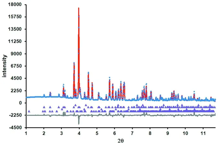
Fig. 1 shows the Rietveld difference plot for the present refinement. The crystal structure of Sr2TiO(Si2O7) is displayed
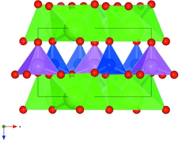
in Fig. 2 and consists of layers of corner-sharing SiO4 and TiO5 polyhedra extending parallel to the ab plane. These layers
are separated along the c axis by layers of distorted SrO6 octahedra.
Experimental
A synthetic sample of Sr-fresnoite was made by melting a stoichiometric mixture of SrCO3, TiO2 and SiO2 to form a
glass. This glass was then quenched to 293 K, reground and then heated for 7 days at 1323 K. A small amount of CeO2
(NIST SRM 674a) standard was added to this powdered sample to act as an internal standard.
Refinement
The powdered sample was loaded into a 0.7 mm diameter quartz capillary, prior to synchrotron X-ray powder diffraction
data collection using the P02.1 high resolution powder diffraction beamline at the PETRA-III synchrotron. The beam on
collected at 293 K out to approx. 11.9°/2θ, the data collection time was 30 s. Powder diffraction data were converted to a
list of 2θ and intensity using FIT2D (Hammersley et al., 1996, Hammersley, 1997). Powder diffraction data in the range
1–11.7°/2θ were used for the Rietveld refinement. Data below 1°/2θ were excluded due to scatter from the beam stop and
as there were no Bragg reflections in this region. Data above 11.7°/2θ were excluded as this corresponded to the edge of
the image plate detector where the Bragg peaks were weaker.
The main Bragg reflections of the powder diffraction pattern could be indexed in space group P4bm with similar lattice
parameters to those of PDF card 39–228 (ICDD, 1989). The unit cell of the incommensurately modulated structure
(Höche et al., 2002) corresponds to a doubled c axis compared to that given on the PDF card. The doubled c axis does not
match with some of the low-angle Bragg reflections for the Sr2TiO(Si2O7) sample used in the present study, therefore this
incommensurate structure was not used for Rietveld refinement. Bragg reflections for three impurity phases could also be
identified in the powder diffraction data. SrTiO3 and SrSiO3 were formed as by-products during preparation.
Initial lattice parameters for the three Sr-containing phases were refined using local software. The CeO2 (NIST SRM
674a) standard was used to calibrate the sample to detector distance. The CeO2 lattice parameter was fixed at 5.4111 Å so
as to calibrate the wavelength as 0.207549 Å.
The P4bm crystal structure of the mineral fresnoite (Ba2TiO(Si2O7); Ochi, 2006) was used as a starting model for the
Rietveld refinement (Rietveld, 1969) of the structure of Sr2TiO(Si2O7). The crystal structures of SrSiO3 (Machida et al.,
1982), SrTiO3 (Mitchell et al., 2000) and CeO2 (Goldschmidt & Thomassen, 1923) were used for the impurity phases in
the refinement. Isotropic atomic displacement parameters were used for all phases. For the Sr2TiO(Si2O7) phase the Si—
O and Ti—O distances in the SiO4 and TiO5 polyhedra were soft-constrained to those for Ba2TiO(Si2O7) (Ochi, 2006).
The Uiso factors for all O sites were constrained to be the same. 81 (1) wt.% of Sr-fresnoite was present in this sample
with 4.6 (3) wt.% CeO2, 7.0 (6) wt.% SrTiO3 and 7.4 (8) wt.% of SrSiO3 present as impurities.
Computing details
Data collection: local software; cell refinement: local software; data reduction: local software; program(s) used to solve
structure: coordinates taken from a related compound; program(s) used to refine structure: FULLPROF
(Rodriguez-Carvajal, 2001); molecular graphics: VESTA (Momma & Izumi, 2008); software used to prepare material for publication:
supplementary materials
sup-3
[image:5.610.130.481.74.305.2]Acta Cryst. (2013). E69, i1
Figure 1
Rietveld difference plot for the multi-phase refinement of Sr2TiO(Si2O7), CeO2, SrTiO3 and SrSiO3. The blue crosses, and
red and black lines show respectively the observed, calculated and difference plots. Calculated Bragg reflection positions
Figure 2
The crystal structure of Sr2TiO(Si2O7). Purple polyhedra show TiO5 units, blue polyhedra show SiO4 units, green
polyhedra show distorted SrO6 units. Green spheres represent Sr atoms, pink spheres represent Ti atoms, blue spheres
represent Si atoms and red spheres represent O atoms.
Distrontium oxidotitanium disilicate
Crystal data Sr2TiSi2O8
Mr = 407.31
Tetragonal, P4bm Hall symbol: P 4 -2ab a = 8.3200 (3) Å c = 5.0239 (2) Å V = 347.77 (2) Å3
Z = 2
Dx = 3.890 (1) Mg m−3
Synchrotron radiation, λ = 0.207549 Å µ = 0.43 mm−1
T = 293 K
Particle morphology: powder white
cylinder, 20 × 0.7 mm
Specimen preparation: Prepared at 1323 K and 100 kPa
Data collection In-house design
diffractometer
Radiation source: Synchrotron Laue DCM diamond(111) & Si(111)
monochromator
Specimen mounting: capillary Data collection mode: transmission Scan method: continuous
supplementary materials
sup-5
Acta Cryst. (2013). E69, i1
Refinement Rp = 0.052
Rwp = 0.073
Rexp = 0.031
RBragg = 0.093
χ2 = 31.068
1476 data points
Excluded region(s): 0-1 and 11.7-12.0 degrees 2θ
Profile function: T-C-H Pseudo-Voigt function 71 parameters
5 restraints
Fractional atomic coordinates and isotropic or equivalent isotropic displacement parameters (Å2)
x y z Uiso*/Ueq
Sr1 0.3282 (2) 0.8282 (2) 0.017 (2) 0.0070 (8)*
Ti1 0.00000 0.00000 0.558 (3) 0.007 (2)*
Si1 0.1305 (6) 0.6305 (6) 0.535 (3) 0.019 (2)*
O1 0.00000 0.50000 0.651 (5) 0.017 (3)*
O2 0.1292 (15) 0.6292 (15) 0.191 (3) 0.017 (3)*
O3 0.2985 (12) 0.5984 (15) 0.678 (3) 0.017 (3)*
O4 0.00000 0.00000 0.209 (3) 0.017 (3)*
Geometric parameters (Å, º)
Sr1—O1i 2.733 (18) Ti1—O3viii 1.961 (12)
Sr1—O2 2.499 (13) Ti1—O3ix 1.961 (13)
Sr1—O2ii 2.676 (13) Ti1—O4 1.75 (2)
Sr1—O2iii 2.676 (13) Si1—O1 1.642 (11)
Sr1—O3iv 2.572 (15) Si1—O2 1.73 (2)
Sr1—O3v 2.572 (14) Si1—O3 1.594 (14)
Ti1—O3vi 1.961 (12) Si1—O3x 1.594 (15)
Ti1—O3vii 1.961 (13)
O3xi—Ti1xii—O3xiii 84.6 (9) O3x—Ti1xii—O4xii 107.9 (12)
O3xi—Ti1xii—O3xiv 144.2 (10) O3xiv—Ti1xii—O4xii 107.9 (12)
O3xiv—Ti1xii—O3xiii 84.6 (8) O1—Si1—O2 110.3 (16)
O3xi—Ti1xii—O4xii 107.9 (12) O1—Si1—O3x 108.0 (9)
O3xiv—Ti1xii—O3x 84.6 (8) O1—Si1—O3 108.0 (10)
O3xiii—Ti1xii—O3x 144.2 (11) O2—Si1—O3 117.1 (15)
O3xiii—Ti1xii—O4xii 107.9 (12) O2—Si1—O3x 117.1 (15)
O3x—Ti1xii—O3xi 84.6 (9) O3x—Si1—O3 95.2 (11)