ISSN Online: 2158-2947 ISSN Print: 2158-2912
DOI: 10.4236/nm.2019.103019 Sep. 23, 2019 247 Neuroscience & Medicine
Environmental Exposure to Lead, Vanadium,
Copper and Selenium: Possible Implications in
the Development of Autism Spectrum
Disorders
A. O. Akinade
1, I. O. Omotosho
1*, I. A. Lagunju
2, M. A. Yakubu
31Department of Chemical Pathology, College of Medicine, University of Ibadan, Ibadan, Nigeria 2Department of Paediatrics, College of Medicine, University of Ibadan, Ibadan, Nigeria
3Department of Environmental Science and Technology, Texas Southern University, Houston, TX, USA
Abstract
Human exposure to toxic metals is on the increase especially in the develop-ing world; this is compounded by the almost unavoidable application of the metals domestically and industrially and their implication in several genetic defects, aging and some chronic illnesses including Autism Spectrum Dis-orders (ASD). This study investigated the concentration of toxic metals (Pb and V) and micro-essential elements (Cu and Se) in children with ASD and controls in Nigeria towards establishing their possible associations with the aetiopathogenesis of ASD. Eight children clinically diagnosed by Paediatric Neurologist and Child Psychiatrist for ASD using DMS-IV and fifteen appar-ently healthy children (age range 2 - 12 years) were recruited as cases and controls respectively. Plasma levels of Pb, V, Cu and Se were analyzed using Induction ICP-MS. Results were analyzed using students t-test. The mean plasma lead and vanadium levels were (7.92 ± 1.30 µg/dl; 1.07 ± 0.22 µg/dl) and (6.83 ± 0.72 µg/dl; 2.59 ± 0.48 µg/dl) in children with ASD and in con-trols respectively. The result showed that blood lead level in ASD was slightly increased but not significant when compared with control (p < 0.433). On the other hand, plasma vanadium concentration in ASD was significantly re-duced (1.07 ± 0.22 µg/dl) when compared with control (2.59 ± 0.48 µg/dl) (P < 0.038). Mean plasma copper was similar in all participants (1.98 ± 0.13, 2.23 ± 0.12) but selenium concentrations were significantly reduced (0.37 ± 0.05 mg/L; 0.57 ± 0.02 mg/L) in ASD relative to controls respectively. Given the physiological functions of vanadium and selenium, the observed reduced le-vels of the two elements in children with ASD may account for the speech How to cite this paper: Akinade, A.O.,
Omotosho, I.O., Lagunju, I.A. and Yakubu, M.A. (2019) Environmental Exposure to Lead, Vanadium, Copper and Selenium: Possible Implications in the Development of Autism Spectrum Disorders. Neuros-cience & Medicine, 10, 247-258.
https://doi.org/10.4236/nm.2019.103019
Received: July 8, 2019 Accepted: September 23, 2019 Published: September 23, 2019
Copyright © 2019 by author(s) and Scientific Research Publishing Inc. This work is licensed under the Creative Commons Attribution International License (CC BY 4.0).
DOI: 10.4236/nm.2019.103019 248 Neuroscience & Medicine
and other neurological dysfunctions of the brain in ASD.
Keywords
Toxic and Essential Metals, Autism Spectrum Disorder, Children, Aetiopathogenesis
1. Introduction
Autism Spectrum Disorders (ASD) are a heterogeneous group of neurodevel-opmental disorders that are behaviourally defined and characterized by impair-ments in communication and social interaction along with restrictive and repeti-tive behaviours beginning in infancy and toddler years [1][2]. Several reasons have been proffered as the pathophysiology of ASD, however, contribution of industrial chemicals and metallic pollutants widely disseminated in the envi-ronment have been suggested as important contributors to the development of this condition [3][4]. The vulnerability of the developing human brain in-utero has also lent credence to its prevalence among children. Recently, interaction between genes and the toxicants has been suggested as basis of genetic modula-tion and the development of ASD [5]. Thus, due to this complex pathophysiol-ogy of ASD, many authors are of the opinion that both genetic and environ-mental factors (and their interactions) are strongly linked in the development of the disorders [6]. The possibility of this assertion has been heightened by the re-cent increase in prevalence of the disorder worldwide.
Specifically, in recent times, research and clinical studies have implicated physiological and metabolic systems that transcend specific organ dysfunction, such as immune dysregulation, inflammation, impaired detoxification, im-paired redox regulation/oxidative stress and energy generation/mitochondrial systems [7][8]. These imply that ASD may arise from, or at least involve, sys-temic physiological abnormalities rather than being a purely central nervous system disorder in a subset of individuals with ASD [9]. It is in this respect that the involvement of trace and toxic metals (which in a developing economy like ours are inevitable pollutants) and their interaction with gene develop-ment especially in subjects that are actively or passively exposed to them be-come important.
Trace elements are chemical micronutrients which are required in small amounts but play a vital role in various physiological and metabolic processes occurring within living tissues [10][11]. There are threshold levels for most of these trace elements; hence, their deficiency or excessive accumulation may cause serious changes in the body leading to disruption of important enzyme ac-tivities [12]. The immature blood-brain barrier, neuronal growth migration and myelination processes that occur on a specific and rapid schedule in a develop-ing foetus may largely facilitate development of ASD in children.
DOI: 10.4236/nm.2019.103019 249 Neuroscience & Medicine
been shown to be harmful to the developing fetus and may be harmful to the human nervous system, even at low levels [13] [14]. Also, several studies have reported that trace metals could easily cross the placenta and affect cognitive development [15] despite its function as a barrier protecting the fetus from toxic metal exposure. The possible interaction between these toxic metals either in modulating the nutritional function of the essential metals or on genetic pairing remains issues in the aetiopathogenesis of ASD. Until recently, the study of po-tential environmental toxicant contributions to the development of ASD has been generally “neglected” [16], hence the need for a clearer look at their con-tribution to the development of ASD especially with the gradual increase in the prevalence of this disorder.
2. Subjects and Methods
Recruitment: A total number of 23 participants were recruited by conveni-ence sampling method for this pilot study between August and November 2015. This comprised of eight (8) children clinically diagnosed for ASD with mean age 5.25 years ± 0.37 and fifteen (15) apparently healthy children with mean age 7.87 years ± 0.89 as controls. All children recruited for this study received routine childhood vaccinations.
Selection: Paediatric Neurologist and Child Psychiatrist clinically selected the subjects after proper evaluations and diagnosis according to Diagnostic and Sta-tistical Manual of Mental Disorder DMS-IV-DR; DMS-5 classification. Children and their parent’s history were taken.
Ethical approval was obtained from the UCH/UI joint Ethical Committee. Informed consent was obtained from each subject through their parents. Inclusion criteria: Clinically diagnosed children with ASD that their parents gave informed consent were recruited for the study.
Exclusion criteria: Participants that were suffering from liver or kidney dis-ease, anemia, or current treatment for iron deficiency, progressive neurological disorders, or epilepsy were excluded from the study.
Method: History of all children in the study was taken covering: parent so-cio-economic status, pregnancy history, developmental milestones, dietary his-tory, and environmental exposure factor. History of major childhood illnesses and immunizations were taken and clinical examination of all body systems with special emphasis on neurological examination was performed by a Neu-ro-paediatrician. All autistic children were subjected to a full clinical child psy-chiatric evaluation for diagnosis of autistic spectrum disorder and exclusion of other psychiatric disorders according to Diagnostic and Statistical Manual of Mental Disorders based on DSM-IV-TR; DSM-5 [17][18].
DOI: 10.4236/nm.2019.103019 250 Neuroscience & Medicine
centrifuge, manufactured in England) to obtain plasma which was promptly se-parated into another clean plane bottle. All samples were kept frozen at -20oC
until they were ready for analysis. They were analyzed for Cu, Se, V and Pb using Induction Coupled Plasma-Mass Spectrometry (ICP-MS).
Statistical analysis: Appropriate in-built software in the instrument was used to calculate the result of the analysis with inclusion of standards and controls. The results were converted to SI units from part per billion (ppb) using appro-priate conversion factors.
Data were reviewed, coded, tabulated and analyzed using statistical package for social science (SPSS 20.0)
Mean standard error (MEAN ± S.E) was used to express descriptive statistics of the results while student t-test was used to access the statistical significance of the difference between the two groups. P value of 0.05 was regarded significant.
[image:4.595.211.536.423.702.2]3. Results
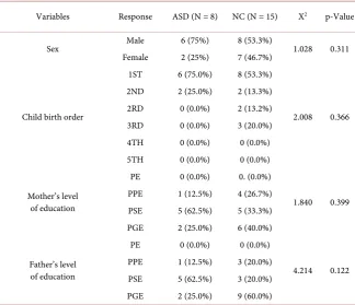
Table 1 showed gender percentage distribution; 6 boys (75%) and 2 girls (25%)
constituted the ASD group while 8 boys (53.3%) and 7 girls (46.7%) constituted the neurotypical (control) group. The result showed that 6 (75.0%) of the ASD group were 1st born among the siblings. It was revealed from the results that the parents of the two groups belong to the same socio-economic status.
Table 1. Comparison of % gender distribution and socio-economic grouping of the two groups.
Variables Response ASD (N = 8) NC (N = 15) X2 p-Value
Sex Male 6 (75%) 8 (53.3%) 1.028 0.311
Female 2 (25%) 7 (46.7%)
Child birth order
1ST 6 (75.0%) 8 (53.3%)
2.008 0.366
2ND 2 (25.0%) 2 (13.3%)
2RD 0 (0.0%) 2 (13.2%)
3RD 0 (0.0%) 3 (20.0%)
4TH 0 (0.0%) 0 (0.0%)
5TH 0 (0.0%) 0 (0.0%)
Mother’s level of education
PE 0 (0.0%) 0. (0.0%)
1.840 0.399
PPE 1 (12.5%) 4 (26.7%)
PSE 5 (62.5%) 5 (33.3%)
PGE 2 (25.0%) 6 (40.0%)
Father’s level of education
PE 0 (0.0%) 0 (0.0%)
4.214 0.122
PPE 1 (12.5%) 3 (20.0%)
PSE 5 (62.5%) 3 (20.0%)
PGE 2 (25.0%) 9 (60.0%)
DOI: 10.4236/nm.2019.103019 251 Neuroscience & Medicine
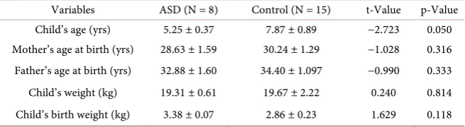
Table 2. Comparison of biodata variables in ASD and control (mean ± S.E.).
Variables ASD (N = 8) Control (N = 15) t-Value p-Value
Child’s age (yrs) 5.25 ± 0.37 7.87 ± 0.89 −2.723 0.050
Mother’s age at birth (yrs) 28.63 ± 1.59 30.24 ± 1.29 −1.028 0.316
Father’s age at birth (yrs) 32.88 ± 1.60 34.40 ± 1.097 −0.990 0.333
Child’s weight (kg) 19.31 ± 0.61 19.67 ± 2.22 0.240 0.814
Child’s birth weight (kg) 3.38 ± 0.07 2.86 ± 0.23 1.629 0.118
[image:5.595.211.541.243.320.2]*significant at p < 0.05.
Table 3. Comparison of trace and toxic elements levels in ASD and control (mean ± S.E.).
Variables ASD (N = 8) Control (N = 15) t-Value p-Value
Copper (mg/L) 1.98 ± 0.13 2.23 ± 0.12 −1.290 0.211
Selenium (mg/L) 0.37 ± 0.05 0.57 ± 0.02 −5.316 0.000*
Lead (µg/dl) 7.92 ± 1.30 6.83 ± 0.72 0.799 0.433
Vanadium (µg/dl) 1.07 ± 0.22 2.59 ± 0.48 −2.219 0.038*
*significant at p < 0.05.
Table 2 showed the biodata of participants. There were no significant differ-ences in the children’s age, mother’s age at birth, father’s age at birth, children’s birth weight and children weight between ASD and control. It may be said that there were no disparities in recorded biodata of the two groups.
Table 3 showed the comparison of levels of Pb, V, Cu and Se in ASD and
control groups. The result showed there were no significant differences in the mean levels of Cu (1.98 ± 0.13 mg/L; 2.23 ± 0.12) and Pb (7.92 ± 1.30 µg/dl; 6.83 ± 0.72 µg/dl) in ASD and controls respectively (p < 0.211; p < 0.433). However, Se level was significantly reduced in ASD when compared with the control (0.37 ± 0.05 mg/L and 0.57 ± 0.02 mg/L) respectively (p < 0.000). Also, plasma vana-dium level was significantly reduced in ASD compared to controls (1.07 ± 0.22 µg/dl and 2.59 ± 0.48 µg/dl) respectively (p < 0.038).
4. Discussion
DOI: 10.4236/nm.2019.103019 252 Neuroscience & Medicine
The study was designed to determine the plasma levels of V, Cu, Se and Pb in ASD and based on their biochemical roles, to compare with controls in order to assess their possible contribution to the disorder. The increased blood Pb level (though not significant) found in this study may be pathognomonic of the dis-ease. This may be the basis of the impaired cognitive and behavioural deficits characteristic of this condition. This may be facilitated by the increased sensitiv-ity of the brain to lead exposure [24]. Lead has been severally reported to dis-place other elements like calcium and magnesium from most of their reactive sites through several processes. The pathological effect especially when present in a prolonged and excessive amount may contribute to the development of ASD. Its effect inducing cognitive and behavioral deficits in children and adult resulting in a distinct neurological effect, with different brain targets and modes of action has been severally documented [25] [26]. Hence, the possible delete-rious effect of air-borne lead in children compared to adults because of the higher respiratory and metabolic activities of the former may have also facili-tated the development of ASD in these children. Previous studies done on hair lead level reported increased lead levels in ASD compared to controls [27][28] [29] just as it has been observed in this study that blood lead level in ASD was higher than in controls. This was also in agreement with other studies that re-ported increased blood levels in ASD [30][31]. Secondly, since calcium has also been reported to be selectively displaced by lead when the latter is in excess; the increased lead level in this study may be precipitated by the hypocalcaemia ob-served in children with ASD which was reported earlier by this team [30]. Nutri-tionally, children with ASD have been reported to have low poor eating habits with attendant low nutritional compliments. It may therefore be possible that reduced essential elements level in ASD children may also exacerbate the devel-opment of autism especially in an environmentally prone setting.
DOI: 10.4236/nm.2019.103019 253 Neuroscience & Medicine
to increase in free radical activity in the body [34]. Studies on this element have been very scanty especially in humans.
The major source of exposure to vanadium for the general population is food. The major report on this element has been on its insulin-like action in-vivo. Vanadium in the form of vanadate and vanadyl sulfate has been shown to im-prove the effect of insulin in diabetic animals; artificially induced diabetes in rats can be reversed by vanadate [35]. Large doses also affect serum fat and choles-terol levels, though more research in this area is needed. Biochemically, the pos-sibility that vanadium may play a role in the regulation of Na+/K+-exchanging
ATPase, phosphoryl-transfer enzymes, adenylate cyclase and protein kinases has been reported. The possible role of the vanadyl ion as an enzyme cofactor and its roles in hormone, glucose, lipid, bone and tooth metabolism have also been dis-cussed [36]. No specific biochemical function has yet been identified for vana-dium in higher animals. However, the recent discovery of vanavana-dium-activated enzymes in lower forms of life lends credence to the view that vanadium has similar roles in higher animals. In this study, plasma level of this essential ele-ment was found to be low in children with ASD. The level was significantly low in comparison to that of control children. Based on what has been reported in lower animals, the essentiality of this element for normal growth and develop-ment is not in doubt. Hence, the observed reduced level in children with ASD may be associated with underdevelopment of the cognitive functions of the brain in this group of neurodevelopmental disorder. Biochemically, this may be due to its role in Na+/K+ ATPase and Phosphoryl enzyme transfer activities which are
all linked to calcium homeostasis making growing children more vulnerable es-pecially in sensitive organs like in their brain.
DOI: 10.4236/nm.2019.103019 254 Neuroscience & Medicine
disorder [39]. Although findings in this study were contrary to previous studies that reported elevated copper levels in ASD compared with control [40] [41] [42], this work was in agreement with the report of Craciun et al. that reported reduced Cu level in children with ASD [43].
Selenium is an essential trace metal needed in the production of antioxidants enzymes. As Selenoproteins, they are essential for brain development, redox control, and preventing and reversing oxidative damage in the brain and neu-roendocrine tissues [44]. This important redox property of Se may have been compromised by the reduced Se level observed in ASD children in this study. The reduced selenium level found in this study may have contributed to the pa-thophysiology of ASD especially since glutathione is a very prominent antioxi-dant in the neuroendocrine tissues of the brain. Glutathione is known to be a major redox buffer in the trans-sulfuration pathway in the neuroendocrine tis-sue [45], this process ensures replication of the redox form of the antioxidant which contributes to the transmission of impulse along the nerves. It may there-fore be inferred that a reduced level of Se greatly hampers this mechanism and may largely account for some of the neuro-behavioural changes associated with ASD. Control of intracellular oxidative tone and findings of increased oxidative damage in children with ASD may be indicative of disruptions of selenoenzyme activities resulting from reduced selenium levels. This finding agreed with pre-vious studies that reported reduced Se levels in analysis done on hair and red blood cells in their studies [46][47][48].
5. Conclusion
The hallmark of ASD in children in this environment may be indicated by a de-ficiency in the essential trace elements-V, Cu and Se with a concurrent increase in Pb especially in environmentally vulnerable children. Abnormality in the le-vels of these elements may be good biomarkers of ASD especially in environ-mentally vulnerable children with or without the presence of the known clinical symptoms of ASD.
6. Limitation
The small sample size was a limitation in this study, a larger sample size may re-veal more. Also, control of environmental pollution is difficult in this part of the world; this may be due to level of public health awareness.
Conflicts of Interest
The authors declare no conflicts of interest regarding the publication of this pa-per.
References
[1] WHO (2012) In World Health Organization, Geneva.
DOI: 10.4236/nm.2019.103019 255 Neuroscience & Medicine
[3] Grandjean, P. (2013) Only One Chance. How Environmental Pollution Impairs Brain Development and How to Protect the Brains of the Next Generation. Oxford University Press, New York.
https://doi.org/10.1093/acprof:oso/9780199985388.001.0001
[4] Grandjean, P. and Perez, M. (2008) Development Neurotoxicity: Implications of Methylmercury Research. International Journal of Environment and Health, 2, 417-428.https://doi.org/10.1504/IJENVH.2008.020933
[5] Rice, D. and Barone, S.Jr. (2000) Critical Periods of Vulnerability for the Develop-ing Nervous System: Evidence from Humans and Animal Models. Environmental Health Perspectives, 108, 511-533.https://doi.org/10.1289/ehp.00108s3511
[6] Blaurock-Busch, E., Amin, O.R. and Rabah, T. (2011) Heavy Metals and Trace Ele-ments in Hair and Urine of a Sample of Arab Children with Autistic Spectrum Dis-order. Maedica (Buchar), 6, 247-257.
[7] Blaurock-busch, E., Amin, O.R., Dessoki, H.H. and Rabah, T. (2012) Toxic Metals and Essential Elements in Hair and Severity of Symptoms among Children with Autism. Maedica: A Journal of Clinical Medicine, 7, 38-48.
https://doi.org/10.1016/S0924-9338(12)74444-9
[8] Rossignol, D.A. and Frye, R.E. (2012) A Review of Research Trends in Physiological Abnormalities in Autism Spectrum Disorders: Immune Dysregulation, Inflamma-tion, Oxidative Stress, Mitochondrial Dysfunction and Environmental Toxicant Exposures. Molecular Psychiatry, 17, 389-401.https://doi.org/10.1038/mp.2011.165
[9] Ming, X., Brimacombe, M., Chaaban, J., Zimmerman-Bier, B. and Wagner, G.C. (2008) Autism Spectrum Disorders: Concurrent Clinical Disorders. Journal of Child Neurology, 23, 6-13.https://doi.org/10.1177/0883073807307102
[10] Herbert, M.R. (2005) Autism: A Brain Disorder or a Disorder That Affects the Brain. Clinical Neuropsychiatry, 2, 354-379.
[11] Prashanth, L., Kattapagari, K.K., Chitturi, R.T., Baddam, V.R. and Prasad, L.K. (2015) A Review on Role of Essential Trace Elements in Health and Disease. Journal of Dr. NTR University of Health Sciences, 4, 75-85.
https://doi.org/10.4103/2277-8632.158577
[12] Bhattacharya, P.T., Misra, S.R. and Hussain, M. (2016) Nutritional Aspects of Es-sential Trace Elements in Oral Health and Disease: An Extensive Review. Scientifi-ca, 2016, Article ID: 5464373.https://doi.org/10.1155/2016/5464373
[13] Skalny, A.V. (2014) Bioelements and Bioelementology in Pharmacology and Nutri-tion: Fundamental and Practical Aspects. In: Atroshi, F., Ed., Pharmacology and Nutritional Intervention in the Treatment of Disease, InTech, Rijeka, 225-241.
https://doi.org/10.5772/57368
[14] Rahbar, M.H., Samms-Vaughan, M., Dickerson, A.S., Loveland, K.A., Ardjo-mand-Hessabi, M., Bressler, J., Shakespeare-Pellington, S., Grove, M.L. and Boer-winkle, E. (2015) Factors Associated with Blood Lead Concentrations of Children in Jamaica. Journal of Environmental Science and Health, Part A, 50, 529-539. [15] National Scientific Council on the Developing Child (2006) Early Exposure to Toxic
Substances Damages Brain Architecture. Working Paper No. 4. Center on the De-veloping Child at Harvard University, Cambridge.
[16] Julvez, J. and Grandjean, P. (2009) Neurodevelopmental Toxicity Risks Due to Oc-cupational Exposure to Industrial Chemicals during Pregnancy. Industrial Health, 47, 459-468.https://doi.org/10.2486/indhealth.47.459
En-DOI: 10.4236/nm.2019.103019 256 Neuroscience & Medicine
vironmental Contributions to Autism: Provocative Clues and False Leads. Mental Retardation and Developmental Disabilities Research Reviews, 10, 292-302.
https://doi.org/10.1002/mrdd.20043
[18] American Psychiatric Association (2000) Diagnostic and Statistical Manual of Mental Disorders, (DSM-5®) 2013 Text Revision. 4th Edition, American Psychiatric Association, Washington DC.
[19] Froehlich, W. and Fung, L.K. (2012) Autism, Comorbidity in Psychiatry, Social Be-havior, Pervasive Developmental Disorder, Addiction.
[20] CDC (Centers for Disease Control and Prevention) (2014) Prevalence of Autism Spectrum Disorders—Autism and Developmental Disabilities Monitoring Network, 14 Sites, United States, 2008. MMWR Surveillance Summaries, 61, 1-19.
[21] Lagunju, I.A., Bella-Awusah, T.T. and Omigbodun, O.O. (2014) Autistic Disorder in Nigeria: Profile and Challenges to Management. Epilepsy & Behavior, 39C, 126-129.https://doi.org/10.1016/j.yebeh.2014.08.020
[22] Baxter, A.J., et al. (2015) The Epidemiology and Global Burden of Autism Spectrum Disorders. Psychological Medicine, 45, 601-613.
https://doi.org/10.1017/S003329171400172X
[23] Modabbernia, A., Velthorst, E. and Reichenberg, A. (2017) Environmental Risk Factors for Autism: An Evidence-Based Review of Systematic Reviews and Me-ta-Analyses. Molecular Autism, 8, 13.https://doi.org/10.1186/s13229-017-0121-4
[24] Ronald, A. and Hoekstra, R.A. (2011) Autism Spectrum Disorders and Autistic Traits: A Decade of New Twin Studies. American Journal of Medical Genetics Part B: Neuropsychiatric Genetics, 156, 255-274.https://doi.org/10.1002/ajmg.b.31159
[25] Bouchard, M.F., Sauvé, S., Barbeau, B., Legrand, M., Brodeur, M.È., et al. (2011) In-tellectual Impairment in School-Age Children Exposed to Manganese from Drink-ing Water. Environmental Health Perspectives, 119, 138-143.
https://doi.org/10.1289/ehp.1002321
[26] Lanphear, B.P., Hornung, R., Khoury, J., Yolton, K., Baghurst, P., et al. (2005) Low-Level Environmental Lead Exposure and Children’s Intellectual Function: An International Pooled Analysis. Environmental Health Perspectives, 113, 894-899.
https://doi.org/10.1289/ehp.7688
[27] El Baz Mohamed, F., et al. (2015) Assessment of Hair Aluminum, Lead, and Mer-cury in a Sample of Autistic Egyptian Children: Environmental Risk Factors of Heavy Metals in Autism. Behavioural Neurology, 2015, Article ID: 545674.
https://doi.org/10.1155/2015/545674
[28] Adams, J.B., Baral, M., Geis, E., et al. (2009) The Severity of Autism Is Associated with Toxic Metal Body Burden and Red Blood Cell Glutathione Levels. Journal of Toxicology, 2009, Article ID: 532640.https://doi.org/10.1155/2009/532640
[29] Geier, D.A., King, P.G., Sykes, L.K. and Geier, M.R. (2008) A Comprehensive Re-view of Mercury Provoked Autism. Indian Journal of Medical Research, 128, 383-411.
[30] Omotosho, I.O., Akinade, A.O. and Lagunju, I.A. (2018) Calcium and Magnesium Levels Are Down-Regulated in Nigeria Children with Autism Spectrum Disorder and Cerebral Palsy. Neuroscience and Medicine, 9, 159-170.
https://doi.org/10.4236/nm.2018.93016
DOI: 10.4236/nm.2019.103019 257 Neuroscience & Medicine
[32] Mason, L.H., Harp, J.P. and Han, D.Y. (2014) Pb Neurotoxicity: Neuropsychological Effects of Lead Toxicity. BioMed Research International, 2014, Article ID: 840547.
https://doi.org/10.1155/2014/840547
[33] Brookes, P.S., Yoon, Y., Robotham, J.L., Anders, M.W. and Sheu, S.-S. (2004) Cal-cium, ATP, and ROS: A Mitochondrial Love-Hate Triangle. American Journal of Physiology, 287, C817-C833.https://doi.org/10.1152/ajpcell.00139.2004
[34] Mutter, J., Naumann, J., Schneider, R., Walach, H. and Haley, B. (2005) Mercury and Autism: Accelerating Evidence. Neuroendocrinology Letters, 26, 439-446. [35] McNeill, J.H., Yuen, V.G., Hoveyda, H.R. and Orvig, C. (1992) Bis(maltolato)oxo-
vanadium(IV) Is a Potent Insulin Mimic. Journal of Medicinal Chemistry, 35, 1489-1491. https://doi.org/10.1021/jm00086a020
[36] Rehder, D. (2013) Vanadium. Its Role for Humans. Metal Ions in Life Sciences, 13, 139-169.https://doi.org/10.1007/978-94-007-7500-8_5
[37] Madsen, E. and Gitlin, J.D. (2007) Copper and Iron Disorders of the Brain. Annual Review of Neuroscience, 30, 317-337.
https://doi.org/10.1146/annurev.neuro.30.051606.094232
[38] Davis, J.M. and Svendsgaard, D.J. (1990) Nerve Conduction Velocity and Lead: A Critical Review and Meta-Analysis. In: Johnson, B.L., Ed., Advances in Neurobeha-vioural Toxicology, Lewis Publishers, Chelsea, 353-376.
[39] Mattie, M.D., McElwee, M.K. and Freedman, J.H. (2008) Mechanism of Cop-per-Activated Transcription: Activation of AP-1, and the JNK/SAPK and p38 Signal Transduction Pathways. Journal of Molecular Biology, 383, 1008-1018.
https://doi.org/10.1016/j.jmb.2008.08.080
[40] Scott, R.J., Murrough, J.W., Han, M.-H., Charney, D.S. and Nestler, E.J. (2012) Neurobiology of Resilience Trace Elements after Dietary Exposure to Toxic Metals.
Biological Trace Element Research, 23, 25-53.
[41] Russo, A.J. and Devito, R. (2011) Analysis of Copper and Zinc Plasma Concentra-tion and the Efficacy of Zinc Therapy in Individuals with Asperger’s Syndrome, Pervasive Developmental Disorder Not Otherwise Specified (PDD-NOS) and Aut-ism. Biomark Insights, 6, 127-133.https://doi.org/10.4137/BMI.S7286
[42] Priya, L. and Geetha, A. (2011) Level of Trace Elements (Copper, Zinc, Magnesium and Selenium) and Toxic Elements (Lead and Mercury) in the Hair and Nail of Children with Autism. Biological Trace Element Research, 142, 148-158.
https://doi.org/10.1007/s12011-010-8766-2
[43] Crăciun, E.C., Bjørklund, G., Tinkov, A.A., Urbina, M.A., Skalny, A.V., Rad, F. and Dronca, E. (2016) Evaluation of Whole Blood Zinc and Copper Levels in Children with Autism Spectrum Disorder. Metabolic Brain Disease, 31, 887-890.
https://doi.org/10.1007/s11011-016-9823-0
[44] Laura, R.J., Deth, R.C. and Ralston, N.V.C. (2014) Potential Role of Selenoenzymes and Antioxidant Metabolism in Relation to Autism Etiology and Pathology. Autism Research and Treatment, 2014, Article ID: 164938.
https://doi.org/10.1155/2014/164938
[45] James, S.J., Cutler, P., Melnyk, S., Jernigan, S., Janak, L., Gaylor, D.W. and Neu-brander, J.A. (2004) Metabolic Biomarkers of Increased Oxidative Stress and Im-paired Methylation Capacity in Children with Autism. The American Journal of Clinical Nutrition, 80, 1611-1617.https://doi.org/10.1093/ajcn/80.6.1611
DOI: 10.4236/nm.2019.103019 258 Neuroscience & Medicine
Autism. Maedica, 7, 38-48.https://doi.org/10.1016/S0924-9338(12)74444-9
[47] Priya, M.D. and Geetha, A. (2010) Level of Trace Elements (Copper, Zinc, Magne-sium and Selenium) and Toxic Elements (Lead and Mercury) in the Hair and Nail of Children with Autism. Biological Trace Element Research, 142, 148-158.
[48] Jory, J. and Woody, R. (2008) Red-Cell Trace Minerals in Children with Autism McGinnis. American Journal of Biochemistry and Biotechnology, 4, 101-104.