ISSN Online: 2160-0406 ISSN Print: 2160-0392
DOI: 10.4236/aces.2019.92016 Apr. 23, 2019 204 Advances in Chemical Engineering and Science
Drug Loading on Microneedles
Meng-Hsuan Hsiao, Hsiu-Feng Ye, Ta-Jo Liu, Jane Wang
*Department of Chemical Engineering, National Tsing Hua University, Taiwan
Abstract
An experimental study was carried out to investigate the amount of drugs loaded on microneedles. The microneedles were made with poly (lactic acid). Aqueous poly (vinyl alcohol) solutions were prepared as drug solutions. Two drug loading approaches, i.e., dropping and dipping, were examined. It was found that capillary number is the only relevant dimensionless group for the two methods. For the dropping approach, dried drugs will spread near the bottom of a microneedle patch provided the surface tension is low. As for the dipping approach, both a single microneedle and an array of nine micro-needles were examined. For a single microneedle, high capillary rises before pulling and pulling speed are two key factors to increase the drug loading vo-lume. For an array of microneedles, the effect of capillary rise owing to the interaction between microneedles would increase the drug loading volume several times higher than a single microneedle of the same dimension.
Keywords
Microneedle, Drug Loading, Dropping, Dipping, Capillary Number, Capillary Rise
1. Introduction
Microneedles (MNs) were first developed by Henry et al. [1] as a new and effec-tive drug delivery system to transport drugs into human bodies in a painless way, since MNs can penetrate human skin but are short enough not to affect the nerve system. Up until now, there are approximately four hundred technical ar-ticles studying the different aspects on how to deliver drugs effectively by mi-croneedles [2].
The definition of a microneedle is not very strict; most MNs are in the solid form with hundreds of needles on a small patch of sizes in the neighborhood of 1 to 4 cm2, but a single needle with a diameter much smaller than the conventional injection tip can be called a microneedle [2].
How to cite this paper: Hsiao, M.-H., Ye, H.-F., Liu, T.-J. and Wang, J. (2019) Drug
Loading on Microneedles. Advances in
Chemical Engineering and Science, 9, 204-222.
https://doi.org/10.4236/aces.2019.92016
Received: January 14, 2019 Accepted: April 20, 2019 Published: April 23, 2019
Copyright © 2019 by author(s) and Scientific Research Publishing Inc. This work is licensed under the Creative Commons Attribution International License (CC BY 4.0).
DOI: 10.4236/aces.2019.92016 205 Advances in Chemical Engineering and Science As of now, there are four types of microneedles, i.e., solid, coated, dissolvable and hollow microneedles [3] [4], and each type has its advantages and draw-backs. The existing works have been focusing heavily on the materials that can be used to make MNs [4], the shapes and geometries of MNs for better insertion into human bodies [5] [6] [7] [8], but most importantly the selection of a suita-ble drug that can be combined with MNs to effectively deliver drugs into human body. Currently, vaccines such as enterovirus 71 [9], influenza [10] [11], hepati-tis B [12], bacillus Calmette-Guérin (BCG) [13], human immunodeficiency virus (HIV) [14], human papillomavirus (HPV) [15], recombinant adenovirus (rADV) [16], diphtheria [17], and drugs such as insulin [18] [19], human growth hormone (hGH) [20], lidocaine [21] have been considered as drugs suitable for the combination with MNs, just to name a few here. Interested readers may refer to several review articles for more details [2] [3] [22].
There are multiple ways to deliver drugs into human body through MNs; drugs can be mixed with FDA approved materials, such as biodegradable poly-mers, including chitosan [23], poly (vinylpyrrolidone) (PVP) [24] [25], carbox-ymethyl cellulose (CMC) [19], poly(vinyl)alcohol (PVA) [24] [26], and then molded and dried to make MNs [27] [28], or drugs can be maintained in liquid form and encapsulated in a hollow space of the MN [29] [30] or injected into an array of hollow MNs [31]. Another approach is to make MNs first, then through different methods, such as ink jet printing [32] [33] [34], spray coating [16] [35]
or dip coating [21] [36] [37] to load drugs on the surface of MNs.
To develop MNs as a useful means for drug delivery, MNs have to be strong enough to penetrate human skin, and the solubility of MNs must be well un-derstood and controlled to make sure that drugs can be diffused into blood ves-sels and function as expected. Most importantly, the amount of drugs entering human body has to be accurately evaluated for curing or controlling illness ef-fectively. It is easier to control the amount of drugs by mixing the drugs with the materials that are used to make MNs, particularly if MNs are made in two steps and drugs can be distributed on the tip region of MNs [23] [38].
Since most drugs and vaccines cannot resist high temperature, once they are mixed with the materials to make MNs, the drying process cannot be carried out at high temperature. Freeze drying or drying below 20˚C is common, but may take several days to obtain dried MNs. One simple way that can be applied to reduce the drying time is MNs that do not contain any drugs or vaccines are made first with materials such as PLA [39] [40] and PLGA [40] [41], since the chosen materials to make MNs are mostly polymers and other additives such as starch [28] [42], trehalose [20], β-cyclodextrin [43] [44] and sodium alginate
DOI:10.4236/aces.2019.92016 206 Advances in Chemical Engineering and Science for this purpose [47]. However, one uncertainty of this approach is the difficul-ties in estimating the precise drug loading quantity and the topology of the drugs loaded on the surface of MNs after drying.
The present study aims to examine two different but simple approaches to load drug solutions on the surface of MNs. The first approach is dropping; which implies that drops of drug solutions are deposited on the top of a micro-needle patch (MNP). The drug solution will cover the entire MNP, then followed by a drying process. Another approach is dipping; where MNs are dipped into a drug solution pool for a moment, then the MNs with drug solution on the sur-face will be lifted and dried. Gill and Prausnitz examined the dip coating process and found that the amount of loaded drug was influenced by solution viscosity, surface tension and wettability [36], which are identical as the study on dip coating operation from the fluid mechanical point [48]. Authors from 3M also had previously shown a number of photos on drugs loaded on MNs with re-peated dip coating operations [21]. In the recent few years, more and more works involve the application of MNs toward the delivery of various drugs be-came available, but the fabrication methods vary drastically [49] [50] [51]. The objective of the present study is to analyze the general effect of critical drug solu-tion characters to enable precise control over the drug loading quantity, as well as the topology of the dried drugs loaded on the surface of MNs.
2. Experimental Work
Several MNs made by poly(lactic acid) (PLA) (L-lactide: D, L-lactide = 70:30, inherent viscosity 0.56 dL/g), purchased from Green Square Materials Inc. (Tai-wan) were used for the drug loading experiment. A metal mold made by Win Coat Co. (Taiwan) was used to produce several polydimethylsiloxane (PDMS; Sylgard 184) molds, which have low surface energy and it is much easier to re-move MNs from the PDMS molds. Three different types of PDMS molds were made to conduct the dropping and dipping experiments separately. For drop-ping, the size of the PDMS mold is 0.035 × 0.035 m with 578 (17 × 34) conical needles on the mold. The height of each needle is 6 × 10⁻⁴ m, the base diameter is 3 × 10⁻⁴ m, and the distance between two tips of the adjacent needles is 6 × 10⁻⁴ m. As for dipping, both a single conical needle mold and a 0.04 × 0.04 m mold with an array of 1 × 9 conical needles were made. The needle height is 1.2 × 10⁻³ m, the needle base diameter is 600 µm and the distance between two needle tips is 9 × 10⁻⁴ m. It is noted that the needles for the dipping experiment are larger than those for dropping because a flow visualization technique was applied to record the dipping process, and it is easier to observe the fluid motion with larger needles.
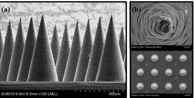
DOI: 10.4236/aces.2019.92016 207 Advances in Chemical Engineering and Science PDMS mold was heated at 150˚C and under 6.67 Pa for 5 hours, and then the MNs were dried and removed from the PDMS mold. As soon as the fabrication process was completed, the digital camera and scanning electron microscope (SEM) images of the needles were taken to evaluate the needle quality. Needle fracture force tests were also applied to make sure the needles could reach the designated strength.
Aqueous PVA solutions were made as the testing solution to study the amount of drugs loaded on the surface of MNs. PVA (MW = 20 - 30 kDa, hy-drolysis = 98% - 99%, Chang Chun Co., Taiwan) were dissolved in DI water to make solutions of different viscosities. A fluorocarbon surfactant (Zonyl® FSO, DuPont) was added to the PVA solutions to adjust the surface tension. In order to evaluate the drug loaded on the needle surface, a small amount of fluorescent tracer dye rhodamine 6G (R4127, Sigma) was added into the solutions. Viscosi-ties of PVA solutions were measured by a viscometer (MCR302, Anton Paar, Graz, Austria) at room temperature and surface tensions of the PVA solution were determined by a surface tensiometer (CBVP-A3, Kyowa Interface Science, Japan). The contact angles of PVA solutions on the PLA surface were analyzed by a contact angle meter (FTA 1000B, First Ten Angstroms, USA).
For the dropping experiment, a MNP of size 0.025 × 0.025 m was set up and a certain amount of PVA solution was dropped onto this MNP. If the viscosity of the PVA solution was too high, the liquid drop would take a very long time to level off. We adopted an easy approach by flooding the MNP with 1.5 ml PVA solutions, such that the liquid level was slightly above the needle tips. A visuali-zation technique was applied to record the side view of the slow drying process of the PVA solutions dropped onto the MNP.
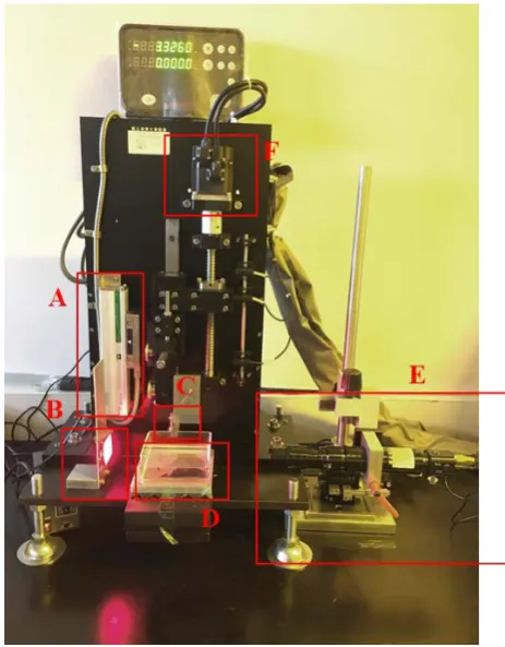
As for dipping, it is necessary to control the penetration and lifting speeds so as to evaluate the amount of PVA solution being carried away from the pool and coated on the surface of MNs. A dip coating device was built as shown in Figure 1; a flow visualization device was attached to record the dip coating process for quantitative analysis later. Dipping on a single conical needle was studied first. Then an array of 1 × 9 conical needles was examined later. The amount of a dried drug loaded on MNs was difficult to determine due to its small size. To evaluate the amount of drugs loaded on the needle surfaces accurately, a calibra-tion curve of fluorescent emission intensity vs. rhodamine 6G concentracalibra-tion was developed first. After a needle was lifted up, it was soaked immediately into a reservoir filled with 3 ml DI water for 20 minutes until the model drug was fully dissolved in the water. Finally, the fluorescent emission intensity of the solution was calculated from the calibration curve, then the amount of drugs loaded on the needle surface could be calculated.
3. Results and Discussions
DOI:10.4236/aces.2019.92016 208 Advances in Chemical Engineering and Science
Figure 1. The dip coating apparatus. (A) Optical linear scale.
(B) Lighting source. (C) Microneedle loading platform. (D) Fluid reservoir. (E) Fluid visualization system. (F) Motor.
Dropping implies that the liquid drops of a drug solution are deposited on a MNP and the drug solution will spread and cover the whole surface of a MNP before drying commences. However, it may not be easy for drug solutions, espe-cially viscous ones, to spread in a short period of time evenly on a flat surface.
Huppert [52] found that the spreading time t of an axisymmetric viscous liq-uid drop can be presented in the following equation:
( )
( )
1/8
1/8
3 K t 0.894 t
gS ν
=
(1)
here
ν
µ
ρ
≡ is the kinematic viscosity, g is the gravitational force, S represents
DOI: 10.4236/aces.2019.92016 209 Advances in Chemical Engineering and Science then start recording how the drug solution is dried under room temperature (25˚C) to find the final topology of the dried drug on the surface on a MNP. The total amount of drug loaded on a MNP can be estimated with the available solid content of the drug solution deposited on the MNP.
The photos of the MNs used in the dropping test are shown in Figure 2. Vari-ations of the diameters and heights of each microneedle are around ±10−5 m.
The tipdiameter of each microneedle is around 1.5 10 ~ 2.5 10× −5 × −5 m.
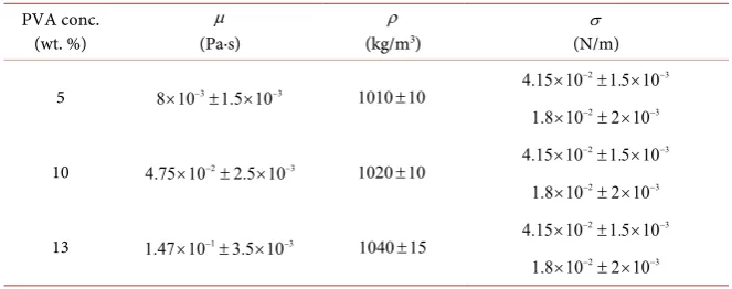
Prausnitz found that surface tension and viscosity are two of the most critical parameters on drug loading, [36] here we also examined the effects of the two parameters. The physical properties of the testing solutions for the dropping test are listed in Table 1. The viscosity and surface tension were varied and the ef-fects of the two parameters were examined. Densities of these solutions are around 1030 to 1060 kg/m3. Note that rhodamine 6G was added to each of the drug solution for flow visualization and the concentration is 2000 µL/mg. The important effects of surface tension and viscosity imply that capillary number Ca is the only critical dimensionless group in this study. Ca is defined as follows:
Ca µV σ
≡ (2)
here µ is the fluid viscosity,
σ
is the surface tension and V is the characte-ristic velocity. The charactecharacte-ristic velocity V can be estimated asdH V
dt
≡ (3)
here H is the distance between the needle tip and the lowest central point of the meniscus after the loaded drug solution is completely dried. For example, if the fluid viscosity is 10−2 Pa·s, surface tension is 4.15 10× −2 N/m, it takes nearly 6
hours to dry the liquid, andH is around 4.6 10× −4 m after drying. Since it takes
[image:6.595.211.538.540.705.2]hours to dry the drug solution at room temperature, V defined in Equation (3) is in the neighborhood of O (10−6), which implies that Ca is very small and the surface tension should be the dominant factor.
DOI:10.4236/aces.2019.92016 210 Advances in Chemical Engineering and Science
Table 1. Physical properties of test solutions for the dropping experiment.
PVA conc. (wt. %)
µ (Pa·s)
ρ (kg/m3)
σ (N/m)
5 8 10× −3±1.5 10× −3 1010 10±
2 3
4.15 10× − ±1.5 10× −
2 3
1.8 10× − ± ×2 10−
10 4.75 10× −2±2.5 10× −3 1020 10±
2 3
4.15 10× − ±1.5 10× −
2 3
1.8 10× − ± ×2 10−
13 1.47 10× −1±3.5 10× −3 1040 15±
2 3
4.15 10× − ±1.5 10× −
2 3
1.8 10× − ± ×2 10−
There are five sets of recorded data with photos showing the variations of the topology of the drug solution surface during the drying process. The photos of the dried drug surfaces are shown in Figure 3. The key points for observation are the point Pf that the dry drug material pinned to the needle surface, and the depth of the dried drug surface represented by H. Data of Pf and H are also given in Figure 3.
We observed that when the drying started, the level of the drug solutions would be lowered gradually owing to solvent evaporation, and the tips of the microneedles would emerge and a meniscus would form between needles when viewing from the front side. The three-phase contact point between the drug so-lution and the needle surface was also lowered gradually as drying proceeded until a certain point Pf was reached, then the liquid meniscus would be pinned at two ends on the needle surface, but the central part would be more and more curved until drying was complete. The pinned point Pf represents the highest point that the drug is attached to the microneedle, the position of Pf together with the depth H of the drug meniscus would affect the rate and the amount of the drug diffusing into human bodies.
Several comments can be made after examining the results in Figure 3. Com-paring between the photos of Cases (A) with Case (B), it is evident that for a more viscous drug solution, Pf appears to be higher, owing to the fact that sol-vent evaporation will increase the fluid viscosity and a more viscous liquid tends to move slower and dry faster. Note that the solid contents of Case (A) and (B) are different and the total amount of dried drugs in the MNP are also different. Once a surfactant was added and the surface tension of the drug solution was reduced, the results shown in Cases (C), (D) and (E) indicate that Pf would move down and the effects of viscosity appear to be less important. Values of H are similar for these three cases, which also verifies the assumption that Ca is small and the effect of surface tension is more critical.
DOI: 10.4236/aces.2019.92016 211 Advances in Chemical Engineering and Science may be necessary to detect the amount diffusing into human body for a particu-lar drug. This is beyond the scope of the present study. The important findings here are the effects of viscosity and surface tension on the topology of the loaded drug.
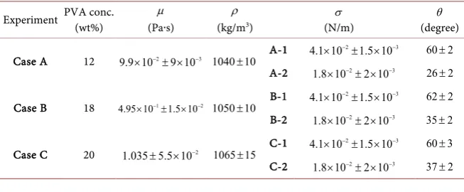
As for the dip coating experiment, several test solutions were prepared. The physical properties of these solutions are given in Table 2. Three pulling speeds, i.e., V =10 ,10 , and 10−4 −3 −2 m/s were set up for the experiments. The
concen-tration of the trace rhodamine 6G was kept the same as the previous dropping experiment.
The experiment on a single microneedle was performed first. Initially the mi-croneedle was lowered just to touch the surface of the drug solution, then the dip coating experiment commenced by penetrating the microneedle into the solu-tion pool at the speed of 10−5 m/s. The penetration depth was set up to be
6
4 10× − m, which is around 1 3 of the total needle height. The microneedle
[image:8.595.234.516.416.538.2]was then pulled up with three different speeds as specified above. The drug solu-tion was attached to the microneedle and a liquid bridge was formed, then the bridge would break up when as the needled was pulled up and the drug solution would form a donut ring around the microneedle surface. The volume of the ring will determine how much of the drugs will adhere to the microneedle sur-face. The position and the shape of the ring after drying will influence the amount of drugs and amount of time it takes the drugs to be dissolved in human bodies within a specified period of time.
Figure 3. Photos of dried drug surfaces for the dropping experiment. Red
circles are the highest attached points Pf.
Table 2. Physical properties of test solutions for the dipping experiment.
Experiment PVA conc. (wt%) (Paµ∙s) (kg/mρ 3)
σ (N/m)
θ (degree)
Case A 12 9.9 10× −2± ×9 10−3 1040 10± A-1
2 3
4.1 10× − ±1.5 10× − 60 2±
A-2 1.8 10× −2± ×2 10−3 26 2±
Case B 18 4.95 10× −1±1.5 10× −2 1050 10± B-1
2 3
4.1 10× − ±1.5 10× − 62 2±
B-2 1.8 10× −2± ×2 10−3 35 2±
Case C 20 1.035 5.5 10± × −2 1065 15± C-1
2 3
4.1 10× − ±1.5 10× − 60 3±
C-2 1.8 10× −2± ×2 10−3 37 2±
(A) μ = 10−2 (Pa∙s)
σ = 4.15 × 10⁻² (N/m)
(B) μ = 10−2 (Pa∙s)
σ = 4.15 × 10⁻² (N/m)
(C) μ = 10−2 (Pa∙s)
σ = 4.15 × 10⁻² (N/m)
(D) μ = 10−2 (Pa∙s)
σ = 4.15 × 10⁻² (N/m)
(E) μ = 10−2 (Pa∙s)
σ = 4.15 × 10⁻² (N/m)
T = 6 (hr)
H
(µm) 280 460 420 460 460
Pf
[image:8.595.211.538.600.735.2]DOI:10.4236/aces.2019.92016 212 Advances in Chemical Engineering and Science Comparison between two single microneedles under the same dipping and drying condition is the displayed in Figure 4; the difference is that the viscosity of one solution is
µ
=0.1 Pa·s and another isµ
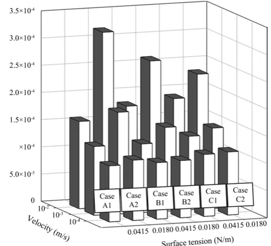
=1 Pa·s. The photos in Fig-ure 4(a) and Figure 4(b) indicate that the capillary rise for the dilute solution was higher. The photos of the rings formed after the liquid bridges were broken on the two microneedle surface are shown in Figure 4(c) and Figure 4(d). It is noted that more drug solution was attached to the microneedle surface for the case with a lower viscosity, and the upper wetting line position was also higher. The initial height of capillary rise might have contributed to the volume attached to the microneedle surface. The shapes of the dried rings on the microneedles are displayed in Figure 4(e) and Figure 4(f); the volumes of the rings shrunk owing to solvent evaporation, gravitational force also pulls the liquid down. However, it is interesting to note that the top and bottom wetting line positions that define the rings improves drastically through the drying process.There are six sets of test solutions labeled as Cases A-1, A-2, B-1, B-2, C-1 and C-2 in Table 2. The effects of the three critical parameters, i.e., surface tension, viscosity and pulling speed on the wet drug loading volume (DLV) are displayed in Figure 5. It is noted that at the lowest pulling speed V =10−4 m/s, there is not
[image:9.595.251.495.435.681.2]much difference for the six cases as shown at the front row of the data in Figure 5. However, as the pulling speed increases, the drug loading amount increases accordingly, the case with the lower surface tension and lower viscosity has a larger drug loading amount. The effect is more significant as the data at the rear row in Figure 5 indicated. It is obvious that as the pulling speed increases, de-creasing both surface tension and viscosity can increase the drug loading volume.
Figure 4. The shapes of the attached drug solution ring before and after drying
DOI: 10.4236/aces.2019.92016 213 Advances in Chemical Engineering and Science
Figure 5. Drug loading volume (DLV) as functions of surface tension,
pulling velocity and fluid viscosity.
It is necessary to analyze the effects of other factors, such as inertial or gravita-tional force on the drug loading amount. The analysis can be focused on three dimensionless groups, i.e., Reynolds number Re, Stokes number St and capillary number Ca. Re and St are defined as follows:
Re
ρ
Vµ
≡ (4)
2
g St
V ρ
µ
≡ (5)
The characteristic length has to be defined first. In the present study, is defined as:
ξ
Ψ ≡
(6)
here, Ψ is the total drug solution attached to the microneedle surface, and
ξ
is the wetted surface. For example, if the fluid viscosity is 1 Pa∙s, surface tension is 1.8 10× −2 N/m and the pulling speed is 10−2 m/s, we found that the liquid
drug volume Ψ on the microneedle surface was around 1.95 10× −11 m3, while the wetted surface
ξ
of the ring bounded by two wetting lines was around7
5.08 10× − m2, thus ≡3.84 10× −5 m.
Values of these dimensionless groups for one representative case are given in
DOI:10.4236/aces.2019.92016 214 Advances in Chemical Engineering and Science Generally speaking, DLV goes up as Ca increases, however, there are three data points A, B and C that are somewhat out of the track. A least-square fitting of the data points excluding these three data points lead to
Ca
DLV =α β (7)
with R2 =0.944,
α
= ×2 10 and−4β
=0.118. Note that for dip coating withCa 1, the analytical solution of film thickness h for flat substrate is [48] 1
6
Ca
[image:11.595.209.537.225.697.2]h∝ (8)
Table 3. Values of the three dimensionless groups in the dipping experiment.
Ca=µσV
1
µ = (Pa·s); σ =0.018 (N/m)
2 10
V= − (m/s)
1 5.56 10× −
Re ρV
µ =
1
µ= (Pa·s); σ =0.018 (N/m)
4 10
V= − (m/s) 5
3.84 10−
= ×
4 4 10× −
2
g St
V
ρ µ =
1
µ = (Pa·s); σ =0.018 (N/m)
4 10
V= − (m/s) 5
3.84 10−
= ×
3 1.505 10× −
Figure 6. Correlation between DLV and Ca for the dipping experiment. Excluding Points
A, B, and C, the correlation coefficient (R2) is 0.9044 with equation y = 0.0002 × 0.118.
V (m/s) μ (Pa∙s) σ (N/m)
A 10−3 10−1 1.8 × 10⁻²
B 10−2 10−1 1.8 × 10⁻²
DOI: 10.4236/aces.2019.92016 215 Advances in Chemical Engineering and Science Therefore, the dependence of DLV on Ca is somewhat close to the conven-tional dip coating operation for flat films. As for the three data points A, B and C, it is noted that the three cases correspond to low surface tension (σ =1.8 10× −2 N/m), higher pulling speed and lower viscosity. Point B has the
highest pulling speed (V =10−2 m/s) and lowest viscosity (
µ
=10−1Pa·s), and DLV is the highest among all data points.
It is noted that the presented dip coating is not a steady-state operation, once the needle penetrates into the drug solution pool, the capillary rise appears im-mediately. After the operation switches from penetration to pulling up, the amount of drug solution attached on the microneedle surface can be quite dif-ferent for each case. In addition to surface tension and viscosity that would in-fluence the capillary rise, the effect of wetting angle can also be critical. The data of wetting angle θ in Table 2 indicate that if the surface tension is maintained
at 4.15 10× −2 N/m, varying the fluid viscosity only influences the wetting angle
slightly, once the surface tension is lowered to 1.8 10× −2 N/m, the wetting angle
drops sharply and for the case with the lowest fluid viscosity, and wetting angles
θ is the smallest. The wetting angle θ will also influence the liquid climbing, or capillary rise of the drug solution. Figure 7 displays the photos of capillary rise for several cases before pulling up. Apparently for the cases with high sur-face tension σ =4.15 10× −2 N/m, the capillary rise is not significant. However,
the three cases with lower surface tension σ =1.8 10× −2 N/m clearly
demon-strate that significant capillary rise would appear. Lower fluid viscosity would increase the capillary rise, but the effect is less significant than that of surface tension. It is concluded that lowering the surface tension, fluid viscosity and wetting angle can increase the capillary rise, which will lead to an increase of the drug loading amount, and surface tension is clearly the dominant factor. The pulling speed is also an important factor. Increasing the pulling speed with the case of high capillary rise would increase DLV substantially.
As for an array of microneedles, the speed and height of capillary rise are much more significant than those of a single needle. For a dilute solution of vis-cosity
µ
=10−1 Pa·s, the drug solution will climb up to the roots of themicro-needles instantly. Figure 8 demonstrates how fast the drug solution can climb up to one-half of the microneedle height. As the fluid viscosity increases, the time for the drug solution to reach the roots will also be increased. For the case with
1 10
µ
= −Pa·s, it took 1 minute to reach one-half of the microneedle height, while for a solution with
µ
=1 Pa·s, the time to reach one-half of themicro-needle height is around 2.5 minutes. It is also noted that the front of the capillary rise is not flat. The central part of the drug solution climbs faster than the two sides.
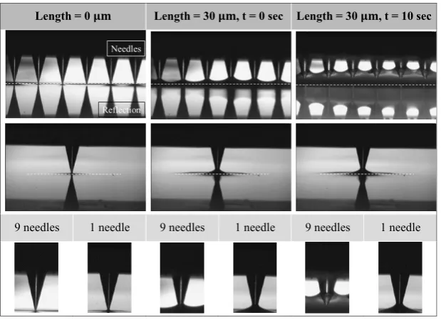
A comparison of capillary rise between a single microneedle and an array of microneedles is shown in Figure 9. The initial conditions are the same, after pe-netrating 30 μm deep into the drug solution, remaining stationary for 10 seconds and then the microneedles were lifted up at 10−1 m/s. It is noted that in
DOI:10.4236/aces.2019.92016 216 Advances in Chemical Engineering and Science penetration depth was only 3 10× −5 m because if the penetration depth was too
[image:13.595.247.501.234.448.2]large, rapid capillary rise would lead the drug solution to reach the roots of the microneedle instantly. The photos in Figures 9(a)-(c) show the three stages of capillary rise between the two cases. It is clear that the interaction between neighboring microneedles can cause a substantial capillary rise at high speeds for an array of microneedles. Figure 10 demonstrates the drug loaded amount for these two cases with three different pulling speeds. The DLV of the three cases for a single microneedle appear to be similar, and apparently the amount on each microneedle in the array is much higher than a single microneedle. For the case with a higher pulling speed, DLV also increase substantially.
Figure 7. Capillary rise of PVA solutions at various concentration
for a single microneedle.
[image:13.595.210.538.494.704.2]DOI: 10.4236/aces.2019.92016 217 Advances in Chemical Engineering and Science
Figure 9. Comparison of capillary rise between a single microneedle and an array
of microneedles.
Figure 10. Comparison of DLV between a single microneedle and
an array of microneedles.
4. Conclusions
We have analyzed two approaches for drug loading on microneedles. Dropping is a simple way to apply drug solution on microneedle patches. For a solution with high viscosity and surface tension, the solution will retain on the upper part of the microneedle surface after drying. Once the surface tension of the drug so-lution is lowered, all the drug soso-lutions will stay at the root area of the micro-needle patches, no matter how the difference of viscosity is. This situation may not be favorable for drug delivery into human body.
Dipping is a better method to retain drug solution on the microneedle surfac-es. The microneedles would first penetrate into a drug solution pool and then the microneedles were being pulled up. The drug solution that retained on the
Length = 0 𝛍𝛍𝛍𝛍 Length = 30 𝛍𝛍𝛍𝛍, t = 0 sec Length = 30 𝛍𝛍𝛍𝛍, t = 10 sec
[image:14.595.248.499.337.511.2]DOI:10.4236/aces.2019.92016 218 Advances in Chemical Engineering and Science microneedle surface would form a donut ring. For a single needle, the volume of the donut ring, or the drug loading amount depends on the initial capillary rise before the pulling operation and capillary number. Surface tension is the domi-nant parameter on the capillary rise. The dependence of DLV on the capillary number is similar to that of flat film provided the initial capillary rise is not sig-nificant. High capillary rise would increase the DLV substantially, and higher pulling speed is also an enhancing factor.
As for an array of microneedle, the interaction between microneedles would create a surprisingly fast capillary rising effect. This phenomenon is more signif-icant for drugs solutions with low viscosity and surface tension. Comparison between the results of a single microneedle with an array of microneedles im-plies that owing to the fast and high capillary rise, the DLV for an array of mi-croneedles can be several times higher than a single microneedle.
We have made an attempt to simulate the dip coating operation for a single and an array of microneedles with some commercial software packages. Owing to the complexity of the system, the numerical predictions on the drug loading amount are much lower than those of experimental findings, although the simi-lar trends are observed. As the effect of viscosity and surface tension of drug so-lution is clearly shown through this work, the experimentally recorded drug loading amount will be useful toward practical applications of microneedles, es-pecially in MN fabrications and drug dosage control.
More details towards the understanding on the physics of the drug loading process, such as prediction of the dynamic wetting lines and capillary interac-tions between microneedles, are necessary to better predict the drug loading amount theoretically.
Conflicts of Interest
The authors declare no conflicts of interest regarding the publication of this pa-per.
References
[1] Henry, S., McAllister, D.V., Allen, M.G. and Prausnitz, M.R. (1998) Microfabricated Microneedles: A Novel Approach to Transdermal Drug Delivery. Journal of Phar-maceutical Sciences, 87, 922-925. https://doi.org/10.1021/js980042+
[2] Kim, Y.-C., Park, J.-H. and Prausnitz, M.R. (2012) Microneedles for Drug and Vac-cine Delivery. Advanced Drug Delivery Reviews, 64, 1547-1568.
https://doi.org/10.1016/j.addr.2012.04.005
[3] Vrdoljak, A. (2013) Review of Recent Literature on Microneedle Vaccine Delivery Technologies. Vaccine: Development and Therapy, 3, 47-55.
https://doi.org/10.2147/VDT.S34682
[4] Indermun, S., Luttge, R., Choonara, Y.E., Kumar, P., du Toit, L.C., Modi, G. and Pillay, V. (2014) Current Advances in the Fabrication of Microneedles for Trans-dermal Delivery. Journal of controlled release, 185, 130-138.
https://doi.org/10.1016/j.jconrel.2014.04.052
Inser-DOI: 10.4236/aces.2019.92016 219 Advances in Chemical Engineering and Science
tion of Microneedles into Skin: Measurement and Prediction of Insertion Force and Needle Fracture Force. Journal of Biomechanics, 37, 1155-1163.
https://doi.org/10.1016/j.jbiomech.2003.12.010
[6] Falo Jr., L.D., Erdos, G. and Ozdoganlar, O.B. (2014) Dissolvable Microneedle Ar-rays for Transdermal Delivery to Human Skin Google Patents.
[7] Gittard, S.D., Chen, B., Xu, H., Ovsianikov, A., Chichkov, B.N., Monteiro-Riviere, N.A. and Narayan, R.J. (2013) The Effects of Geometry on Skin Penetration and Failure of Polymer Microneedles. Journal of Adhesion Science and Technology, 27, 227-243. https://doi.org/10.1080/01694243.2012.705101
[8] Kochhar, J.S., Quek, T.C., Soon, W.J., Choi, J., Zou, S. and Kang, L. (2013) Effect of Microneedle Geometry and Supporting Substrate on Microneedle Array Penetra-tion into Skin. Journal of Pharmaceutical Sciences, 102, 4100-4108.
https://doi.org/10.1002/jps.23724
[9] Zhu, Z., Ye, X., Ku, Z., Liu, Q., Shen, C., Luo, H., Luan, H., Zhang, C., Tian, S. and Lim, C. (2016) Transcutaneous Immunization Via Rapidly Dissolvable Microneedles Protects against Hand-Foot-and-Mouth Disease Caused by Enterovirus 71. Journal of Controlled Release, 243, 291-302. https://doi.org/10.1016/j.jconrel.2016.10.019 [10] Koutsonanos, D.G., Vassilieva, E.V., Stavropoulou, A., Zarnitsyn, V.G., Esser, E.S.,
Taherbhai, M.T., Prausnitz, M.R., Compans, R.W. and Skountzou, I. (2012) Deli-very of Subunit Influenza Vaccine to Skin with Microneedles Improves Immunoge-nicity and Long-Lived Protection. Scientific Reports, 2, Article Number: 357.
https://doi.org/10.1038/srep00357
[11] Weldon, W.C., Zarnitsyn, V.G., Esser, E.S., Taherbhai, M.T., Koutsonanos, D.G., Vassilieva, E.V., Skountzou, I., Prausnitz, M.R. and Compans, R.W. (2012) Effect of Adjuvants on Responses to Skin Immunization by Microneedles Coated with In-fluenza Subunit Vaccine. PLoS ONE, 7, e41501.
https://doi.org/10.1371/journal.pone.0041501
[12] Qiu, Y., Guo, L., Zhang, S., Xu, B., Gao, Y., Hu, Y., Hou, J., Bai, B., Shen, H. and Mao, P. (2016) DNA-Based Vaccination against Hepatitis B Virus Using Dissolving Microneedle Arrays Adjuvanted by Cationic Liposomes and Cpg Odn. Drug Deli-very, 23, 2391-2398.
[13] Hiraishi, Y., Nandakumar, S., Choi, S.-O., Lee, J.W., Kim, Y.-C., Posey, J.E., Sable, S.B. and Prausnitz, M.R. (2011) Bacillus Calmette-Guerin Vaccination Using a Mi-croneedle Patch. Vaccine, 29, 2626-2636.
https://doi.org/10.1016/j.vaccine.2011.01.042
[14] Pattani, A., McKay, P.F., Garland, M.J., Curran, R.M., Migalska, K., Cassidy, C.M., Malcolm, R.K., Shattock, R.J., McCarthy, H.O. and Donnelly, R.F. (2012) Micro-needle Mediated Intradermal Delivery of Adjuvanted Recombinant Hiv-1 Cn54gp140 Effectively Primes Mucosal Boost Inoculations. Journal of Controlled Release, 162, 529-537. https://doi.org/10.1016/j.jconrel.2012.07.039
[15] Kines, R.C., Zarnitsyn, V., Johnson, T.R., Pang, Y.-Y.S., Corbett, K.S., Nicewonger, J.D., Gangopadhyay, A., Chen, M., Liu, J. and Prausnitz, M.R. (2015) Vaccination with Human Papillomavirus Pseudovirus-Encapsidated Plasmids Targeted to Skin Using Microneedles. PLoS ONE, 10, e0120797.
https://doi.org/10.1371/journal.pone.0120797
[16] Vrdoljak, A., McGrath, M.G., Carey, J.B., Draper, S.J., Hill, A.V., O’Mahony, C., Crean, A.M. and Moore, A.C. (2012) Coated Microneedle Arrays for Transcutane-ous Delivery of Live Virus Vaccines. Journal of Controlled Release, 159, 34-42.
https://doi.org/10.1016/j.jconrel.2011.12.026
DOI:10.4236/aces.2019.92016 220 Advances in Chemical Engineering and Science
D.J., Kersten, G. and Bouwstra, J.A. (2009) Microneedle Arrays for the Transcuta-neous Immunization of Diphtheria and Influenza in Balb/C Mice. Journal of Con-trolled Release, 136, 71-78.https://doi.org/10.1016/j.jconrel.2009.01.025
[18] Narayan, R.J. (2014) Transdermal Delivery of Insulin via Microneedles. Journal of Biomedical Nanotechnology, 10, 2244-2260.https://doi.org/10.1166/jbn.2014.1976
[19] Veiseh, O. and Langer, R. (2015) Diabetes: A Smart Insulin Patch. Nature, 524, 39-40.https://doi.org/10.1038/524039a
[20] Lee, J.W., Choi, S.O., Felner, E.I. and Prausnitz, M.R. (2011) Dissolving Micro-needle Patch for Transdermal Delivery of Human Growth Hormone. Small, 7, 531-539.https://doi.org/10.1002/smll.201001091
[21] Zhang, Y., Brown, K., Siebenaler, K., Determan, A., Dohmeier, D. and Hansen, K. (2012) Development of Lidocaine-Coated Microneedle Product for Rapid, Safe, and Prolonged Local Analgesic Action. Pharmaceutical Research, 29, 170-177.
https://doi.org/10.1007/s11095-011-0524-4
[22] Arya, J. and Prausnitz, M.R. (2016) Microneedle Patches for Vaccination in Devel-oping Countries. Journal of Controlled Release, 240, 135-141.
https://doi.org/10.1016/j.jconrel.2015.11.019
[23] Chen, M.-C., Huang, S.-F., Lai, K.-Y. and Ling, M.-H. (2013) Fully Embeddable Chitosan Microneedles as a Sustained Release Depot for Intradermal Vaccination.
Biomaterials, 34, 3077-3086.https://doi.org/10.1016/j.biomaterials.2012.12.041
[24] Ito, Y., Hagiwara, E., Saeki, A., Sugioka, N. and Takada, K. (2006) Feasibility of Mi-croneedles for Percutaneous Absorption of Insulin. European Journal of Pharma-ceutical Sciences, 29, 82-88.https://doi.org/10.1016/j.ejps.2006.05.011
[25] Sullivan, S.P., Koutsonanos, D.G., del Pilar Martin, M., Lee, J.W., Zarnitsyn, V., Choi, S.-O., Murthy, N., Compans, R.W., Skountzou, I. and Prausnitz, M.R. (2010) Dissolving Polymer Microneedle Patches for Influenza Vaccination. Nature Medi-cine, 16, 915-920.https://doi.org/10.1038/nm.2182
[26] Yang, S., Feng, Y., Zhang, L., Chen, N., Yuan, W. and Jin, T. (2012) A Scalable Fa-brication Process of Polymer Microneedles. International Journal of Nanomedicine, 7, 1415-1422.
[27] Lee, I.-C., He, J.-S., Tsai, M.-T. and Lin, K.-C. (2015) Fabrication of a Novel Partial-ly Dissolving PoPartial-lymer Microneedle Patch for Transdermal Drug Delivery. Journal of Materials Chemistry B, 3, 276-285.https://doi.org/10.1039/C4TB01555J
[28] Ling, M.-H. and Chen, M.-C. (2013) Dissolving Polymer Microneedle Patches for Rapid and Efficient Transdermal Delivery of Insulin to Diabetic Rats. Acta Bioma-terialia, 9, 8952-8961.https://doi.org/10.1016/j.actbio.2013.06.029
[29] Davis, S.P., Martanto, W., Allen, M.G. and Prausnitz, M.R. (2005) Hollow Metal Microneedles for Insulin Delivery to Diabetic Rats. IEEE Transactions on Biomedi-cal Engineering, 52, 909-915.https://doi.org/10.1109/TBME.2005.845240
[30] Prausnitz, M.R. (2004) Microneedles for Transdermal Drug Delivery. Advanced Drug Delivery Reviews, 56, 581-587.https://doi.org/10.1016/j.addr.2003.10.023
[31] Martin, F.E. and Evans, J.D. (2007) Device for Manipulating a Needle or Abrader Array and Method of Use, Editor Editors. Google Patents.
[32] Boehm, R.D., Daniels, J., Stafslien, S., Nasir, A., Lefebvre, J. and Narayan, R.J. (2015) Polyglycolic Acid Microneedles Modified with Inkjet-Deposited Antifungal Coatings. Biointerphases, 10, Article ID: 011004.https://doi.org/10.1116/1.4913378
DOI: 10.4236/aces.2019.92016 221 Advances in Chemical Engineering and Science
https://doi.org/10.1016/j.mattod.2014.04.027
[34] Uddin, M.J., Scoutaris, N., Klepetsanis, P., Chowdhry, B., Prausnitz, M.R. and Douroumis, D. (2015) Inkjet Printing of Transdermal Microneedles for the Delivery of Anticancer Agents. International journal of pharmaceutics, 494, 593-602.
https://doi.org/10.1016/j.ijpharm.2015.01.038
[35] McGrath, M.G., Vrdoljak, A., O’Mahony, C., Oliveira, J.C., Moore, A.C. and Crean, A.M. (2011) Determination of Parameters for Successful Spray Coating of Silicon Microneedle Arrays. International Journal of Pharmaceutics, 415, 140-149.
https://doi.org/10.1016/j.ijpharm.2011.05.064
[36] Gill, H.S. and Prausnitz, M.R. (2007) Coating Formulations for Microneedles.
Pharmaceutical Research, 24, 1369-1380.
https://doi.org/10.1007/s11095-007-9286-4
[37] Ameri, M., Kadkhodayan, M., Nguyen, J., Bravo, J.A., Su, R., Chan, K., Samiee, A. and Daddona, P.E. (2014) Human Growth Hormone Delivery with a Microneedle Transdermal System: Preclinical Formulation, Stability, Delivery and Pk of Thera-peutically Relevant Doses. Pharmaceutics, 6, 220-234.
https://doi.org/10.3390/pharmaceutics6020220
[38] Chen, M.-C., Ling, M.-H. and Kusuma, S.J. (2015) Poly-Γ-Glutamic Acid Micro-needles with a Supporting Structure Design as a Potential Tool for Transdermal De-livery of Insulin. Acta Biomaterialia, 24, 106-116.
https://doi.org/10.1016/j.actbio.2015.06.021
[39] Park, J.-H., Choi, S.-O., Seo, S., Choy, Y.B. and Prausnitz, M.R. (2010) A Micro-needle Roller for Transdermal Drug Delivery. European Journal of Pharmaceutics and Biopharmaceutics, 76, 282-289.https://doi.org/10.1016/j.ejpb.2010.07.001
[40] Park, J.-H., Allen, M.G. and Prausnitz, M.R. (2005) Biodegradable Polymer Micro-needles: Fabrication, Mechanics and Transdermal Drug Delivery. Journal of Con-trolled Release, 104, 51-66.https://doi.org/10.1016/j.jconrel.2005.02.002
[41] Park, J.-H., Allen, M.G. and Prausnitz, M.R. (2006) Polymer Microneedles for Con-trolled-Release Drug Delivery. Pharmaceutical Research, 23, 1008-1019.
https://doi.org/10.1007/s11095-006-0028-9
[42] Hong, X., Wu, Z., Chen, L., Wu, F., Wei, L. and Yuan, W. (2014) Hydrogel Micro-needle Arrays for Transdermal Drug Delivery. Nano-Micro Letters, 6, 191-199.
https://doi.org/10.1007/BF03353783
[43] Chen, W., Wang, C., Yan, L., Huang, L., Zhu, X., Chen, B., Sant, H.J., Niu, X., Zhu, G. and Yu, K. (2014) Improved Polyvinylpyrrolidone Microneedle Arrays with Non-Stoichiometric Cyclodextrin. Journal of Materials Chemistry B, 2, 1699-1705.
https://doi.org/10.1039/C3TB21698E
[44] Wendorf, J.R., Ghartey-Tagoe, E.B., Williams, S.C., Enioutina, E., Singh, P. and Cleary, G.W. (2011) Transdermal Delivery of Macromolecules Using Solid-State Biodegradable Microstructures. Pharmaceutical Research, 28, 22-30.
https://doi.org/10.1007/s11095-010-0174-y
[45] Demir, Y.K., Akan, Z. and Kerimoglu, O. (2013) Characterization of Polymeric Mi-croneedle Arrays for Transdermal Drug Delivery. PLoS ONE, 8, e77289.
https://doi.org/10.1371/journal.pone.0077289
[46] Demir, Y.K., Akan, Z. and Kerimoglu, O. (2013) Sodium Alginate Microneedle Ar-rays Mediate the Transdermal Delivery of Bovine Serum Albumin. PLoS ONE, 8, e63819.https://doi.org/10.1371/journal.pone.0063819
DOI:10.4236/aces.2019.92016 222 Advances in Chemical Engineering and Science
Chang, M.-W. and Ahmad, Z. (2015) Microneedle Coating Techniques for Trans-dermal Drug Delivery. Pharmaceutics, 7, 486-502.
https://doi.org/10.3390/pharmaceutics7040486
[48] Landau, L. (2012) Dragging of Liquid by a Moving Plato. Dynamics of Curved Fronts, 141.
[49] Duarah, S., Sharma, M. and Wen, J. (2019) Recent Advances in Microneedle-Based Drug Delivery: Special Emphasis on Its Use in Paediatric Population. European Journal of Pharmaceutics and Biopharmaceutics, 136, 48-69.
https://doi.org/10.1016/j.ejpb.2019.01.005
[50] Jin, X., Zhu, D.D., Chen, B.Z., Ashfaq, M. and Guo, X.D. (2018) Insulin Delivery Systems Combined with Microneedle Technology. Advanced Drug Delivery Re-views, 127, 119-137.https://doi.org/10.1016/j.addr.2018.03.011
[51] Kumar, P., Choonara, Y.E., du Toit, L.C. and Pillay, V. (2018) Therapeutic Applica-tions and Pharmacoeconomics of Microneedle Technology Au Richter-Johnson, Jolanda. Expert Review of Pharmacoeconomics & Outcomes Research, 18, 359-369.
https://doi.org/10.1080/14737167.2018.1485100