doi:10.4236/nm.2011.22014 Published Online June 2011 (http://www.SciRP.org/journal/nm)
Advances in Oligoprotection
Hong Liao, Dixit Sanjaya Mani
National Centre for Drug Screening, China Pharmaceutical University, Nanjing, China. Email: [email protected]
Received April 6th, 2011; revised April 17th, 2011; accepted May 9th, 2011.
ABSTRACT
Oligodendrocytes, the myelinating glial cells of the nervous system, in various disease states display great vulnerability to excitotoxic damage, oxidative stress, and inflammatory cytokines. Besides demyelinating diseases where oligoden-drocyte injury is primarily implicated, damage to them also occurs secondarily in various neuropathies. Oligoprotec-tion should, therefore, be looked into something not just as a means for protecting oligodendrocytes alone but also as a common target of protecting neurons and the neurogliovascular unit as a whole. In this review, we provide a descrip-tion on oligodendrocytes, the reasons for their vulnerability, the evolving new concepts of protecting neurovascular unit and recently neurogliovascular unit; the various diseases where oligodendrocyte injury is implicated with a brief idea on different injury mechanisms of oligodendrocytes. Finally, we present the summary of the drugs that have shown promising results in protecting oligodendrocytes or in protecting white matter as a whole in different in vivo and in vitro models.
Keywords: Oligodendrocytes, Neurovascular Unit, Oxidative Stress, Excitotoxicity, Demyelinating Diseases, Stroke,
Spinal Cord Injury, Neuropharmacology
1. Introduction
Neurobiology has been a field of ever-growing interest with more and more researchers working aimed at pre-venting or curing various neurological diseases like Mul-tiple Sclerosis (MS), Alzheimer’s disease (AD), Parkin-son’s disease (PD). The incidence of these diseases is on the rise with the increasing elderly population. Our high paced life style has increased auto-accidents and the cases of traumatic brain injury (TBI) and spinal cord in-jury (SCI) which leads to loss of lives or debilitating conditions lifelong.
Research focused in central nervous system (CNS) pathologies in the yesteryears focused centrally on neu-ronal mechanisms. However, the concept of neuroprotec-tion alone failed to prove optimal therapeutic strategy hence the neuron-based model has taken a paradigm shift to integrate the non-neuronal cells which maintain the homeostatic environment for neurons. It has widened to embrace much wider concept of Neurovascular unit (NVU) which itself has remolded into Neurogliovascular unit [1,2]. The neurovascular unit has been recently de-fined as a unit that comprises neurons and non-neuronal
cells such as vascular cells (endothelia, pericytes and vascular smooth muscle cells) and glia (astrocytes, mi- croglia and oligodendrocytes), along with an extracellu- lar matrix that maintains the integrity of brain tissue [3]. Also the concept of oligovascular niche has emerged which includes the main cell types comprising white matter—the neuronal axon, oligodendrocyte (myelin), astrocyte, and endothelial cell [1].
Oligodendrocytes (OLGs) are the myelinating glial cells of the CNS. OLGs in the nervous system are analogous to the Schwann cells in the peripheral nervous system in providing support for quick transfer of neu- ronal signals. OLGs not only myelinate axons but also maintain their functional integrity and survival thorough oligodendrocyte specific proteins and/or trophic factor release by secreting soluble factors. Factors like Brain derived neurotrophic factor (BDNF), Neurotrophin-3 (NT-3), Nerve growth factor (NGF), and Platelet derived growth factor (PDGF) are reported as oligodendrocyte- axon signaling mediators [1]. They differ greatly mor- phologically based on their function and subtypes [4].
Myelin sheath, the protective covering of the nerve fi-bers insulating the axons, is responsible for transmission of the electrical signals with high velocity and without the loss of signal strength. The myelin sheath produced by OLGs is up to 600 × surface area and 100 × weight of
soma and therefore their energy requirement is 2-3 times more than other brain cells [5]. These facts make OLGs the most vulnerable cells in the brain coupled with high- est rate of metabolism, high reactive oxygen species (ROS) formation, low levels of antioxidant glutathione; high iron stores for cholesterol and lipid synthesis re- quired for myelin formation; high levels of polyunsatu- rated fatty acids (PUFA) which react with ROS trigger- ing lipid peroxidation [5-7]. OLGs are damaged as a re- sult of adverse environmental signals and stress situa- tions, such as oxidation, which may retard or inhibit myelination [8]. Neurons require assistance of glial cells for maintaining brain homeostasis from extracellular concentrations of ions, neurotransmitters and neurohor- mones for provision of an uninterrupted metabolic sup- port [9]. The denuding of axons following oligodendro- cyte death leads to functional loss of neurons and even- tually their death, moreover, with a single oligodendro- cyte myelinating numerous axons, death of a single oli- godendrocyte exposes multiple neurons to risk [9].
2. Oligodendrocytes in Disease States
2.1. Multiple Sclerosis (MS)
MS is an autoimmune demyelinating disease with a neu-rodegenerative component most commonly diagnosed in patients 20 - 40 years of age [10]. In MS, the immune system attacks the white matter of the brain and spinal cord, causing multifocal demyelination leading to dis- ability and/or paralysis. In acute MS, oligodendrocytes die mainly by apoptosis, however, in chronic MS lesion apoptosis was absent or rare which could because of the rapid removal of apoptotic cells in vivo [11]. Oxidative stress plays an important role in oligodendroglial death in MS [12]. Macrophages from MS patients show increased production of superoxide radicals and H2O2; while CSF
displays significantly high level of NO metabolites, ni- trite and nitrate compared to controls [13]. As the MS lesion formation starts, locally produced ROS induce tight junction and cytoskeletal changes in brain endothe- lial cells which facilitate transendothelial monocyte mi- gration [6]. ROS also has a critical role in myelin phago- cytosis and it has been observed that using ROS inhibi- tors like NADPH oxidase inhibitors, lipoic acid or cata- lase enzyme reduced myelin phagocytosis by macro- phages [6]. Excitatory glutamate levels are increased in acute MS lesions and in normal-appearing white matter in MS patients [14]. In addition, oxidative stress contri- butes to the increase in glutamate concentrations in the extracellular space, as free radicals reduce the efficiency of GluTs [14]. TNF-α is found at high levels in multiple sclerosis (MS) plaques and induces both apoptotic and necrotic death of OLGs and inhibition of caspase-1 and
caspase-3 protects them against TNF-α-induced apop- tosis [11]. Interferon gamma (IFN-γ) produced by lym- phocytes in MS lesions plays a role in apoptosis of oli- godendrcoytes both in vivo and in vitro [11]. TNF-α in- creases the IFN-γ-induced death of OPCs and this effect can be partially suppressed by caspase inhibition [11]. Interleukin-11 is expressed by reactive astrocytes in MS tissue samples along the myelinated border of both active and silent lesions [15].
2.2. Paraventricular Leukomalacia (PVL)
Paraventricular leukomalacia (PVL) is the leading cause of brain damage in preterm infants, prior to 32 weeks of gestation; resulting in cerebral palsy and cognitive im- pairment [16,17]; and occurs due to damage to OPCs and hypomyelination [18]. It involves death of small areas of brain tissue around ventricles, the fluid filled areas of brain; creating holes in the brain [19]. The pathogenesis of OPC injury relates to hypoxia-ischemia and systemic infection/inflammation, common occurrences in prema- ture infants. Three downstream mechanisms have been identified, i.e., microglial activation, excitotoxicity and free radical attack [20].
2.3. Stroke
Stroke, a leading cause of death occurs due to the inter- ruption of blood supply to any part of the brain owing to the blockade or bursting of blood vessels, hence classi- fied as ischemic and hemorrhagic stroke [21]. Immedi- ately following interrupted blood supply to brain, the injury process initiates, if recovery of normal blood flow is not achieved within a short period of time, death of cells within the ischemic core begins [22]. Stroke dis- rupts the blood brain barrier (BBB) which in turn affects stroke progression and neuroregeneration [23]. Ischemia, in stroke, is known to cause cell death mainly through the oxidative stress and excitotoxicity. Energy deprivation in stroke leads to neuronal death, axonal dysfunction and loss of OLGs which are very sensitive to transient oxy- gen and glucose deprivation. It has been seen that OLGs in immature state are even more sensitive to ischemic injury in mature forms. These cells may release gluta- mate under ischemic conditions by reverse functioning of their glutamate transporter (GluTs) [14]. During ischemia/ reperfusion of brain tissue, oxidative stress sets in with a rise in Hydrogen Peroxide (H2O2) levels for about 1hr
reaching up to 175 μM (approx.) [14]. Also, increased TNF-mRNA is seen 3 hr after stroke in transient Middle cerebral artery occlusion (MCAO) models in rats [23].
like tau. Thereby, they weaken the structural integrity of axons and also impair axonal mechanisms for antero- grade and retrograde transport [25].
2.4. Spinal Cord Injury (SCI)
Spinal cord injury (SCI) refers to the damage to the spi- nal cord resulting in permanent neurological deficit. It is caused by direct injury or indirectly from damage to the surrounding bones, tissues, or blood vessels resulting from auto accidents, falls, sports injuries and other acci- dents. It is a very tragic case with higher prevalence in young and otherwise healthy individuals, thus creating physical, emotional, and economic burdens on both suf- ferer and society [26]. The loss of axonal transmission of sensory and motor nerves occurs distal to point of SCI, leading to multiple health problems such as recurrent kidney stones, urinary tract infection, pressure sores, and cardiac and respiratory dysfunction [27]. Acute SCI in- volves mechanical primary and biochemical secondary mechanism of injury; primary due to blunt cord compre- ssion owing to dislocation of spinal cord or bone frag- ments; biochemical factors in hemorrhagic necrotic tis- sue causing secondary damage to spinal cord [28]. SCI causes strong up-regulation of genes involved in tran- scription, inflammation and cell death [29]. Excitotoxic- ity, oxidative stress, lipid peroxidation/free radical injury, inflammatory factors all seem to play parts worsening the homeostasis of the brain and causing cell death [28].
2.5. Leukodystrophy
Leukodystrophies are rare diseases appearing during in- fancy or childhood and characterized by a progressive degeneration of the white matter due to imperfect growth or development of the myelin sheath [30]. Myelin is a complex framework made up of at least ten different chemicals. The leukodystrophies are a group of disorders caused by genetic defects in how myelin produces or metabolizes its chemicals components [4]. Each type of leukodystrophy results from the defect in the gene that controls one of the chemicals causing defects in enzymes such as arylsulphatase A (Metachromatic Leukodystro- phy), aspartoacylase (Canavan disease), galactosylcera- midase (Krabbe disease) or in enzymes responsible for breaking down phytanic acid (Refsum’s disease), defects in a fatty acid transfer protein (Adrenoleukodystrophy), or defects in the myelin protein PLP1 (Pelizaeus-Merz- bacher disease) [31].
2.6. Alzheimer’s Disease (AD)
AD is a neurodegenerative disease with stealth onset that progresses relatively rapidly with the parenchymal depo- sition of beta amyloid (Aβ) in the form of senile/neuritic plagues and the neurofibrillatory tangles (NFT) resulting
in the loss of neurons and synapses [32]. The white mat- ter lesions and myelin abnormalities are common in the AD patients [33]. A is known to induce apoptosis of ma- ture OLGs which can be attenuated by antioxidants. A also causes oxidative stress by upregulation of inducible nitric oxide synthase (iNOS) in mature OLGs through a TNF-α-mediated ceramide pathway [32]. Some cases of AD are caused by mutation of presenilin-1 gene. It has been seen that glutamate-mediated excitotoxicity in OLGs is potentiated by presenilin-1 mutation [31].OLGs that differentiate and myelinate later in life produce thinner myelin sheaths around axons and these neurons have been observed to be more susceptible to the damage in AD [32]. The transentorrhinal region and nearby neo- cortical association areas are the late-myelinating regions known; these areas are also the seat for early Aβ deposits and myelin breakdown [5].
2.7. Schizophrenia (SZ)
Schizophrenia is a complex mental disorder of unknown etiology, usually surfacing in the young adulthood, which carries a genetic link [34]. Neuroimaging and microarray studies have provided evidence for myelin and oligoden- drocyte abnormalities in SZ. As evidenced from electron microscopy, OLGs are the most severely affected cells in SZ undergoing dystrophy, necrosis and apoptosis [35]. Decreased expression of myelin and oligodendrocyte- related genes is seen in postmortem samples obtained from prefrontal cortex regions of schizophrenic patients, suggesting OLG loss or disruption at functional levels [36]. Six myelin-related genes specific to OLGs have decreased expression levels in schizophrenia. OLGs are decreased in number in the superior frontal gyrus of schizophrenics. Absence of myelin associated glycopro- tein (MAG) genes may be the link in schizophrenia since MAG-knockout mice develops as a mice model for the disease [37]. Abnormal glutamate homeostasis is consid- ered one of the leading pathological mechanisms of schi- zophrenia and this also puts OLGs at increased risk for excitototic damage [38]. Abnormalities in the white mat- ter have been observed; however, so far it is not clear whether it is a cause or a consequence of the disease [32].
2.8. Other Diseases
Oligodendrocytes are also injured in a variety of other diseases like Progressive Multifocal Leukoencephalopa- thy [7], Post-Infectious Encephalitis [39], and also fol- lowing radiation injury [40], etc.
3. Different Mechanisms of Injury to
Oligodendrocytes
in the brain undergo injury in different disease states by various mechanisms like oxidative stress, excitotoxicity, and inflammatory cytokines. Developing therapeutic approaches to OLG injuries and related diseases needs better understanding of the underlying mechanism by which damage occurs to these cells. The mechanism of injury in neurons which has been researched for long may not fit in case of OLGs and hence should be consid- ered separately. The injury mechanism and mediators causing the injury in disease states are often intertwined with one type eliciting the other in response. Various mechanisms of injury to OLGs has been summed up and presented below in Figure 1.
3.1. Oxidative Stress
OLGs on exposure to reactive oxygen species (ROS) causes formation of lipid peroxides which further react with proteins leading to oxidative modifications of amino acid residue. Also the protein aggregates are formed and protein fragmentation occurs [41], and also leads to DNA fragmentation and finally apoptosis of cells. H2O2, a ma-
jor ROS, is normally produced in the brain in small amounts. Its toxicity is due to its reaction with transition metals (iron-mediated Fenton reaction) to form hydroxyl radicals, which can further react with amino acids, nu- cleic acids, phospholipids and sugars, eventually causing cell death [42].
3.2. Excitotoxicity
[image:4.595.82.507.354.608.2]Oligodendrocytes injury and death occurs on exposure to excitotoxic agents like glutamate and extraceullular ATP [14]. Mature OLGs undergo necrosis on exposure to kainite through the Alpha-amino-3-hydroxy-5-methyl-4- isoxazolepropionic acid (AMPA), and kainite (KA) re-ceptors; recently N-methyl-D-aspartate (NMDA) have also been known to play role in OLG pathology [43,44]. Oka et al. was the first to provide evidence that OLGs are highly vulner able to glutamate using the primary cultures. The oligodendroglial toxicity is mediated by a transporter-related mechanism involving the inhibition of cystine uptake, which results in glutathione depletion and cellular vulnerability to toxic-free radicals. Glutamate
Figure 1. Oligodendrocyte injury mechanisms. Schematic representation portrays different mechanisms of injury to oli-godendrocytes. Hypoxia/ischemia, excitotoxicity, oxidative stress and cytokines are considered to be important players exert-ing cytotoxic effects to the oligodendrocytes. Glutamate mediated excitotoxicity occurs directly through activation of AMDA and KA receptors, and indirectly by causing intracellular depletion of cysteine and glutathione, thereby making them more prone to oxidative injury. Hypoxia/ischemia causes a direct injury to axons and glia causing TNF-α release (inflammatory cytokine) and indirectly increases excitotoxicity; increases mitochondrial stress. TNF-α causes apoptosis and necrosis in oli-godendrocyte by caspase related pathways and indirectly by increasing intracellular glutamate concentration by interrupting astrocytes uptake. H2O2 produced in higher levels from injured microglia increases ROS production which proceeds to cause
can also cause glial death indirectly by inducing the re- lease of toxic agents like TNF-α from microglia [14].
3.3. Inflammatory Cytokines
Cytokines, a family of proteins related to immune system, are known to cause oligodendrocyte injury accompany-ing autoimmunological pathology; studies have mainly focused on TNF-α, IFN-γ and Interleukin-1β (IL-β). TNF-α produced by immune system cells and by neurons, astrocytes and microglia. It induces both apoptotic and necrotic death of OLGs and inhibition of caspase-1 and caspase-3 protects them against TNF-α-induced apop- tosis [11]. Mutations or breakages in mitochondrial DNA, damage to oligodendrocyte mitochondria or inhibition of oxidative metabolism could also deplete the supply of energy for biosynthesis of the components of mature membrane sheets and/or trigger oligodendroglial degen- eration [45]. TNF-α regulates glutamate transport capac-ity in astrocytes, which leads to the accumulation of glu-tamate, increasing excitotoxic damage to OLGs and neuron [46]. TNF-α pro-apoptotic actions on OLGs are attributed to decreased IGF-1 expression and activity which can be rescued for external application of IGF-1 both in vivo and in vitro [11]. TNF-α may directly dam-age OLGs by forming peroxides of the long-chain fatty acids in lipids or via generation of ceramide which is cytotoxic to OLGs [47]. IFN-γ produced by lymphocytes in MS lesions plays a role in apoptosis of oligoden- drcoytes both in vivo and in vitro. TNF-α increases the IFN-γ induced death of OPCs and caspase inhibition par-tially suppresses it [11]. IL-1β, a prominent microglial pro-inflammatory cytokine adversely affects oligoden-drocyte survival. It is a potent regulator of the production of various matrix metalloproteinase (MMP) members which are implicated in demyelination and axonal loss in CNS diseases [48].
4. Oligoprotectants and White Matter
Protecting Agents
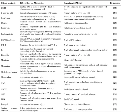
Research on oligoprotectants has been going on for a long time, since oligopathy has long been noted in de- myelinating diseases like multiple sclerosis, periven- tricular leukomalacia (PVL), acute disseminated enceph- alomyelitis (ADEM). Later on, with the focus on pro- tecting neurovascular unit in stroke and other neurologi- cal diseases more researches have been carried out aimed at protecting oligodendrocytes. Hereby, a few drugs and factors that have proved useful in protecting OLGs and white matter as a whole using different models are dis- cussed, a summary of which can be seen in Table 1.
4.1. Antibacterial Agent—Minocycline
Minocycline, a second-generation tetracycline, has shown
beneficial effects in a wide range of acute neurological injuries. In rodent brain ischemic models, it showed anti-inflammatory effects by inhibiting immune media- tors such as microglia. It has shown to attenuate hypoxia/ ischemia-induced white matter injury in the neonatal rat though report of directly protecting OLGs against ische- mic stress in adult rodent stroke model is missing. It has also been seen that delayed minocycline treatment re- duced long-term functional deficits as well as white mat- ter injury in ET-1-induced rat ischemia model. Despite reports from animal models clinical trial using mino cy- cline in Amyotrophic lateral sclerosis (ALS) patients, failed to show efficacy, this could be attributed to the long term use of minocycline, a powerful metallopro- teinase inhibitor. It has been suggested that long-term suppression of metalloproteinases may be detrimental for neurovascular homeostasis [49].
4.2. Antioxidants and Free Radical Scavengers
Antioxidants and free radical scavengers have proved beneficial to OLGs which show a great vulnerability to oxidative stress. Body’s endogenous component and various synthetic drugs have been tried for use in pre- venting damage to OLGs and a few of them which proved beneficial have been listed below.
4.2.1. Endogenous Component-CoenzymeQ10 (CoQ10)
CoQ10 an endogenous component of the mitochondrial electron transport chain is known to attenuate the per- oxidation of lipids and protect neurons against free radi- cal in both in vitro primary cell culture and experi mental animals. It was shown that it can protect OLGs against TNF-α using primary culture of rat glial cells. In the same study, CoQ10 restored the numbers of mature mye- lin basic protein-positive cells and the ability of the OLGs to form membrane sheets [47]. However, there is published case of an in vivo model of focal and global ischemia in rats where CoQ10 failed to protect brain [50].
4.2.2. Synthetic Drugs
Various synthetic drugs in the market possessing anti- oxidant activity are being screened for their effectiveness in protecting the OLGs in different injury models. Here, we have listed some drugs that have proved to be useful in the researches aimed at protecting OLGs and white matter injury.
4.2.2.1. Ebselen
Ebselen is a synthetic seleno-organic drug with glutath- ione peroxidase like activity [51]. Imai et al. using rat transient ischemia models showed that ebselen reduced axonal damage and oligodendrocyte pathology [24]. 4.2.2.2. Edaravone
Table 1. Oligoprotectants and white matter protecting agents.
Oligoprotectants Effects Bserved-Mechanism Experimental Model References
CNTF Inhibits TNF-α-induced apoptotic death of oligodendrocyte precursor cell In vitroline systems of oligodendrocyte precursor cell [54]
C enzyme Q10 Protects oligodendrocytes against TNF-injury Primary cultures of rat glial cells [47]
Cord blood Protects striatal white matter tracts protects mature oligodendrocytes in culture in vivo; In vivo oxygen and glucose deprivation model model; primary oligodendrocyte cultures [55]
Ebselen Reduces axonal damage and oligodendrocyte pathology Rat transient ischemia models [24]
Edaravone Decreases oligodendrocyte loss and attenuates white matter lesions Rat chronic hypoperfusion model [52]
Erythropoietin
Increases oligodendrogenesis, recovery of injured white matter and improved neurological function recovery
Neonatal hypoxic-ischemic injury in rats [56]
HSP90 inhibitors Protects OPCs and adult oligodendrocytes against HSP90 autoantibodies in MS In vitro OPC cultures [57]
IGF-1 Decreases the pro-apoptotic actions of TNF-α In vitro and in vivo systems [11]
IL-11 Potentiates oligodendrocyte survival and maturation; myelin formation In vitro human cell cultures; rodent co-culture studies [15]
Linomide Prevents oligodendrocytes damage by suppressing nitric oxide production in microglia and astrocytes Primary cultures of rat glial cells [58]
Melatonin Reduces oxidative damage in neurons and oligodendrocytes Mouse MCAO model [59]
Memantine Attenuated white matter injury, reduced ischemic damage to mature and precursor oligodendrocytes in brain slices
Rat model of periventricular malacia and ischemia;
patch clamp system [60]
Methylprednisolone Selectively inhibits oligodendrocyte but not neuronal death Staurosporine or AMPA model of injury through glucocorticoid receptor [53]
Minocycline Attenuates white matter injury In neonatal hypoxia/ ischemia induced [49]
NAC
Restores the number of MBP positive cells; restores ability of oligodendrocytes to form membranous sheets
In vitro primary rat cell culture; neuroblastoma
cultures [47]
NBQX Reduces white matter injury and improve locomotor function Rat ischemic spinal cord model [24]
NGF Protects oligodendrocytes from TNF-α-mediated cytotoxicity Primary cultures of rat oligodendrocytes [61]
PBN Protects both oligodendrocytes and axons from ischemic insults Rat MCAO model [60]
Ramipril Attenuates white matter injury Chronic hypoperfusion ishcemia [62]
Sodium channel blockers
Offers protection to white matter against
anoxia-induced injury In vitro rat optic nerve preparation [63] List of different drugs that showed promise in protecting the Oligodendrocytes or white matter as a whole, the mechanism of their protection, experimental models they were tested in along with the appropriate references.
shown to decrease the loss of OLGs and attenuate white matter lesions through endothelial protection and free radical scavenging effects in a chronic hypoperfusion model in rats. Randomized clinical trial setting has also shown significant functional improvement in acute ischemic stroke patients [52].
4.2.2.3. N-Acetyl Cysteine (NAC)
NAC is a free radical scavenger and an antioxidant that can modulate gene transcription and apoptosis. Cammer using in vitro primary rat cell culture showed that NAC was able to restore the numbers of mature myelin basic protein-positive cells and the ability of the OLGs to form
membrane sheets following studies that it protected neu- roblastoma cultures against oxidative stress and cytotox- icity [47].
4.2.2.4. Methylprednisolone
ner in staurosporine or AMPA model of injury through glucocorticoid receptor dependent mechanism; however, same dose failed to prevent the neurons [53].
4.2.2.5. Phenyl-N-tert-butyl-nitrone (PBN)
PBN a free radical scavenger has proved to be useful in preventing oligodendrocyte damage in various models. Using rat MCAO model, Irving et al. demonstrated that a free radical scavenger PBN reduced injury to the OLGs through the reduced number of tau-positive OLGs in the sub-cortical white matter of the ischemic hemisphere in PBN treated rats. Lin et al. using hypoxia-ischemia model in the neonatal rat brain displayed PBN treatment protected both OLGs and axons from ischemic insults. In the same model, it was seen that PBN also inhibits up- regulation of inflammatory cytokines such as IL-1β, TNF-α and inducible NOS mRNA expression [60]. 4.2.2.6. Ramipril
Ramipril, an angiotensin converting enzyme inhibitor, has shown to attenuate white matter lesions against chro- nic hypoperfusion ischemia owing to its free radical sca- venging effect in rat models [62].
4.3. Cord Blood
It has been shown that infusion of human umbilical cord blood cells protects striatal white matter tracts in vivo and directly protects mature primary oligodendrocyte cultures from oxygen glucose deprivation (OGD) [55].
4.4. Endogenous Factors
Different endogenous factors secreted in animals have positive influences in OLGs. The PDGF, basic-fibroblast growth factor (b-FGF), and IGF-1 are known to influence oligodendrocyte lineage cell proliferation and survival [64]. A few of these endogenous factors have shown to afford protection to the OLGs against different stress conditions. The promising results from endogenous fac- tors have been listed below.
4.4.1. Ciliary Neurotrophic Factor (CNTF)
CNTF, a trophic factor found in astrocytes, has shown to inhibit TNF-α apoptotic death of oligodendrocytes in an
in vitro model. It plays a role in survival of OLGs during development and may help in developing treatment strategies aimed at demyelinating diseases [54].
4.4.2. Insulin-Like Growth Factor-1 (IGF-1)
IGF-1 is known for its neuroprotective effects and is taken as a promising candidate for neuromuscular dis- eases [65]. It plays an immense role in OLGs right from the developmental stage, enhanching OPC survival to maturation and stimulating myelination [64,66,67]. IGF- 1 also known as the survival factor for OLGs [68], when applied externally has shown to decrease the pro-apo ptotic actions of TNF-α [11].
4.4.3. Interleukin-11 (IL-11)
IL-11, an astrocyte-derived which is localized in the ac- tive and silent lesions of MS, is linked with increase in OLG number and remyelination in the early stages. It potentiates oligodendrocyte survival and maturation, and myelin formation. In vitro human cell cultures have shown that immature OLGs express IL-11Rα receptors and IL-11 has been linked with enhanced oligodendro- cyte survival and maturation. Rodent CNS co-culture studies have linked it to increased myelination, thus im- plicating IL-11 in oligodendrocyte viability, maturation, and myelination [15].
4.4.4. Nerve Growth Factor (NGF)
NGF has long been known for causing neurite outgrowth and hence the name nerve growth factor. It protects OLGs from TNF-α mediated cytotoxicity. Nerve growth factor also prevented the tumor necrosis factor-induced loss of mitochondrial membrane potential [61].
4.5. Glutamate Receptor Antagonist
Glutamate excitotoxicity related death of oligodendro- cyte is seen in many disease states and thus exploiting the receptor antagonists have proved to be beneficial in different experiments. The antagonists of AMPA/KA and NMDA receptor types associated with glutamate signal- ing have shown significant reduction of OLG death in hypoxia/ischemia models. Hence, they may prove bene- ficial targets for prevention of ischemic stroke and re- lated OLG deaths.
4.5.1. AMPA/KA Antagonist—NBQX
The AMPA/kainate receptor mechanism has recently been implicated in white matter injury following white matter ischemia. NBQX, an AMPA/kainate receptor an- tagonist, has shown to reduce ischemia-induced white matter injury and improve locomotor function in a rat ischemic spinal cord model [24].
4.5.2. NMDA Antagonist—Memantine
Memantine, an uncompetitive NMDA receptor antago- nist, is a licensed drug for moderate-to-severe Alzhei- mer’s disease in US and EU. Memantine attenuated white matter injury in a rat model of periventricular leu- komalacia and may also be protective to white matter in ischemia. It has been reported that memantine reduced ischemic damage to mature and precursor OLGs in brain slices following assessment by patch-clamp system [60].
4.6. Hormones
considered to be vital for survival of OLGs and it is known to trigger the maturation process of OLGs [69].
4.6.1. Erythropoietin
The hormone erythropoietin made by the kidney cells under hypoxic conditions acts on stem cells in the bone marrow to increase the production of red blood cells. Erythropoietin mediated neuroprotection against neonatal hypoxic-ischemic brain injury has been known since 2003. It promotes differentiation and maturation of OLGs and is also expected to affect the ability of OLGs to promote myelin repair in normal and damaged CNS [70]. Increased oligodendrogenesis, recovery of injured white matter and improved neurological function recov- ery by delayed and repeated administration of erythro- poietin was seen after neonatal hypoxic-ischemic brain injury in rats although the delayed administration of erythropoietin did not decrease infarct volume [56].
4.6.2. Melatonin
Melatonin, a naturally occurring hormone, has been in use for the treating sleep related disorders. It has been found to reduce oxidative damage in neurons and OLGs through antioxidant and radical scavenging activity as seen in an experiment using mouse middle cerebral ar- tery occlusion (MCAO) model using amyloid precursor protein (APP) and microtubule associated protein Tau-1 as markers for evaluating post-ischemic disrupted axonal flow and oligodendrocyte pathology respectively through immunohistochemical studies. It is known to reduce in- farct volumes and enhance neurobehavioral and electro- physiological recoveries [59].
4.7. HSP90 Inhibitors
Heat shock protein 90 (HSP90) present in OPCs surface has been known to form autoantibodies in Multiple Sclerosis. HSP90 inhibitors radicicol and 17-allylamino- 17-demethoxygeldanamycin (17-AAG) at non-cytotoxic doses target cell-surface HSP90 in OPCs and through competitive inhibition of receptors prevent HSP90-anti- body-induced OPC death thus protecting them against antibody attack. They also have shown protective effect on adult OLGs [57].
4.8. Immunomodulator (Synthetic)-Linomide
Bao et al. using primary cultures of rat glial cells showed that a synthetic immunomodulator linomide prevents OLGs damage by suppressing nitric oxide production in microglia and astrocytes [58].
4.9. Sodium Channel Blockers
Ischemic injury in the gray matter is associated with ex- citatory amino acid neurotransmitters release, and in the white matter is associated with intracellular sodium ac-
cumulation. The effect of sodium channel blocking drugs phenytoin, carbamazepine and diazepam on anoxia-in- duced injury in CNS white matter in vitro rat optic nerve preparation has shown to offer protection against anoxic injury at concentrations much lower than that required for treating epilepsy [63]. Sodium channel blockers rep- resent an attractive therapeutic target because of their ability to simultaneously interfere indirectly with several calcium sourcing pathways [71].
5. Conclusions
Oligodendrocytes are most vulnerable cells of the brain with highest energy requirement, high iron stores, and low glutathione content; implicated primarily in demye- linating diseases and secondarily in neuropathies. The maintenance of OLGs at functional level is necessary for proper functioning of neuronal pathways, the loss of which may lead to multiple functional loss, axonal de- generation and/or neuronal death. Therefore, the neuro- centric view of neuroprotection has widened to embrace the glial cells. In order to get along with neuroprotection, which has been the central focus for long, much work needs to be done on finding ways of protecting the glial cells. For this, a better understanding of the mechanisms by which glial cells death ensues in various disease states is utmost important. A better understanding of the injury mechanisms of OLGs should be beneficial as we move along the path of protecting the long shadowed glial cells hand in hand with neuroprotection as the concept of the neurogliovascular unit flourishes.
REFERENCES
[1] K. Arai and E. H. Lo, “Oligovascular Signaling in White Matter Stroke,” Biological & Pharmaceutical Bulletin, Vol. 32, No. 10, 2009, pp. 1639-1644.
doi:10.1248/bpb.32.1639
[2] M. Bastide, T. Ouk, F. Plaisier, O. Pétrault, S. Stolc and R. Bordet, “Neurogliovascular Unit after Cerebral Ische-mia: Is the Vascular Wall a Pharmacological Target,”
Psychoneuroendocrinology, Vol. 32, Supplement 1, No. 1, 2007, pp. S36-S39.
[3] B.V. Zlokovic, “Neurodegeneration and the Neurovascu-lar Unit,” Nature Medicine, Vol. 16, No.12, 2010, pp. 1370-1371. doi:10.1038/nm1210-1370
[4] N. Baumann and D. Pham-Dinh, “Biology of Oli-goden-Drocyte and Myelin in the Mammalian Central Nervous System,” Physiological Reviews, Vol. 81, No. 2, 2001, pp. 871-927.
[5] G. Bartzokis, “Age-Related Myelin Breakdown: A De-velop-Mental Model of Cognitive Decline and Alz-heimer’s Disease,” Neurobiology of Aging, Vol. 25, No. 1, 2004, pp. 5-18. doi:10.1016/j.neurobiolaging.2003.03.001
Patho-genesis,” Biochimica et Biophysica Acta, Vol. 1812, No. 2, 2010, pp. 141-150.
[7] M. Bradl and H. Lassmann, “Oligodendrocytes: Biology and Pathology,” Acta Neuropathologica, Vol. 119, No. 1, 2010, pp. 37-53. doi:10.1007/s00401-009-0601-5
[8] C. Richter-Landsberg and U. Vollgraf, “Mode of Cell Injury and Death after Hydrogen Peroxide Exposure in Cultured Oligodendroglia Cells,” Experimental Cell Re- search, Vol. 244, No. 1, 1998, pp. 218-229.
doi:10.1006/excr.1998.4188
[9] M. Nedergaard, J. J. Rodríguez and A. Verkhratsky, “Glial Calcium and Diseases of the Nervous System,”
Cell Calcium, Vol. 47, No. 2, 2010, pp. 140-149.
doi:10.1016/j.ceca.2009.11.010
[10] D. Zieve, “Multiple Aclerosis,” Pubmed Health, 2010. http://www.ncbi.nlm.nih.gov/pubmedhealth/PMH000174 7/
[11] C. A. Tegla, C. Cudrici, V. Rus, T. Ito, S. Vlaicu, A. Singh and H. Rus, “Neuroprotective Effects of the Com-plement Terminal Pathway during Demyelination: Im-plica-tions for Oligodendrocyte Survival,” Journal of Neuroimmunology, Vol. 213, No. 1-2, 2009, pp. 3-11.
doi:10.1016/j.jneuroim.2009.06.006
[12] A. Jana and K. Pahan, “Oxidative Stress Kills Human Primary Oligodendrocytes via Neutral Sphingomyelinase: Implications for Multiple Sclerosis,” Journal of Neuro-immune Pharmacology, Vol. 2, No. 1, 2007, pp. 184-193.
doi:10.1007/s11481-007-9066-2
[13] M. Vanmeeteren, J. Hendriks, C. Dijkstra and E. Vantol, “Dietary Compounds Prevent Oxidative Damage and Ni-tric Oxide Production by Cells Involved in Demyelinating Disease,” Biochemical Pharmacology, Vol. 67, No. 5, 2004, pp. 967-975. doi:10.1016/j.bcp.2003.10.018
[14] C. Matute, E. Alberdi, M. Domercq, M.-V. Sánchez- Gómez, A. Pérez-Samartín, A. Rodríguez-Antigüedad, and F. Pérez-Cerdá, “Excitotoxic Damage to White Mat-ter,” Journal of Anatomy, Vol. 210, No. 6, 2007, pp. 693-702. doi:10.1111/j.1469-7580.2007.00733.x
[15] Y. Zhang, C. Taveggia, C. Melendez-Vasquez, S. Ein-heber, C. S. Raine, J. L. Salzer, C. F. Brosnan and G. R. John, “Interleukin-11 Potentiates Oligodendrocyte Sur-vival and Maturation, and Myelin Formation,” The Jour-nal of Neuroscience, Vol. 26, No. 47, 2006, pp. 12174- 12185. doi:10.1523/JNEUROSCI.2289-06.2006
[16] S. A Back, N. L. Luo, N. S. Borenstein, J. M. Levine, J. J. Volpe and H. C. Kinney, “Late Oligodendrocyte Pro-genitors Coincide with the Developmental Window of Vulnerability for Human Perinatal White Matter Injury,”
The Journal of Neuroscience, Vol. 21, No. 4, 2001, pp. 1302-1312.
[17] M. V. Johnston, A. Ishida, W. N. Ishida, H. B. Matsushita, A. Nishimura and M. Tsuji, “Plasticity and Injury in the Developing Brain,” Brain & Development, Vol. 31, No. 1, 2009, pp. 1-10. doi:10.1016/j.braindev.2008.03.014
[18] Y. Pang, L. Campbell, B. Zheng, L. Fan, Z. Cai and P. Rhodes, “Lipopolysaccharide-Activated Microglia Induce Death of Oligodendrocyte Progenitor Cells and Impede
Their Development,” Neuroscience, Vol. 166, No. 2, 2010, pp. 464-475.
doi:10.1016/j.neuroscience.2009.12.040
[19] K. G. Lee, “Periventricular Leukomalacia,” Pubmed Health, 2009.
http://www.ncbi.nlm.nih.gov/pubmedhealth/PMH0004493/ [20] J. J. Volpe, H. C. Kinney, E. J. Frances,and P. A. Rosenberg, “The Developing Oligodendrocyte: Key Cel-lular Target in Brain Injury in the Premature Infant,” In-ternational Journal of Developmental Neuroscience, Vol. 29, No. 4, 2011, pp. 423-440.
[21] D. B. Hoch, “Stroke,” Pubmed Health, 2010.
http://www.ncbi.nlm.nih.gov/pubmedhealth/PMH0001740/ [22] A. S. Hazell, “Excitotoxic Mechanisms in Stroke: An
Up-Date of Concepts and Treatment Strategies,” Neuro-chemistry International, Vol. 50, No. 7-8, 2007, pp. 941-953. doi:10.1016/j.neuint.2007.04.026
[23] W. Pan and A. J. Kastin, “Tumor Necrosis Factor and Stroke: Role of the Blood-Brain Barrier,” Progress in Neurobiology, Vol. 83, No. 6, 2007, pp. 363-374.
doi:10.1016/j.pneurobio.2007.07.008
[24] D. Dewar, S. M. Underhill and M. P. Goldberg, “Oli-godendrocytes and Ischemic Brain Injury,” Journal of Cerebral Blood Flow & Metabolism, Vol. 23, No. 1, 2003, pp. 263-274. doi:10.1097/00004647-200303000-00001
[25] E. H. Lo, T. Dalkara and M. A. Moskowitz, “Mechanisms, Challenges and Opportunities in Stroke,” Nature Reviews Neuroscience, Vol. 4, No. 5, 2003, pp. 399-415.
doi:10.1038/nrn1106
[26] D. Zieve, “Spinal Cord Trauma,” Pubmed Health, 2010. http://www.ncbi.nlm.nih.gov/pubmedhealth/PMH0002061/ [27] R. Talac, J. A. Friedman, M. J. Moore, L. Lu, E. Jabbari,
A. J. Windebank, B. L. Currier and M. J. Yaszemski, “Animal Models of Spinal Cord Injury for Evaluation of Tissue Engineering Treatment Strategies,” Biomaterials, Vol. 25, No. 9, 2004, pp. 1505-1510.
doi:10.1016/S0142-9612(03)00497-6
[28] G. D. Carlson and C. Gorden, “Current Developments in Spinal Cord Injury Research,” The Spine Journal, Vol. 2, No. 1, 2002, pp. 116-128.
doi:10.1016/S1529-9430(01)00029-8
[29] Y. H. Ahn, G. Lee and S. K. Kang, “Molecular Insights of the Injured Lesions of Rat Spinal Cords: Inflammation, Apoptosis, and Cell Survival,” Biochemical and Bio-physical Research Communications, Vol. 348, No. 1, 2006, pp. 560-570. doi:10.1016/j.bbrc.2006.07.105
[30] “Leukodystrophies,” Medline Plus, 2011.
http://www.nlm.nih.gov/medlineplus/leukodystrophies.html. [31] R. Káradóttir and D. Attwell, “Neurotransmitter
Recep-tors in the Life and Death of Oligodendrocytes,” Neuro-science, Vol. 145, No. 4-5, 2007, pp. 1426 -1438. [32] B. D. Butts, C. Houde and H. Mehmet,
[33] A. D. Roth, G. Ramírez, R. Alarcón and R. V. Bernhardi, “Oligodendrocytes Damage in Alzheimer’s Disease : Beta Amyloid Toxicity and Inflammation,” Biological Re-search, Vol. 38, No. 1, 2005, pp. 381-387.
[34] D. B. Merrill, “Schizophrenia,” Pubmed Health, 2010. http://www.ncbi.nlm.nih.gov/pubmedhealth/PMH0001925/ [35] N. A. Uranova, V. M. Vostrikov, O. V. Vikhreva, I. S.
Zimina, N. S. Kolomeets and D. D. Orlovskaya, “The Role of Oligodendrocyte Pathology in Schizophrenia,”
The International Journal of Neuropsychopharmacology, Vol. 10, No. 4, 2007, pp. 537-545.
doi:10.1017/S1461145707007626
[36] P. R. Hof, V. Haroutunian, C. Copland, K. L. Davis and J. D. Buxbaum, “Molecular and Cellular Evidence for an Oligodendrocyte Abnormality in Schizophrenia,” Neuro-chemical Research, Vol. 27, No. 10, 2002, pp. 1193-1200.
doi:10.1023/A:1020981510759
[37] D. Segal, J. R. Koschnick, L. H. A. Slegers and P. R. Hof, “Oligodendrocyte Pathophysiology: A New View of Schizophrenia,” The International Journal of Neuropsy-chopharmacology, Vol. 10, No. 4, 2007, pp. 503-511.
doi:10.1017/S146114570600722X
[38] A. Verkhratsky and V. Parpura, “Recent Advances in (Patho) Physiology of Astroglia,” Acta Pharmacologica Sinica, Vol. 31, No. 1, 2010, pp. 1044-1054.
doi:10.1038/aps.2010.108
[39] C. Mihai and B. Jubelt, “Post-Infectious Encephalomye-litis,” Current Neurology and Neuroscience Reports, Vol. 5, No. 1, 2005, pp. 440-445.
doi:10.1007/s11910-005-0031-2
[40] K. Sano, K. Morii, M. Sato, H. Mori and R. Tanaka, “Ra-diation-Induced Diffuse Brain Injury in the Neonatal Rat Model-Radiation-Induced Apoptosis of Oligodendro-cytes,” Neurologia Medico-Chirurgica, Vol. 40, No. 10, 2000, pp. 495-499. doi:10.2176/nmc.40.495
[41] A. Ernst, A. Stolzing, G. Sandig and T. Grune, “Anti- Oxidants Effectively Prevent Oxidation-Induced Protein Damage in OLN 93 Cells,” Archives of Biochemistry and Biophysics, Vol. 421, No. 1, 2004, pp. 54-60.
doi:10.1016/j.abb.2003.10.008
[42] H. Mann, M. T. McCoy, J. Subramaniam, H. V. Remmen, and J. L. Cadet, “Overexpression of Superoxide Dismu-tase and Catalase in Immortalized Neural Cells: Toxic Effects of Hydrogen Peroxide,” Brain Research, Vol. 770, No. 1, 1997, pp. 163-168.
doi:10.1016/S0006-8993(97)00768-3
[43] C. Matute, “Oligodendrocyte NMDA Receptors: A Novel Therapeutic Target,” Trends in Molecular Medicine, Vol. 12, No. 7, 2006, pp. 289-292.
doi:10.1016/j.molmed.2006.05.004
[44] E. A. Leuchtmann, A. E. Ratner, R. Vijitruth, Y. Qu and J. W. Mcdonald, “AMPA Receptors are the Major Media-tors of Excitotoxic Death in Mature Oligodendrocytes,”
Neurobiology of Disease, Vol. 14, No. 1, 2003, pp. 336-348. doi:10.1016/j.nbd.2003.07.004
[45] W. Cammer and H. Zhang, “Maturation of Oligodendro-cytes is More Sensitive to TNF-α than is Survival of
Pre-cursors and Immature Oligodendrocytes,” Journal of Neuroimmunology, Vol. 97, No. 1, 1999, pp. 37-42.
doi:10.1016/S0165-5728(99)00045-4
[46] W. Chadwick, T. Magnus, B. Martin, A. Keselman, M. P. Mattson and S. Maudsley, “Targeting TNF-α Receptors for Neurotherapeutics,” Trends in Neurosciences, Vol. 31, No. 10, 2008, pp. 504-511.
doi:10.1016/j.tins.2008.07.005
[47] W. Cammer, “Protection of Cultured Oligodendrocytes against Tumor Necrosis Factor-α by the Antioxidants Coenzyme Q10 and N-acetyl Cysteine,” Brain Research Bulletin, Vol. 58, No. 6, 2002, pp. 587-592.
doi:10.1016/S0361-9230(02)00830-4
[48] J. L. Takahashi, F. Giuliani, C. Power, Y. Imai and V. W. Yong, “Interleukin-1β Promotes Oligodendrocyte Death through Glutamate Excitotoxicity,” Annals of Neurology, Vol. 53, No. 5, 2003, pp. 588-595. doi:10.1002/ana.10519
[49] T. Y. Yune, J. Y. Lee, G. Y. Jung, S. J. Kim, M. H. Jiang, Y. C. Kim, Y. J. Oh, G. J. Markelonis and T. H. Oh, “Minocycline Alleviates Death of Oligodendrocytes by Inhibiting Pro-nerve Growth Factor Production in Micro-glia after Spinal Cord Injury,” The Journal of Neurosci-ence, Vol. 27, No. 29, 2007, pp. 7751-7761.
doi:10.1523/JNEUROSCI.1661-07.2007
[50] H. Li, G. Klein, P. Sun and A. M. Buchan, “CoQ10 Fails to Protect Brain against Focal and Global Ischemia in Rats,” Brain Research, Vol. 877, No. 1, 2000, pp. 7-11.
doi:10.1016/S0006-8993(00)02609-3
[51] Y. Nakamura, Q. Feng, T. Kumagai, K. Torikai, H. Ohi-gashi, T. Osawa, N. Noguchi, E. Niki and K. Uchida, “Eb-selen, a Glutathione Peroxidase Mimetic Seleno-Organic Compound, as a Multifunctional Antioxidant,” The Jour-nal of Biological Chemistry, Vol. 277, No. 4, 2002, pp. 2687-2694. doi:10.1074/jbc.M109641200
[52] Y. Ueno, N. Zhang, N. Miyamoto, R. Tanaka, N. Hattori and T. Urabe, “Edaravone Attenuates White Matter Le-sions through Endothelial Protection in a Rat Chronic Hypoperfusion Model,” Neuroscience, Vol. 162, No. 2, 2009, pp. 317-327.
doi:10.1016/j.neuroscience.2009.04.065
[53] J.-M. Lee, P. Yan, Q. Xiao, S. Chen, K.-Y. Lee, C. Y. Hsu and J. Xu, “Methylprednisolone Protects Oligoden-drocytes but Not Neurons after Spinal Cord Injury,” The Journal of Neuroscience, Vol. 28, No. 12, 2008, pp. 3141-3149. doi:10.1523/JNEUROSCI.5547-07.2008
[54] J. C. Louis, E. Magal, S. Takayama and S. Varon, “CNTF Protection of Oligodendrocytes against Natural and Tu-mor Necrosis Factor-Induced Death,” Science, Vol. 259, No. 5095, 1993, pp. 689-692.
doi:10.1126/science.8430320
[55] D. D. Rowe, C. C. Leonardo, A. A. Hall, M. D. Sha-haduzzaman, L. A. Collier, A. E. Willing and K. R. Pen-nypacker, “Cord Blood Administration Induces Oli-godendrocyte Survival through Alterations in Gene Ex-pression,” Brain Research, Vol. 1366, No. 1, 2010, pp. 172-188. doi:10.1016/j.brainres.2010.09.078
Mat-ter Protection by Erythropoietin: An Emerging MatMat-ter in the Treatment of Neonatal Hypoxic-Ischemic Brain In-jury,” Stroke, Vol. 41, No. 11, 2010, p. e595.
doi:10.1161/STROKEAHA.110.590844
[57] C. Cid and A. Alcazar, “Protection of Oligodendrocyte Precursor Cells by Low Doses of HSP90 Inhibitors in Cell Culture,” Experimental Neurology, Vol. 225, No. 7, 2010, pp. 29-33. doi:10.1016/j.expneurol.2009.11.017
[58] B. G. Xiao, X. F. Bail, G. X. Zhang, G. Hedlund and H. Link, “Linomide-Mediated Protection of Oligodendro- cytes is Associated with Inhibition of Nitric Oxide Pro-duction and IL-1β Expression in Lewis Rat Glial Cells,”
Neuroscience Letters, Vol. 249, No. 1, 1998, pp. 17-20.
doi:10.1016/S0304-3940(98)00371-1
[59] E.-J. Lee, M.-Y. Lee, H.-Y. Chen, Y.-S. Hsu, T.-S. Wu, S.-T. Chen and G.-L. Chang, “Melatonin Attenuates Gray and White Matter Damage in a Mouse Model of Transient Focal Cerebral Ischemia,” Journal of Pineal Research, Vol. 38, No. 1, 2005, pp. 42-52.
doi:10.1111/j.1600-079X.2004.00173.x
[60] K. Arai and E. H. Lo, “Experimental Models for Analysis of Oligodendrocyte Pathophysiology in Stroke,” Experi-mental & Translational Stroke Medicine, Vol. 1, No. 6, 2009.
[61] R. Takano, S. Hisahara, K. Namikawa, H. Kiyama, H. Okano and M. Miura, “Nerve Growth Factor Protects Oli-godendrocytes from Tumor Necrosis Factor-α-In- duced Injury through Akt-Mediated Signaling Mecha-nisms,” The Journal of Biological Chemistry, Vol. 275, No. 21, 2000, pp. 16360-16365.
doi:10.1074/jbc.M910419199
[62] J.-S. Kim, I. Yun, Y. B. Choi, K.-S. Lee and Y.-I. Kim, “Ramipril Protects from Free Radical Induced White Matter Damage in Chronic Hypoperfusion in the Rat,”
Journal of Clinical Neuroscience, Vol. 15, No. 2, 2008, pp. 174-178. doi:10.1016/j.jocn.2006.12.003
[63] R. Fern, B. R. Ransom, P. K. Stys and S. G. Waxman, “Pharmacological Protection of CNS White Matter during Anoxia: Actions of Phenytoin, Carbamazepine and Di-azepam,” The Journal of Pharmacology and Experimen-tal Therapeutics, Vol. 266, No. 3, 1993, pp. 1549-1555.
[64] W. H. Lagarde, R. Benjamin, A. T. Heerens, P. Ye, R. I. Cohen, B. M. Moats-Staats and A. J. D’ercole, “A Non-Transformed Oligodendrocyte Precursor Cell Line, OL-1, Facilitates Studies of Insulin-Like Growth Factor-I Signaling during Oligodendrocyte Development,” Inter-national Journal of Developmental Neuroscience, Vol. 25, No. 2, 2007, pp. 95-105.
doi:10.1016/j.ijdevneu.2006.12.006
[65] A. Musarò, G. Dobrowolny and N. Rosenthal, “The Neu-roprotective Effects of a Locally Acting IGF-1 Isoform,”
Experimental Gerontology, Vol. 42, No. 1-2, 2007, pp. 76-80. doi:10.1016/j.exger.2006.05.004
[66] P. Ye and A. J. D’Ercole, “Insulin-Like Growth Factor I Protects Oligodendrocytes from Tumor Necrosis Fac-tor-α-Induced Injury,” Endocrinology, Vol. 140, No. 7, 1999, pp. 3063-3072. doi:10.1210/en.140.7.3063
[67] F. A. McMorris, T. M. Smith, S. DeSalvo and R. W. Fur-lanetto, “Insulin-Like Growth Factor I/Somatomedin C: A Potent Inducer of Oligodendrocyte Development,” Pro- ceedings of the National Academy of Sciences of the United States of America, Vol. 83, No. 3, 1986, pp. 822-826. doi:10.1073/pnas.83.3.822
[68] J. L. Mason, S. Xuan, I. Dragatsis, A. Efstratiadis and J. E. Goldman, “Insulin-Like Growth Factor (IGF) Signaling through Type 1 IGF Receptor Plays an Important Role in Remyelination,” The Journal of Neuroscience, Vol. 23, No. 20, 2003, pp. 7710-7718.
[69] S. A. Jones, D. M. Jolson, K. K. Cuta, C. N. Mariash and G. W. Anderson, “Triiodothyronine is a Survival Factor for Developing Oligodendrocytes,” Molecular and Cel-lular Endocrinology, Vol. 199, No. 1-2, 2003, pp. 49-60.
doi:10.1016/S0303-7207(02)00296-4
[70] M. Sugawa, Y. Sakurai, Y. Ishikawa-Ieda, H. Suzuki and H. Asou, “Effects of Erythropoietin on Glial Cell Devel-opment; Oligodendrocyte Maturation and Astrocyte Pro-liferation,” Neuroscience Research, Vol. 44, No. 4, 2002, pp. 391-403. doi:10.1016/S0168-0102(02)00161-X
[71] P. K. Stys, “White Matter Injury Mechanisms,” Current Molecular Medicine, Vol. 4, No. 2, 2004, pp. 113-130.