NITRATE TRANSPORT AND ASSIMILATION IN
ASPERGILLUS NIDULANS
Naureen Akhtar
A Thesis Submitted for the Degree of PhD
at the
University of St Andrews
2012
Full metadata for this item is available in
Research@StAndrews:FullText
at:
http://research-repository.st-andrews.ac.uk/
Please use this identifier to cite or link to this item:
http://hdl.handle.net/10023/3093
This item is protected by original copyright
Nitrate transport and assimilation in
Aspergillus nidulans
Naureen Akhtar
This thesis is submitted in partial fulfilment for the degree of PhD
at the
University of St Andrews
i Candidate's declarations
I, Naureen Akhtar, hereby certify that this thesis, which is approximately 58,000 words in length, has been written by me, that it is the record of work carried out by me and that it has not been submitted in any previous application for a higher degree.
I was admitted as a research student in November, 2008 and as a candidate for the degree of PhD in November, 2008; the higher study for which this is a record was carried out in the University of St Andrews between 2008 and 2012.
Date …...… Signature of candidate ……...…
Supervisor's declarations
I hereby certify that the candidate has fulfilled the conditions of the Resolution and Regulations appropriate for the degree of PhD in the University of St Andrews and that the candidate is qualified to submit this thesis in application for that degree.
ii Permission for electronic publication
In submitting this thesis to the University of St Andrews I understand that I am giving permission for it to be made available for use in accordance with the regulations of the University Library for the time being in force, subject to any copyright vested in the work not being affected thereby. I also understand that the title and the abstract will be published, and that a copy of the work may be made and supplied to any bona fide library or research worker, that my thesis will be electronically accessible for personal or research use unless exempt by award of an embargo as requested below, and that the library has the right to migrate my thesis into new electronic forms as required to ensure continued access to the thesis. I have obtained any third-party copyright permissions that may be required in order to allow such access and migration, or have requested the appropriate embargo below.
The following is an agreed request by candidate and supervisor regarding the electronic publication of this thesis:
Access to all of printed copy but embargo of all of electronic publication of thesis for a period of two years on the following grounds:
publication would preclude future publication; publication would be in breach of law or ethics
iii Acknowledgment
I wholeheartedly thank my supervisor, Dr. Jim Kinghorn, whose encouragement, supervision and support from the preliminary to the final level enabled me to develop an understanding of the subject. I am indebted to Dr. Kinghorn for his enormous amount of help, teaching and valuable advices throughout my research work. I appreciate his support and help for reviewing my chapters with enthusiasm and for providing constructive feedback. I have learnt much from him and I believe without his guidance and encouragement this thesis would not have been completed or written. I also warmly appreciate the generosity of Dr. Kinghorn regarding his efforts in solving my personal problems, completing forms on my behalf and providing reference letters whenever needed.
My special gratitude goes to Dr. Shiela Unkles, my co-supervisor, who has supported me throughout my research work with patience and knowledge. I cannot find words to express my thanks to Dr. Unkles for her efforts to train me in the field of Molecular Biology that was absolutely new to me when I came to St. Andrews University. She has taught me, both consciously and un-consciously, how good experimental Biology is done. I acknowledge all her input, time and ideas for me during my research work. Her encouraging personal guidance has provided a good basis for the present thesis. I hope that one day I would become as good an advisor to my students as Dr. Unkles has been to me.
In my daily work I have been blessed with a friendly and cheerful group of fellows, Eugenia Karabika, Vicky Symington and Amy Clarke. Thanks go to them for their support and friendship throughout and I wish them all success in their future careers and in life generally. Further help was given by others during particular sections of the research and these individuals are acknowledged at the end of the relevant chapter.
It gives me great pleasure to acknowledge the support and help from Professor Alyson Tobin and Professor Richard Abbott and for their supportive discussion during their continuous monitoring of my research progress. Special thanks go to Dr. Roger Griffiths and Professor Tobin for their support in finding finances when I was really in deep trouble.
iv My special appreciation goes to my parents, brothers and sisters for their love and care. I would like to express my heartiest thanks to my mother-in-law for her affection and looking after my sons in Pakistan during my absence.
The School of Biology, University of St. Andrews has provided the support and equipment I have needed to produce and complete this thesis. A special acknowledgement goes to the staff of the Sir Harold Mitchell Building especially Mr. Harry Hodge, Ms. Lianne Baker and Mrs. Maureen Cunningham for their continuous assistance.
I gratefully acknowledge funds provided by the Higher Education Commission (HEC) of Pakistan and University of the Punjab Lahore (Pakistan) that made it possible for me to come to the UK for PhD studies. I also acknowledge the partial financial support from the University of St. Andrews, The Charles Wallace Pakistan Trust, British Federation of Women Graduates (BfWG) and The Leche Trust UK.
Finally but foremostly I offer my humble gratitude to Allah Almighty for His all kindness and blessings.
Thanks to all.
Naureen Akhtar
v Table of contents
Page No.
Abstract...1
Chapter One General Introduction………..………..2-32 1.1 Nitrogen...2
1.2 Nitrate...3
1.3 Cellular transport systems...3
1.3.1 ABC superfamily...4
1.3.2 Major facilitator superfamily (MFS) ...4
1.4 Nitrate transport systems...5
1.4.1 Nitrate transport systems in plant model, Arabidopsis thaliana...6
1.4.2 Nitrate transport systems in bacteria...7
1.5 Application of the aspergilli to biological and medical research...7
1.5.1 Aspergillus nidulans - a model eukaryotic organism...8
1.6 Nitrate transport systems in A. nidulans...9
1.6.1 General characteristics of NrtA protein...11
1.6.2 The NrtB nitrate transporter...12
1.7 Nitrite transport by A. nidulans...13
1.8 Nitrate assimilation ...15
1.9 Dissimilatory nitrate reduction...16
1.9.1 Fungal denitrification ...17
1.9.2 Fungal ammonium fermentation...17
1.10 Nitrate assimilation and A. nidulans...18
1.11 Molybdenum cofactor biosynthesis in A. nidulans...19
1.12 Structures and mechanisms of membrane transporter proteins...22
1.13 Cysteine scanning mutagenesis...26
1.13.1 Cysteine scanning mutagenesis of LacY...26
1.13.2 Cysteine scanning of NrtA...27
1.14 Thiol cross-linking of double cysteine mutants...27
1.14.1 Thiol-cross-linking studies in LacY...28
1.15 Membrane protein crystallisation...29
1.15.1 Detergent screening for membrane protein crystallisation...29
1.15.2 Solubility screening of protein detergent complex...30
vi Chapter Two
Materials and methods………...………33-56
2.1 Introduction...33
2.2 Media and supplements for Aspergillus growth...33
2.3 Pathogenicity testing...33
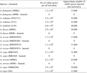
2.3.1 Inoculum preparation and survival assay...34
2.4 Study of the role of novel and uncharacterised genes in A. nidulans nitrate metabolism...35
2.4.1 In silico studies...35
2.4.2 Selection of A. nidulans mutant strains...35
2.4.3 Anaerobic studies...35
2.4.4 Synthesis of gene probes...36
2.4.5 Extraction and gel electrophoresis of total RNA...37
2.4.6 Northern blot analysis and 32P labelling of genes...38
2.4.7 Generation and characterisation of knock-out mutants...39
2.5 Inhibition of growth and net nitrate / nitrite transport in A. nidulans...39
2.5.1 Determination of vitamin auxotrophic markers within mutants...40
2.5.2 Growth tests...40
2.5.3 Net nitrate / nitrite transport assays...40
2.5.4 Determination of kinetic parameters...41
2.6 Cysteine-scanning mutagenesis and thiol cross-linking studies in NrtA...42
2.6.1 Plasmid preparation...42
2.6.2 Preparation of E. coli competent cells...42
2.6.3 Transformation of plasmids in E. coli strain DH5α...42
2.6.4 Preparation of single cysteine mutants...43
2.6.5 Construction of double cysteine mutants...44
2.6.6 Transformation of mutant plasmids in A. nidulans....44
2.6.7 Southern blotting...45
2.6.8 Western blotting and protein expression...47
2.6.9 Thiol cross-linking of double cysteine mutants followed by Factor Xa digestion...49
2.7 Crystallography attempts of prokaryotic nitrate transporters...50
2.7.1 Construction of fusion proteins...50
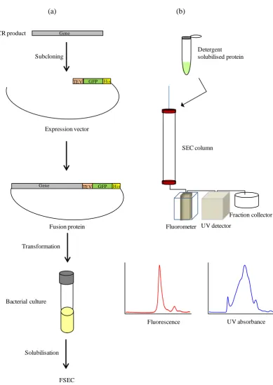
2.7.2 Optimisation of GFP-fused protein expression...51
2.7.3 Protein over-expression trails for E. coli NarU and T. thermophilus NarK1...……….…..….51
2.7.4 Crude membrane preparation from E. coli...52
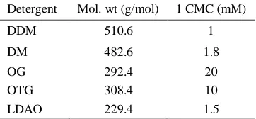
2.7.5 Pre-crystallisation screening of detergent...53
vii
2.7.7 Large scale bacterial protein extraction...55
Chapter Three In vivo pathogenicity studies………....………….57-72 3.1 Introduction……….………..57-60 3.1.1 Health risks associated with Aspergillus species...57
3.1.2 Galleria mellonella, a model host to study fungal pathogenesis...58
3.1.3 Studies of fungal pathogenicity using Galleria mellonella larvae...59
3.2 Objective...60
3.3 Results...61-70 3.3.1 Determination of viable spore number in the fungal inoculum...61
3.3.2 A trial experiment...62
3.3.3 A. nidulans...64
3.3.4 A. flavus...65
3.3.5 A. oryzae...66
3.3.6 A. sojae...67
3.3.7 A. terreus...67
3.3.8 A. niger...69
3.4 Discussion...70-72 3.5 Conclusion...72
Chapter Four Characterisation of newly discovered genes associated with nitrate metabolism…...….……73-96 4.1 Introduction...73-74 4.1.1 Background...73
4.1.2 Discovery of four putative genes for nitrate metabolism...73
4.2 Objective...74
4.3 Results...74-91 4.3.1 In silico analysis...74
4.3.2 Search for cnxL, cnxK, niaB and niaC transcripts...89
4.3.3 Generation and characterisation of knock-out mutants...91
viii Chapter Five
Studies of residue proximity in the NrtA transporter by thiol cross-linking technique…....97-121
5.1 Introduction...97-101
5.1.1 General characteristics of NrtA protein...97
5.1.2 Thiol cross-linking of cysteine residues...98
5.2 Objectives...101
5.3 Results...101-117 5.3.1 Construction of cysteine mutants in the NrtA protein...101
5.3.2 DNA:DNA hybridisation and identification of single copy nrtA mutants...102
5.3.3 Growth testing of transformed strains...103
5.3.4 Protein expression levels in cysteine mutants...103
5.3.5 Studies of cysteine residues proximity by thiol cross-linking...104
5.4 Discussion...117-121 5.5 Conclusion...121
Chapter Six Construction of a library of single cysteine mutants in NrtA transmembrane domains 2 and 8...122-130 6.1 Introduction...122-124 6.1.1 Background...122
6.1.2 Cysteine-scanning mutagenesis...122
6.1.3 Cysteine-scanning mutagenesis of E. coli LacY...122
6.1.4 Cysteine-scanning mutagenesis of some other transporter proteins...123
6.1.5 Cysteine-scanning mutagenesis of A. nidulans NrtA...123
6.2 Objective...124
6.3 Results...124-128 6.3.1 Single cysteine substitutions in Tm 2 and Tm 8 residues in NrtA...124
6.3.2 Identification of single copy transformants...125
6.3.3 Growth tests of single cysteine mutant transformants...126
6.3.4 Protein expression of NrtA single cysteine mutants...128
ix Chapter Seven
Crystallography trials of bacterial nitrate transporters………...….……….131-159
7.1 Introduction...131-132
7.1.1 Background...131
7.1.2 Membrane protein crystallisation...131
7.1.3 Pre-crystallisation screening of detergent………..…..…...132
7.2 Objectives...133
7.3 Results...133-156 7.3.1 GFP based screening of proteins over-expression...133
7.3.2 SDS-PAGE of crude membranes over-expressing E. coli NarU fusion protein...133
7.3.3 Determination of over-expression of some other prokaryotic nitrate transporter proteins...135
7.3.4 Detergent screening for protein solubilisation by FSEC...137
7.3.5 Large scale expression and purification of nitrate transporter fusion proteins...148
7.3.6 Western blot analysis of fusion proteins...155
7.4 Discussion...156-159 7.5 Conclusion...159
Chapter Eight Substrate specificity and inhibition of nitrate and nitrite permeases………...…….160-185 8.1 Introduction...160-162 8.1.1 Nitrate and nitrite transporters...160
8.1.2 Inhibition of nitrate transport...160
8.2 Objective...162
8.3 Results...162-181 8.3.1 Nitrate specificity of NrtA and NrtB proteins...162
8.3.2 Nitrite transport by NrtA and NrtB proteins...174
8.4 Discussion...181-185 8.5 Conclusion...185
Chapter 9
x Appendices...189-204
Appendix I...189-192 Appendix II...193-196 Appendix III...197-200 Appendix IV...201-203 Appendix V...204
References...205-232
List of Figures……….…....233-235
1 Abstract
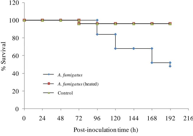
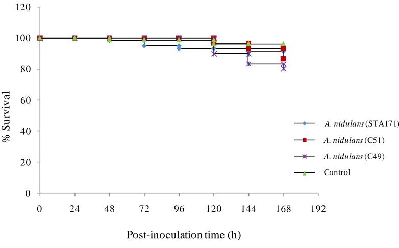
In this study, several aspects of nitrate assimilation and transport have been studied using the filamentous fungus Aspergillus nidulans, which has been shown to be a safe laboratory organism as judged by it’s pathogenicity towards insect larvae. In silico analysis of the A. nidulans genome sequence, identified two putative genes designated cnxL and cnxK that might be involved in molybdenum cofactor biosynthesis as well as two putative nitrate reductases encoding genes niaB and
niaC. All four genes are hitherto unknown. Although many features of these proteins provided clues of
functionality, biochemical and genetical approaches employed in this present study failed to elicit expression of any of these four genes.
A NrtA protein structure model was developed based on residue homology with the Escherichia coli
GlpT a protein, the structure of which has been solved. The results of thiol cross-linking of three double cysteine mutants in four NrtA essential residues, R87, R368, N168 and N459, indicated that the molecular distance between R87 and R368 is ~ 0.4 Å, R368 and N168 ~ 6.2 Å, R87 and N459 is ~ 2.2 Å. Another important observation was the change in the confirmation of Tm 2 and Tm 8 in the presence of nitrate. This shift resulted in an increase of ~ 2 Å gap between the residues R87 and R368. Distances between amino acid residue pairs estimated using such molecular rulers contradicted the NrtA existing model. Cysteine-scanning mutagenesis studies were extended to the generation of a library of single cysteine mutants of NrtA residues spanning Tm 2 and Tm 8. The majority of single cysteine mutants possessed wild type NrtA protein expression levels but unfortunately most were found to be loss-of-function. Consequently, thiol chemistry of this crop of mutants was not perused.
Attempts were also made to overexpress and crystallise the bacterial nitrate transporters. In this regard, bacterial nitrate transporters, NarU (E. coli), Nar (Bacillus cereus), NarK1 and NarK2 (Pseudomonas
aeruginosa) and NarK2 (Thermus thermophilus) fused with GFP were expressed in E. coli and used in
crystallisation trials. Although this approach has proved successful for a number of membrane proteins, unfortunately was not helpful with regard to the purification of any of the above bacterial nitrate transporters to yield protein expression levels required for successful protein crystallography.
Finally, the effects of potential nitrate transport inhibitors were studied on net nitrate transport by NrtA and NrtB proteins of A.nidulans. The results indicated that chlorate had more of an inhibitory effect on NrtA net nitrate transport than that by NrtB. Chlorite and sulphite equally affected net nitrate transport by either NrtA or NrtB proteins while caesium strongly inhibited the net nitrate transport by NrtB transporter.
2 Chapter One
General Introduction
1.1 Nitrogen
Nitrogen, the fifth most abundant element on earth and the key component of biomolecules, is an essential nutrient for all organisms. The major source of nitrogen is in the atmosphere, but here, nitrogen is present in the form of an inert gaseous molecule, N2 that cannot be converted directly to organic nitrogen by living organisms. Therefore, this unavailable form of nitrogen needs to be converted first to an available form of nitrogen, ammonium. This is carried out by the process of nitrogen fixation by abiotic or biotic environmental factors. On the other side of the nitrogen cycle, considerable nitrogen is present in the form of complex organic nitrogenous matter that cannot be utilised directly for example by plants. First such complex molecules must be converted to inorganic forms, mainly ammonium, by degradation systems present particularly in the fungi. The resulting ammonium is oxidised to nitrite and then to nitrate by soil bacteria such as Nitrobacter and Nitrosomonas respectively (Falkowski, 1997; Hayatsu et al., 2008; Canfield et al., 2010; Dechorgnat
et al., 2011). The activity of microorganisms in the nitrogen cycle has been summarised in Figure 1.1.
Figure 1.1: Schematic representation of microbiological processes in the nitrogen cycle.
(1) Nitrogen fixation, (2) bacterial, archaeal and heterotrophic nitrification, (3) bacterial,
archaeal and fungal denitrification, (4) co-denitrification by fungi (5) co-denitrification by
anammox (bacteria that convert nitrite and ammonium into dinitrogen gas under anaerobic
conditions) and (6) ammonium oxidation. This figure and legends has been reproduced
directly from Hayatsu et al. (2008).
3 crucial crops, wheat, maize and rice (Raun and Johnson, 1999). The use of fertilisers not only increases the cost of crop productivity but also nitrogen from unused fertilisers is leached into the water and caused severe environmental and health problems (Good et al., 2004; Galloway et al.,
2008; Diaz and Rosenberg, 2008; Hayatsu et al., 2008). There is a need for an effective management strategy to avoid (or minimise) the negative impacts of nitrogen fertilisers (Galloway et al., 2008). One way to overcome the extensive use of fertilisers is to improve the nitrogen utilisation efficiency by crop plants. This may be achieved by targeting enzymes, transporters or regulatory genes of nitrogen metabolism by molecular or genetic approaches (Fernandez and Galvan, 2008 and references therein).
1.2 Nitrate
Nitrate is the most available inorganic form of nitrogen and is an important nutrient of plants, fungi and bacteria, inhabiting most aerobic soils. The level of nitrate concentration in the soil varies with respect to the season and the activity of various biotic (type or population density of decomposers) and abiotic (type and amount of litter, temperature, humidity etc) environmental factors (Miller et al.,
2007; Unkles et al., 2001; Forde, 2000; Crawford and Glass, 1998).
Nitrate is not only the major source of available nitrogen in the soil but also stimulates the growth of organisms by inducing genes encoding nitrate transporter proteins or enzymes involved in nitrate assimilation (Crawford and Glass, 1998). In addition nitrate also affects plant morphogenic processes for example root development (Zhang and Forde, 1998), root and shoot balance (Scheible et al.,
1997a) and carbon metabolism (Scheible et al., 1997b). Finally the role of nitrate in gametogenesis has been reported in Chlamydomonas reinhardtii (Pozuelo et al., 2000).
Nitrate is the most preferred source of nitrogen that can be assimilated efficiently when sufficient supply of oxygen is available (Takasaki et al., 2004a; Takasaki et al., 2004b; Takaya, 2009). Most of the organisms obtain their nitrogen by reduction of nitrate to ammonium. Plants absorb nitrate from soil solutions by active transport through root cells. From the root cells, nitrate is transported to different parts of the plant in the phloem and then enters into the cells using channels or transporters and is often stored in the vacuoles of cells (Crawford and Glass, 1998).
1.3 Cellular transport systems
4 Channels are pores that open only when substrate is present in the surroundings and allow the movement of substrate down an electrochemical gradient. Carrier proteins are integral membrane proteins that are involved in the movement of molecules by facilitated diffusion or active transport. Active transport of materials is the movement of substances against the concentration gradient and may be (i) primary active transport that uses the energy to translocate the materials against a concentration gradient or (ii) secondary active transport that uses an electrochemical potential gradient of one substance to translocate another substrate (Dahl et al., 2004).
Transmembrane transporter proteins are classified according to the Transporter Classification System (TC) which is based on combined transporter proteins functionality and phylogenetic information. According to the TC system (approved by the International Union of Biochemistry and Molecular Genetics, IUBMB), transporters are grouped into various hierarchical classification levels; class, subclass, superfamily or family, subfamily and finally the specific transporter (www.tcdb.org; Busch and Saier, 2002; Saier et al., 2006). Many transporter proteins belong to one of two major classes of transporters, namely the ATP-binding cassette (ABC) protein superfamily or the major facilitator superfamily (MFS) (Pao et al., 1998).
1.3.1 ABC superfamily
The ABC transporters are primary transporters as they utilise energy generated by ATP hydrolysis and hence allow the movement of materials against a concentration gradient. Downstream of the ATP binding domain(s), LSGGQ, a highly conserved signature sequence of the ABC protein superfamily is present (Szentpetery et al., 2004).
Compared to the MFS, ABC protein is a less studied group of transporter proteins and is involved in the influx of materials in prokaryotes or efflux in both prokaryotes and eukaryotes. The diversity of substrates of ABC transporters range from a single ion to a complete protein (Saier, 2000; Dahl et al.,
2004; Hollenstein et al., 2007). The mechanism of transport by ABC transporters is not well understood. However it is postulated that conformational change due to ATP binding to the protein results in the formation of a translocation pathway, known as ‘powerstrock’. Finally the affinity of the substrate decreases with the protein binding site and substrate releases (Hollenstein et al., 2007).
1.3.2 Major facilitator superfamily (MFS)
5 topological structure. The length of the single peptide chained proteins belonging to this superfamily range from 400 - 600 amino acids and are arranged in 12 transmembrane helixes (Saier, 2000).
Being secondary active transporters, MFS transporter proteins translocate substrates using energy released by the downhill movement of a driver solute to energise the transport of the substrate. MFS proteins may either be (i) uniporters, that transport one type of substrate in one direction driven by a concentration gradient, (ii) symporters (eg. LacY, FucP) that translocate two or more different kinds of substrates in the same direction using electro-chemical gradient of one of the substrate and (iii) antiporters (eg. GlpT) that allow movement of two or more substrates in opposite directions through a membrane (Kaback et al., 2001; Law et al., 2008).
Unique to all MFS members is the MFS signature sequence with in loops between Tm 2 / Tm 3 and Tm 8 / Tm 9. As MFS proteins transport a vast variety of substrate therefore it is obvious that MFS signature sequence does not bind to a substrate. The possible roles of MFS signature described are gating mechanisms, conformational changes and / or solute accessibility (Jessen-Marshall et al., 1995; Pazdernik et al., 1997; Pazdernik et al., 2000).
1.4 Nitrate transport systems
Nitrate and nitrite are charged molecules and therefore should face difficulty in crossing cell membranes at a rapid rate against the membrane potential that is negative inside and also against a concentration gradient. Therefore the nitrate-utilising organisms have developed active transporter proteins for nitrate and nitrite transport (Clegg et al., 2002). Nitrate transporters belong to a sub-family of MFS, the nitrate / nitrite porter sub-family (NNP) that includes nitrate / nitrite transporters from both prokaryotes and eukaryotes (www.tcdb.org; Pao et al,. 1998).
6 1.4.1 Nitrate transport systems in the plant model, Arabidopsis thaliana
Nitrate transport in Arabidopsis thaliana has been studied over a number of years and consequently there is a considerable body of knowledge for this process (Tsay et al., 1993; Tsay et al., 2007; Ho et al., 2009; Krouk et al., 2010b; Tsay et al., 2011). Transport of nitrate from soil to root cells is a highly efficient process in plants. Depending on the concentration of nitrate available, low affinity nitrate transporters NRT1 (operates at >1 mM nitrate) or high affinity nitrate transport systems, NRT2 (functional when concentration of nitrate is <1 mM) are functional (Glass and Siddiqi, 1995; Forde, 2000).
Low and high affinity nitrate transporter proteins in A. thaliana are encoded by the genes belonging to the gene family AtNRT1 and AtNRT2 respectively. Nitrate inducible AtNRT1.1 is a master gene and performs several important roles, (i) encodes a nitrate transporter protein, AtNRT1.1 (Tsay et al.,
1993), (ii) acts as nitrate sensor and activates the expression of other nitrate metabolism related genes
(Ho et al., 2009), (iii) involves in signalling processes that affect root development and seed
germination (Ho and Tsay, 2010) and (iv) represses the lateral root growth by directing the transport of auxin from the roots (Krouk et al., 2010a). The AtNRT1.2 protein, another member of NRT1 protein family in A. thaliana is encoded by the gene AtNRT1.2 (Huang et al., 1999).In contrast to the AtNRT1.1 protein which is an inducible component of the low affinity nitrate transport system, expression of AtNRT1.2 protein is independent of the presence of nitrate and makes the constitutive component of low affinity nitrate transport system.
The A. thaliana AtNRT2 protein family belongs to the high affinity nitrate transport system and takes
up nitrate when the concentration of available nitrate is low (Filleur et al., 2001; Orsel et al., 2004). AtNRT2.1, a member of the NRT2 protein family, carries out 72 % nitrate transport in A. thaliana. However NRT2.1 is unable to function alone and requires another protein NAR2 which has been found to be physically attached with the NRT2.1 to form a 150 kDa tetramer a functional stable form of the NRT2.1 nitrate transporter (Okamoto et al., 2006; Orsel et al., 2006; Yong et al., 2010). A disruption in the NAR2 gene results in complete loss of transport activity by inducible high affinity nitrate transport (Okamoto et al., 2006; Orsel et al., 2006). Such NRT2 and NAR2 complexes have also been reported in other plants, for example barley and rice (Glass, 2009; Ishikawa et al., 2009; Feng et al., 2011).
The AtNRT2.2 a functionally redundant protein of AtNRT2.1 makes a small contribution to nitrate transport but its activity increases when the AtNRT2.1 protein is lost or becomes non-functional (Li et al., 2007). Also within the NRT2 family five other gene members have been reported for A. thaliana,
7 NRT2.3 - NRT2.6 have not yet been characterised for their functional roles (Orsel et al., 2002; Okamoto et al., 2003). The Km value calculated by kinetic analysis for the A. thaliana high affinity nitrate transporters is ~ 50 µM and that of low affinity transporters is ~ 4 mM (Liu et al., 1999; Guo et al., 2002).
1.4.2 Nitrate transport systems in bacteria
Little is known about the nitrate transport mechanisms in bacteria. Moir and Wood (2001) suggested the involvement of ATP-dependent transporter proteins in bacteria that assimilate nitrate, and MFS nitrate transporter proteins, (homologues of NarK protein family) that transport nitrate usually for dissimilation under anaerobic conditions. It is unclear and controversial as to how the NarK protein functions (Moir and Wood, 2001; Jia et al., 2009).
In the eubacterium E. coli, two polytopic homologous redundant nitrate transporter proteins NarK and NarU encoded by the genes narK and narU respectivelyhave been identified. Both NarK and NarU belong to the MFS superfamily and possess 12 transmembrane domains. In E. coli, both NarK and NarU proteins are capable of effluxing nitrate from the cell to support anaerobic growth (Clegg et al.,
2002; Jia et al., 2009). The expression of E. coli NarK protein is induced by nitrate. Actively growing cells of E. coli show high expression of NarK. Similar to other members of narK genes, E. colinarK
also forms a gene cluster with the nitrate reductase encoding gene, narGHJI. In contrast to E. coli
NarK, the highest level of NarU expression was observed when the cells were in stationary growth phase or growing slowly, facing nutrient deficiency (Clegg et al., 2006). E. coli NarK and NarU not only transport nitrate but also catalyse the nitrite uptake and export (Jia et al., 2009).
In the thermophilic bacterium Thermus thermophilus two narK1 and narK2 genes that encode the nitrate transporter proteins NarK1 and NarK2 for anaerobic respiration have been reported. Both
narK1 and narK2 form a gene clustered with the nitrate reductase (Ramirez et al., 2000). Similarly, in
Pseudomonas aeruginosa, a denitrifying bacterium, two genes narK1 and narK2 encode nitrate/nitrite
transporter proteins NarK1 and NarK2 respectively that cluster with nitrate reductase encoding gene,
narGHJI (Sharma et al., 2006).
1.5 Application of the aspergilli to biological and medical research
8
A. flavus (Yu et al., 2005), A. oryzae (Machida et al., 2005), A. nidulans (Galagan et al., 2005) and A.
niger (Pel et al., 2007).
Comparative analysis of A. nidulans genome sequence with the A. fumigatus, a serious human pathogen and A. oryzae, used in food industry to prepare miso, sake and soya sauce, revealed that over 5000 non-coding gene sequences were conserved in all the three Aspergillus species. It was also observed that similar to A. nidulans, A. fumigatus and A. oryzae also have potential sequences for a sexual life cycle although a sexual stage has not been yet observed in A. oryzae. Availability of the genome sequence of the model organism A. nidulans and other widely studied aspergilli has opened new horizons to study genome evolution and gene regulation (Galagan et al., 2005). The free open access to genome sequences of Aspergillus species resulted in diverse research efforts in the fields of transcriptome analysis, proteomics, metabolomics and metabolic modelling in Aspergillus species (Andersen and Nielsen, 2009 and references therein).
For instance, comprehensive research has been carried on proteomics and metabolomics of medically important Aspergillus species (Andersen and Nielsen, 2009 and references therein). In this regard, the aspergilli produce a number of mycotoxins (secondary metabolite) that are not thought to be necessary for their own survival. The most common mycotoxins concerned with human health are aflatoxin, sterigmatocystin, ochratoxin and gliotoxin (www.aspergillus.org.uk; Bennett and Klich, 2003 and references therein). It has also been reported that out of 250 known species of genus
Aspergillus only 40 species cause infection in humans although this figure is increasing (Klich, 2006)
probably with the increase in number of patients with reduced immunity.
The aspergilli (including A. nidulans) are not specialised as human pathogens rather they are opportunistic and are able to cause diseases in immune-suppressed patients (Kim et al., 1997; Latge, 1999; Lucas et al., 1999; Latge, 2001; Khanna et al., 2005; Bennett, 2009). A. fumigatus, A. flavus
and A. terreus are the most significant infection-causing species of genus Aspergillus for humans
(Latge, 2001; Hedayati et al., 2007; Person et al., 2010). Although A. nidulans is generally thought to have no or a very low pathogenicity, a few human A. nidulans infections have actually been reported (Kim et al., 1997; Chakrabarti et al., 2006). Details of human health risk from aspergilli will be discussed further in Chapter 3, Section 3.1.1.
1.5.1 Aspergillus nidulans - a model eukaryotic organism
Aspergillus nidulans, a filamentous Ascomycetes, is a well-established fungal model system and has
9 fungus and can also be induced to carry out heterokaryosis with the formation of heterokaryons. Also genetic transformation of A. nidulans is now routine (Tilburn et al., 1983; Yelton et al., 1984; Ballance and Turner, 1985).
A number of physiological and metabolic processes have been successfully studied using A. nidulans
as a eukaryotic model organism. For example the process of mitosis, the formation of tubulin and microtubules (Oakley, 2003; Szewczyk and Oakley, 2011) and carbon (David et al., 2006; Mogensen
et al., 2006), nitrogen (Cove and Pateman, 1963; Pateman et al., 1964; Pateman et al., 1967; Cove,
1976; Brownlee and Arst, 1983; Unkles et al., 1991; Unkles et al., 2001) and sulphur metabolism. Currently, many laboratories all over the world from the USA to Japan are conducting research into the biology, biochemistry, genetics and applications of A. nidulans. Some of these laboratories are working jointly in international terms to promote and disperse knowledge derived from A. nidulans
(www.broadinstitute.org).
Genetically, A. nidulans is very close to certain medically and industrially important Aspergilli, for example, A. fumigatus, A. flavus, A. niger and A. oryzae (Aspergillus Genome Database, AspGD; www.aspergillusgenome.org). Also it is quite straightforward to induce chemical and site-directed mutations in A. nidulans (Unkles et al., 2004a; Kinghorn et al., 2005 and references therein). All the above features made this fungus very attractive for scientific studies.
1.6 Nitrate transport systems in A. nidulans
A. nidulans has been used extensively to study nitrate transport and metabolism for the last six
decades resulting in our fundamental understanding of this pathway. The first regulatory and structural gene involved in nitrate assimilation of A. nidulans was identified in 1963 by Cove and Pateman. Most of the genetic and biochemical aspects of nitrate assimilation pathway in the lower eukaryotic fungus, A. nidulans are similar to the higher plants so that the information can be transferred to plants. However, on the other hand nitrate reductase activity is required for nitrate transport in fungi but not for plants, highlighting a major difference between fungi and plants at least as shown in Aspergillus and Arabidopsis (Unkles et al., 2004b).
The earlier conventional physiological studies of growth of A. nidulans mutant strains on nitrate characterised the main components of nitrate assimilation, including its regulation and inhibition by chlorate (Cove and Pateman, 1963; Pateman et al., 1964; Pateman et al., 1967; Cove, 1976). The gene
nrtA (originally designated crnA) was identified that encodes a nitrate transporter protein, NrtA in A.
10 Figure 1.2: Life cycle of Aspergillus nidulans.
During the asexual cycle of A. nidulans, unicellular haploid spores, conidia or conidiospores are produced. Upon germination, conidia form haploid homokaryons. Two homokaryon may fuse with each other to form
a heterokaryon. During the parasexual cycle, nuclei of heterokaryons fuse to form a diploid. In the sexual
life cycle, ascospores are formed within the asci which are enclosed in the fruiting body, the cleistothecium.
This figure has been taken directly from Casselton and Zolan (2002). Cleistothecium
Ascus
Ascospore
Meiosis
Ascospore
Ascogenous hypha
Mitotic nuclear division
Haploidization
Hetrokaryon
Unstable but can be
maintained Diploid
homokaryon Sexual cycle
11 It was also observed that mutant strains in the nrtA gene continue to grow on nitrate provided as the sole source of nitrogen. Also the net nitrate uptake kinetics was not straight forward. Both observations leading to the prediction of the presence of there being more than one nitrate uptake system. Subsequently, another nitrate transporter, NrtB, encoding gene in A. nidulans, nrtB was observed, cloned and characterised (Unkles et al., 2001). NrtA and NrtB belong to the high affinity nitrate transporter protein family and mutation in one gene does not affect the growth on nitrate as a sole source of nitrogen whereas the mutant in both genes abolishes the growth on nitrate even in the presence of up to concentrations of 200 mM nitrate. From mutant growth results and BLAST searches, it is suggested that nrtA and nrtB are the only genes involved in the nitrate transport in A.
nidulans, although NrtA and NrtB are regulated in a similar manner and perform the same function.
The rate of transport and affinity of nitrate by NrtA is approximately three times higher than NrtB suggesting ecological plasticity of nitrate transport (Unkles et al., 2001).
Brownlee and Arst (1983) provided fundamental kinetic data for nitrate transport in A. nidulans and described interesting and exciting new facts regarding nitrate transport. The conclusions drawn by Brownlee and Arst were (i) nrtA1 (crnA1) mutant has several fold reduction in net nitrate uptake in conidia and young mycelium but not in older mycelium, (ii) uptake of nitrate by nrtA1 mutant suggested the presence of more than one uptake system, (iii) net nitrate uptake is strongly inhibited by chlorate, a potential structural analogue of nitrate, (iv) uncouplers inhibit nitrate uptake probably due to lack of proton gradient across the membrane, (v) net nitrate uptake and nitrate reductase are sensitive to the metabolic inhibitors, cyanide, azide and N-ethylmaleimide.
1.6.1 General characteristics of NrtA protein
nrtA, encoding NrtA protein, was the first eukaryotic high affinity nitrate transporter gene to be sequenced (Unkles et al., 1991). A 57 kDa (507 amino acids) NrtA protein, TC 2.A.1.8.5 (www.tcdb.org), belongs to the high affinity nitrate transporter family of major facilitator superfamily, MFS (Unkles et al., 1991; Forde, 2000; Unkles et al., 2001). nrtA is located on chromosome VIII forming a gene cluster with niaD (nitrate reductase encoding gene) and niiA (nitrite reductase encoding gene) in a gene order nrtA-niiA-niaD (Tomsett and Cove, 1979; Johnstone et al.,
1990). Similar to other members of the MFS, the NrtA protein contains 12 putative transmembrane domains (Tms). These hydrophobic Tms of NrtA are present in the form of α-helices that pass through the membrane in a zigzag manner and are connected by hydrophilic loops. Both C- and N-terminal ends of NrtA are present most likely inside the cytoplasm. It was also proposed that the Tms form the boundary of hydrophilic cavity for the transport of nitrate (Unkles et al., 1991; Trueman et
12 The MFS signature motif, GXXXNXXGXR, is present in NrtA located near the loop between Tm 2 and Tm 3 with another copy around loop between Tm 8 and Tm 9. The NrtA protein has another conserved motif, AAGXGNXGGG, unique to all members of the high affinity nitrate transporters and referred to as the nitrate signature sequence (NS). Two copies of nitrate signature are present in NrtA protein, NS1 located in Tm 5 (amino acid residues 163-172) and NS2 in Tm 11 (Trueman et al., 1996; Forde, 2000). The occurrence of the two copies of MFS motif and the nitrate signature, one copy of each in the first six Tms and the second in second six Tms in particular positions, support the notion that duplication of the first six Tms took place during the course of evolution (Pao et al., 1998). These two halves of NrtA proteins are joined by a large substantial central loop between Tm 6 and Tm 7 (Kinghorn et al., 2005) (Figure 1.3). However it has also been observed that when only one half of the protein was made (6 Tms) no transport of nitrate was recorded.
Besides conserved MFS and nitrate signature motifs, a number of amino acid residues and motifs are highly conserved between nitrate transporter proteins. Kinghorn and colleagues (2005) identified sixteen highly conserved amino acid residues among 52 nitrate transporters from archaebacteria to plants and A. nidulans NrtA. The residues that were conserved among all 52 protein sequences are R87 (Tm 2), F140 (Tm 5) G157 (Lp Tm 4/ Tm 5), G170 (Tm 5), Y323 (Tm 7) G328 (Tm 7), R368 (Tm 8), G371 (Tm 8), G372 (Tm 8), D376 (loop Tm 8 / Tm 9) and G452 (Tm 11). Among these highly conserved residues, R87 and R368 are essential for substrate binding (Unkles et al., 2004a). Many residue alterations of NrtA identified residues that play an important role in nitrate transport (Kinghorn et al., 2005). Mutational analysis of the nrtA gene helped us to understand its structure and/or function. Whereas NrtA alteration in certain amino acid residues showed wild type growth and NrtA wild type protein expression levels, changes in other residues resulted in loss-of-function. For example when highly conserved arginine residues, R87 (Tm 2) and R368 (Tm 8) were replaced with a number of amino acids (Unkles et al., 2004a; Kinghorn et al., 2005), only a lysine replacement in R386 was tolerated with the mutant R368K having a significant decrease in Km value for nitrate (Unkles et al., 2004a).
1.6.2 The NrtB nitrate transporter
13 transport shows that this protein has Vmax ~ 150 nmol/mg DW/h and the Km ~ 10 µM (Unkles et al., 2001), a very different to NrtA.
Figure 1.3: Putative secondary structure of A. nidulans NrtA.
Yellow dashes enclose the MFS signature motifs and blue surround the nitrate signature sequence.
The residues highlighted in red are highly conserved as predicted by the alignment of 52 available
NrtA homologue sequences. The figure and legends have been adapted from Kinghorn et al. (2005).
1.7 Nitrite transport by A. nidulans
Interestingly, although the double mutant (nrtA, nrtB) results in growth abolition on nitrate as the sole source of nitrogen, the double mutation did not affect growth on nitrite thus suggesting the presence of nitrite transporter(s) and that it is incapable of nitrate transport. The NitA protein, encoded by the
nitA gene, was identified as a sole nitrite transporter in A. nidulans (Km ~ 4.2 µM and Vmax ~ 170 nmol/mg DW/h).
14
Figure 1.4: The amino acid sequence alignment of A. nidulans NrtA and NrtB nitrate transporter proteins.
NrtA ---MDFAKLLVASPEVNPNNRKALTIPVLNPFNTYGRVFFFSWFGFMLAFLSWYAFPPLL 57 NrtB MKPTQVLRLAVAAPDVNPQTRKARSIPVLNPFDLYGRVFFFSWIGFLVAFLSWYAFPPLL 60
:. :* **:*:***:.*** :*******: *********:**::************
NrtA TVTIRDDLDMSQTQIANSNIIALLATLLVRLICGPLCDRFGPRLVFIGLLLVGSIPTAMA 117 NrtB SVTIKKDLHMSQDDVANSNIVALLGTFVMRFIAGPLCDRFGPRLVFVGLLICGAVPTAMA 120 :***:.**.*** ::*****:***.*:::*:*.*************:***: *::*****
NrtA GLVTSPQGLIALRFFIGILGGTFVPCQVWCTGFFDKSIVGTANSLAAGLGNAGGGITYFV 177 NrtB GLVTTPQGLIALRFFVGILGATFVPCQVWCTGFFDKNIVGTANSLAGGFGNAGGGITYFV 180
****:**********:****.***************.*********.*:***********
NrtA MPAIFDSLIRDQGLPAHKAWRVAYIVPFILIVAAALGMLFTCDDTPTGKWSERHIWMKED 237 NrtB MPAIYDSFVHDRGLTPHKAWRVSYIVPFIIIVSIALAMLFTCPDTPTGKWADR--- 233 ****:**:::*:**..******:******:**: **.***** *******::*
NrtA TQTASKGNIVDLSSGAQSSRPSGPPSIIAYAIPDVEKKGTETPLEPQSQAIGQFDAFRAN 297
NrtB -EKTSGQSIVDLSSTPNASSAN-SINISSDEKKAVHPEVTDSEAQVHVRAG-QIESSDA- 289 :.:* .****** .::* .. . .* : *. : *:: : : :* *::: *
NrtA AVASPSRKEAFNVIFSLATMAVAVPYACSFGSELAINSILGDYYDKNFPYMGQTQTGKWA 357 NrtB VIEAPTIKRYLSIALDPSALAVAVPYACSFGAELAINSILGAYYLLNFPLLGQTQSGRWA 349
.: :*: *. :.: :. :::***********:********* ** *** :****:*:**
NrtA AMFGFLNIVCRPAGGFLADFLYRKTNTPWAKKLLLSFLGVVMGAFMIAMGFSDPKSEATM 417
NrtB SMFGLVNVVFRPMGGFIADLIYARTNSVWAKKMWLVVLGLAMSGMAILIGFLDPHRESVM 409 :***::*:* ** ***:**::* :**: ****: * .**:.*..: * :** **: *:.*
NrtA FGLTAGLAFFLESCNGAIFSLVPHVHPYANGIVSGMVGGFGNLGGIIFAIIFRYSHHDYA 477 NrtB FGLVVLMAFFIAASNGANFAIVPHVHPSANGIVSGIVGGMGNFGGIIFAIVFRYNGTQYH 469
***.. :***: :.*** *::****** *******:***:**:*******:***. :*
NrtA RGIWILGVISMAVFISVSWVRPVPKSQMRE 507 NrtB RSLWIIGFIILGCTLFFSWVRPVPKQNH-- 497 *.:**:*.* :. : .********.:
The amino acid sequences of the two proteins encoded by nrtA and nrtB genes were aligned using the European Bioinformatics Institute (EBI) protein alignment tool ClustalW (www.ebi.ac.uk). The numbers on
the right side represent the amino acid residues. The amino acid residue motifs highlighted in blue denote
nitrate signature sequence and those highlighted with yellow are the copies of MFS motif. Conserved
arginine residues (R87 and R368 in NrtA) are highlighted in pink. ‘*’ is used for the conserved residues, ‘:’
15 Recently A. nidulans NitA, the high affinity nitrite transporter and a member of the formate / nitrite transporter (FNT) family has been characterised using the tracer 13NO2¯ (Unkles et al., 2011). Kinetic studies of NitA indicated that the Km is 4.8 ± 8 µM and Vmax 228 ± 49 nmol/mg DW/h for nitrite. Unkles and colleagues also presented two and three dimensional models of NitA protein. In this respect, asparagine residues, N122 (Tm 3) and N246 (Tm 6) are highly conserved in the FNT family while N173 (Tm 4) and N214 (Tm 5b) were found to be present in the 80 % homologues of NitA (Finn et al., 2008). Mutational analysis of conserved asparagine residues N122 (Tm 3), N173 (Tm 4), N214 (Tm 5b) and N246 (Tm 6) revealed that N122 and N246 are irreplaceable. However the replacement of asparagines at position 214 with other negatively charged residue, aspartate resulted in wild type growth. N173 was found to be tolerant to smaller side chain amino acid replacements (Unkles et al., 2011).
1.8 Nitrate assimilation
Fungi play a critical role in the nitrogen cycle in the soil and are able to assimilate both organic (amino acids and nucleotides) and inorganic (nitrate, nitrite and ammonium) nitrogen compounds present in the soil. Fungi have well developed metabolic systems for the assimilation of nitrate (as well as nitrite) to ammonium and then ammonium assimilation to glutamine and glutamate (reviewed in Cove, 1979 and references therein). Once nitrate is transported into the cell by nitrate transporters, it is reduced to nitrite by nitrate reductase and then to ammonium by nitrite reductase. In organisms that perform nitrate assimilation, nitrate induces the expression of nitrate reductase and nitrite reductase encoding genes in fungi and plants (Quesada and Fernandez, 1994; Perez et al., 1997; Krapp et al., 1998; Zhuo et al., 1999; Vidmar et al., 2000; Unkles et al., 2001; Clegg et al., 2002).
The genes encoding the nitrate transporter(s), nitrate reductase and nitrite reductase may be present in operon(s) in prokaryotes (Omata et al., 1993; Lin et al., 1994). In certain lower eukaryotic organisms; for example, Aspergillus nidulans (Johnstone et al., 1990), Hansenula polymorpha and
Chlamydomonas reinhardtii (Perez et al., 1997) the genes are clustered but do not form operons. And
16 Figure 1.5: Secondary and tertiary structure models of Aspergillus nidulans nitrite transporter, NitA.
(a) Secondary structure model of NitA based on crystal structure of E. coli formate transporter, FocA. Tms are rainbow coloured from blue (N-terminal) to red (C-terminal).
Conserved asparagine residues are outlined in black. (b &c) tertiary structure model of NitA
viewed from the plan of membrane (c) same tertiary structure model of NitA showing relative
positions of conserved asparagine residues. Figure and legends have been taken directly from
Unkles et al. (2011).
1.9 Dissimilatory nitrate reduction
Most of the eukaryotic organisms are aerobic and require oxygen for their growth and survival. However, in contrast with higher eukaryotes, some yeasts and filamentous fungi have evolved to anaerobic environments (Tsuruta et al., 1998; Takaya, 2009 and references therein). In such organisms a set of genes is expressed under the hypoxic or anoxic condition to support anaerobic growth and survival (Zitomer and Lowry, 1992; Zhou et al., 2002; Takasaki et al., 2004b). Dissimilation of nitrate usually occurs in filamentous fungi either by denitrification or ammonium fermentation under oxygen stress conditions to produce energy. Fusarium oxysporum (Zhou et al.,
2002) and A. nidulans (Takasaki et al., 2004b) are fungal example that such organisms can respire
a
17 nitrogen in anaerobic environments. Dissimilation of nitrate is unusual for eukaryotes and such processes are typically present in bacteria (Nakahara et al., 1993).
1.9.1 Fungal denitrification
Denitrification originally discovered and characterised in bacteria, is a sequential anaerobic process composed of a series of reduction reactions under hypoxic or low oxygen conditions resulting in generation of ATP. During denitrification, nitrate or nitrite is reduced to gaseous nitrogen such as nitrous oxide (N2O) or dinitogen (N2). The reduction process of nitrate to nitric oxide occurs in mitochondria (Takasaki et al., 2004a; Takasaki et al., 2004b; Takaya, 2009).
The reduction of nitrate in fungal denitrification occurs by ubiquinol-Nar, nitrate reductase. This process takes place in mitochondria and is coupled to ADP phosphorylation. Fungal Nar shares the similar properties with the bacterial Nar, for example inhibition by tungstate, an inhibitor of the molybdemum enzyme, nitrate reductase (Uchimura et al., 2002). However fungal Nar is evolutionary distant from the bacterial Nar and also bacterial Nar is localised in cell membrane. However it is noteworthy that most denitrifying fungi lack Nar and are unable to reduce nitrate. Instead the denitrification substrate for such fungi is nitrite which is reduced to nitric oxide by Nir, nitrite reductase (Tsuruta et al., 1998).The steps involved in fungal denitrification are shown in Figure 1.6. The enzyme that catalyses each step is indicated.
Figure 1.6: Schematic representation of fungal denitrification.
NO3¯ NO2¯ NO N2O
Nitrate (NO3¯) is reduced to nitrite (NO2¯) by nitrate reductase (Nar), the resulting nitrite
is reduced to nitric oxide (NO) by nitrite reductase (Nir), and by nitric oxide reductase
(Nor) finally reduced to nitrous oxide.
1.9.2 Fungal ammonium fermentation
While denitrification occurs under hypoxic conditions, ammonium fermentation takes places in the complete absence of oxygen (Lockington et al., 1997). Zhou et al. (2002) studied enzymes involved in fungal ammonium fermentation. Alcohol dehydrogenase (Adh), aldehyde dehydrogenase (Add) and acetate kinase (Ack) were identified for the oxidation of ethanol to acetate. This catalytic step is coupled with ATP generation and the reduction of nitrate to ammonium. The reaction steps in fungal ammonium fermentation with their catalytic enzymes are summarised in Figure 1.7.
Takasaki and colleagues (2004b) identified ammonium fermentation in A. nidulans. In A. nidulans
during ammonium fermentation ethanol is used as the carbon source. This ethanol oxidation results in the formation of acetate with the generation of ATP (Figure 1.7). It was also observed that the strains
18 mutant in either niaD or niiA gene failed to oxidise the ethanol. Therefore it was concluded that A.
nidulans has adapted dissimilation process utilising assimilatory enzymes.
Figure 1.7: Schematic representation of fungal ammonium fermentation.
Enzymes involved in ethanol fermentation by A. nidulans are alcohol dehydrogenase (Adh), aldehyde dehydrogenase (Add) and acetate kinase (Ack). This figure is adapted from Takaya, (2009).
1.10 Nitrate assimilation and A. nidulans
Aspergillus nidulans has been the host for over five decades to study nitrogen assimilation and such
investigations have contributed significantly to our knowledge regarding the genes, enzymes and transporters involved in nitrogen assimilation (Figure 1.8). In the cytoplasm, nitrate is reduced to nitrite by nitrate reductase (NR) and nitrite reductase (NiR) to ammonium. These two enzymes, NR and NiR, are encoded by the genes niaD and niiA respectively (Cove and Pateman, 1963; Pateman et al., 1964; Pateman et al., 1967; Johnson etal., 1980; Brownlee and Arst, 1983; Millar et al., 2001; Unkles et al., 1997; Unkles et al., 2001; Heck et al., 2002; Unkles et al., 2004b; Wang et al., 2008a; Takaya, 2009; Schinko et al., 2010; Unkles et al., 2011).
Figure 1.8: A simplified illustration of the nitrate assimilation pathway in A. nidulans.
Nitrate permeates into the cell by two transporters NrtA and NrtB encoded by nrtA and nrtB gens respectively. Inside the cell, nitrate is reduced to nitrite by nitrate reductase which is encoded by
niaD gene (Cove and Patemen, 1963) and the resulting nitrite is reduced to ammonia by the gene product of niiA (Pateman et al., 1967), the nitrite reductase. This figure has been adapted from Kinghorn and Unkles, (1994).
NO3¯ NO2¯ NH4
+
NiaD NiiA
NADH NAD+ 3NADH 3NAD+
Ethanol acetaldehyde acetyl-CoA acetate
NAD+ NADH
NAD+ NADH
Ack
ATP Add
Adh
NO3¯ NO3¯ NO2¯ NH4
+ nrtA
nrtB
Permease
niaD niiA
Nitrate reductase
Nitrite reductase
19 Two further genes, nirA and areA are crucial for the regulation of structural genes niaD, niiA, nrtA,
nrtB and nitA of nitrate assimilation. The regulatory gene nirA encodes the NirA protein that is involved in nitrate induction for the expression of niaD, niiA, nrtA, nrtB and nitA genes. The expression of NirA itself is constitutive and therefore does not depend on the presence (or absence) of nitrate (or any other nitrogen source) (Burger et al., 1991; Strauss et al., 1998). The protein AreA, encoded by the regulatory areA gene, is involved in ammonium repression (or nitrogen metabolite repression) and represses niaD, niiA,nrtA, nrtB and nitA expression (and many other genes involved in nitrogen break down pathways) if ammonium or glutamine is present in the medium to conserve energy (Kudla et al., 1990; Bloom et al., 1992; Strauss et al., 1998; Schinko et al., 2010 and references therein).
The NirA protein encoded by nirA gene exerts positive regulatory control mechanism. The nirA
binding sequence is CTCCGTGG in structural gene promoters (Punt et al., 1995). The positively acting AreA protein encoded by a regulatory areA gene binds to GATA-binding sequences (Punt et al., 1995; Marzluf, 1997 and references therein).
1.11 Molybdenum cofactor biosynthesis in A. nidulans
The molybdenum cofactor is the essential and common component of several enzymes that catalyses one of a number of biochemical reactions in carbon, nitrogen and sulphur metabolism. Molybdenum cofactor is ubiquitous from bacteria to humans (Rajagopalan, 1996; Heck et al., 2002; Schwarz et al.,
2009).
For the first time, in A. nidulans, such a cofactor was reported by Cove and Pateman in 1963 (also see Pateman et al., 1964) when they isolated a number of nitrate reductase defective mutants and observed that certain mutants simultaneously lost xanthine dehydrogenase activity. These results were interpreted by the presence of a common cofactor for nitrate reductase and xanthine dehydrogenase and the strains referred to as ‘cnx’ mutants.
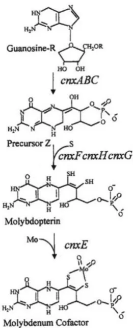
A number of genes, cnxABC, cnxE, cnxF, cnxG and cnxH were identified in A. nidulans by conventional genetic analysis (reviewed by Cove, 1979 and references therein). After the advent of DNA sequencing methods, and with biochemical reference in certain cases to E. coli, the A. nidulans
20 protein CnxJ has also been reported associated with regulation of the level of molybdenum cofactor in
A. nidulans (Arst et al., 1982 and references therein).
A. nidulans CnxE (encoded by cnxE gene) consists of two domains, one has high similarity with the
E. coli protein MoeA and other one with MogA (Millar et al., 2001; Heck et al., 2002). TheCnxE
protein has homologues in Drosophila melanogaster (cinnamon), rat (gephyrin) and Arabidopsis
thaliana (Cnx1). Similar to CnxE, all orthologues have fused MogA-like and a MoeA-like domains.
The orientation of MogA-like and MoeA-like domains is reverse in Cnx1 as compared to CnxE, cinnamon and gephyrin (Stallmeyer et al., 1995; Stallmeyer et al., 1999; Heck et al., 2002; Schwarz
et al., 2009). The illustration in Figure 1.10 represents the comparison of molybdenum cofactor
[image:32.595.228.359.316.637.2]biosynthesis genes and steps involved in the different groups of organisms.
Figure 1.9: Schematic overview of molybdenum cofactor biosynthesis pathway in A. nidulans.
21 Figure 1.10: Comparison of molybdenum cofactor biosynthesis pathways in E. coli, plants and
humans.
This is a generalised scheme of molybdenum cofactor (MoCo) synthesis pathway. In eukaryotes, MoCo
synthesis is a four steps pathway. In E. coli (prokaryotes) an additional step occurs in which with the addition of GTP to MoCo, molybdenum guanine dinucleotide (MGD) is formed. Proposed (first) step and partially
characterised (second) step intermediates are indicated in parenthesis. Different proteins that catalyse the each
step are highlighted with different colours while similar colour is applied to proteins / domains involved in
nucleotide transfer. Homologous protein nomenclature is as, cnx, plants; Mo, E. coli and MOCS, gephyrin. The grey box encloses the separate pathway of molybdopterin (MPT) synthesis. The figure and legends were
22 1.12 Structures and mechanisms of membrane transporter proteins
Uhl and Hartig, (1992) used the term ‘transporter explosion’ for the increasing knowledge of transporter proteins and these words proved to be accurate with the availability of sequenced genomes of a number of organisms. Phylogenetic analysis demonstrated the independent evolution of membrane transporter proteins (Saier, 2000; Busch and Saier, 2002). However the available crystal structures of MFS membrane transporter proteins show that they share the same architecture (Abramson et al., 2004). If the folding pattern of a protein of unknown crystal structure is the same with that of a protein with known structure, a three dimensional model of protein can be generated on the basis of known protein structure as the template (Dahl et al., 2004).
In comparison with water soluble proteins, the number of membrane proteins that have been crystallised so far is low because of two main reasons (i) their hydrophobic nature and (ii) low expression in nature. Recent advances in protein overexpression techniques have been encouraging. However crystallisation of membrane proteins due to overexpression has been successful in bacteria.
Purification and crystallisation of membrane proteins is especially challenging. Only a few membrane transporter proteins from MFS group have been crystallised. The proteins for which crystal structures are available include E. coli lactose transporter, LacY in inward facing conformation (Abramson et al., 2003; Guan et al., 2007; Chaptal et al., 2011), antiporter of inorganic phosphate and glycerol-3-phosphate, GlpT of E. coli (Huang et al., 2003), the multidrug transporter protein, EmrD also in occluded state (Yin et al., 2006). The crystal structure of E. coli FucP, the fucose/H+ symporter has also been solved in an outward open conformation (Dang et al., 2010). The crystal structure of PepTSo, the mammalian homologue of the peptide transporter protein in Shewanella oneidensis has solved in occluded state (Newstead et al., 2011). Finally the two dimensional structure of
Oxalobacter formigenes oxalate-formate transporter, OxlT (Hirai et al., 2002).
From the crystal structures available, it was obvious that members of MFS proteins share similar even super-imposable structures. Another architectural feature is the presence of a central hydrophilic pore that harbours the substrate binding site. Access to this central cavity alternates from the outside to the inside of the cell and vice versa, is made possible by flexible movement of the transmembrane domains. This mechanism of substrate translocation in and out of the cell is called the alternating access model and is illustrated in Figure 1.11 (Huang et al., 2003; Abramson et al., 2004; Law et al.,
23 Figure 1.11: The alternating access model of substrate translocation.
Schematic representation of the alternating access model for a MFS antiporter. (a-e) represent the
stages of translocation of two different substrates moving in opposite directions. The N-terminal
six Tms bundle of the membrane protein is indicated with blue coloured barrels while C-terminal
half of transporter protein is represented as red barrels. Both N- and C-terminal halves are
connected by the cytoplasmic loop. The two different substrates are represented as small black
and yellow circles. (a) the C- and N-terminal bundles move away from each other and open
towards periplasmic side, (b) substrate molecule (black) enters in the central cavity and both
halves of protein close the central cavity from the periplasmic side (c) both bundles now open
towards cytoplasmic side and the substrate enters in the cytoplasm, (d) the other substrate (yellow)
enters in the central cavity, (e) Tm bundles close the cavity from the cytoplasmic side and (e) the
cavity opens towards the periplasm to transport the substrate out of the cell. This figure is based
on Huang et al. (2003) and Abramson et al. (2004).
The lactose permease (LacY) catalyses the coupled translocation or symport of β-galactosidase and
H+ in E. coli. LacY is encoded by gene LacY, the first membrane transport protein encoding gene that
was DNA sequenced (Buchel et al., 1980). LacY, the most extensively studied MFS protein has proved to be a model to study transport mechanisms in other member proteins of this superfamily. The LacY contains 417 amino acids with a 46 kDa molecular weight.
Crystal structures of (i) wild type LacY (Guan et al., 2007) and mutant LacY (Abramson et al., 2003; Mirza et al., 2006; Chaptal et al., 2011) has been determined (Figure 1.12). These crystal structures of LacY show that it consists of 12 α-helical transmembrane domains arranged in two pseudo-symmetrical bundles, N-terminal and C-terminal bundles, each with six helices. These N- and C- terminal bundles surround a large central cavity. Most of the residues involved in the sugar binding (E126 and R144) are present in the N-terminal bundle and residues important for the translocation of H+ (E269 and R302, H322 and E325) are present in the C-terminal bundle (Abramson et al., 2003).
Glycerol-3-phosphate is an antiporter (GlpT) of inorganic and organic phosphate while the energy provided by an inorganic phosphate ion gradient. The 3.3Å crystal structure of GlpT (Figure 1.13) in
Periplasm
Cytoplasm
M
e
m
b
ra
n
e


