Review Article
The efficacy of volar locking plates and external
fixation for patients with unstable distal
radial fractures: a meta-analysis
Shuangwei Qu1,2*, Binfei Zhang1*, Kun Shang1, Pengfei Wang1, Xing Wei1, Yan Zhuang1, Kun Zhang1
1Honghui Hospital, Xi’an Jiaotong University, Xi’an, Shaanxi Province, People’s Republic of China; 2Xi’an Medical
University, Beilin District, Xi’an, Shaanxi Province, People’s Republic of China. *Co-frst authors.
Received September 22, 2017; Accepted September 12, 2018; Epub March 15, 2019; Published March 30, 2019
Abstract: Questions/Purposes: In this meta-analysis, we compared the use of volar locking plates (VLPs) with
ex-ternal fixation (EF) for unstable distal radial fractures. Materials and methods: A systematic review of the literature was conducted to identify randomized controlled trials (RCTs) using VLPs and EF in patients with unstable distal
radial fractures. Trials were performed before July 2017 and retrieved using MEDLINE, Embase, and the Cochrane Library databases. The meta-analysis was performed using STATA version 12.0. Results: Ten RCTs with a total of 932
patients were included. Of these, 451 were in the VLP group and 481 in the EF group. The VLP group had a higher
Disabilities of the Arm, Shoulder and Hand score at 3 months (P<0.001) and 6 months (P<0.001); the scores were comparable for the groups at 12 months (P=0.166). Range of motion was better in the VLP group at 3 months.
No difference between the groups was seen at 6 and 12 months. Grip strength in the VLP group was significantly greater than that in the EF group at 3 months (P<0.001); no difference between the groups was seen at 6 (P=0.526) and 12 months (P=0.507). There was no significant difference in reoperation and complication rates between the
two procedures. Conclusions: We found that VLP may have improved functional recovery in the early period after
surgery, but patients undergoing either VLP or EF showed similar levels of functional recovery at a later stage. The
study was limited by the heterogeneity of the trials and should be interpreted with caution. Multicenter RCTs of higher quality and with larger sample numbers are needed.
Keywords: Distal radial fractures, external fixation, volar plate, meta-analysis
Introduction
Imaging findings in patients with unstable dis -tal radial fractures include angular displace-ment >10°, radial shortening >5 mm, and articular surface step-off >2 mm. Lateral radio-graphs over the midline may show a comminut-ed fracture in a volar view and a dorsal cortical
comminuted fracture. Good reduction is diffi -cult to achieve in these fractures.
Restoration of the articular surface, stable fixa -tion, and early wrist motion are crucial factors in the treatment of patients with unstable distal radial fractures [1]. Several techniques can be used to manage unstable fractures of the distal radius, such as closed reduction with percuta-neous Kirschner wires [2], pins and plaster,
closed reduction with external fixation (EF) [3], and internal fixation (IF) with plates [4]. IF is the
most common method used, especially with
volar locking plates (VLPs) [5]. EF is another
option. However, extensive dissection of soft tissue around the fracture zone compromises the biological environment for fracture healing and increases the risk of nonunion or malunion. The incidence of complications is reportedly 9.7-15%, with tendon problems and complex regional pain syndrome being most prevalent [6-9]. VLPs offer a biomechanical advantage when treating patients with an unstable distal
radius [10]. Unlike open surgery, EF is a closed,
minimally invasive method in which the
frac-tured bone is not exposed to direct view [3]. EF
use is better than EF use for unstable distal
radial fractures is unclear.
Therefore, we performed a meta-analysis to
compare the results of open reduction and IF with VLPs versus EF in patients with complex
fractures of the wrist combining serious me- taphyseal comminution and articular involve-ment. The outcomes of interest included clini-cal, functional, and radiological results, and complication rates.
Materials and methods
Literature search
We searched MEDLINE (1966 to July 2017), Embase (1974 to July 2017), and the Cochrane Library (through issue 6 of 12, 2017) databas-es using the keywords “radius”, “fracturdatabas-es”,
“external fixation”, “EF”, and “plate”. Retrieval
dates included the time from database cre-ation to July 2017.
Inclusion criteria
Inclusion criteria were as follows: randomized controlled trial (RCT); participants with unsta-ble distal radial fractures; patients and controls
who underwent EF or volar plating; primary
endpoints of Disabilities of the Arm, Shoulder and Hand (DASH) score, range of motion, or
generation, allocation concealment, blinding, incomplete outcome data, freedom from selec-tive reporting, freedom from other bias, base-line balance between groups, no funding sup-port, and valid sample size estimation.
Statistical methods
Relative risk (RR) and weighted mean differ-ence (WMD) were used for effect size, with 95%
confidence intervals (95% CIs). The
Mantel-Haenszel (M-H) or inverse variance (I-V) was used as the statistical method. We assessed heterogeneity with I2 statistics. During quan- titative synthesis, a fixed-effects model was
employed when heterogeneity was low (I2<50%,
P>0.1). When heterogeneity was high (I2>50%,
P<0.1), subgroup analysis was performed to explore possible sources of heterogeneity, or a random-effects model was used. We also used endpoint substantive knowledge as a factor in the selection model. P<0.05 was considered a
statistically significant difference. STATA 12.0
version (STATA Corporation, College Station, TX, USA) was used to perform the analysis.
Results
Process for selecting trials
Most of the 243 potential eligible citations Figure 1. Flow chart of studies in
-cluded in the meta-analysis.
Visual Analogue Scale (VAS) scores, complications, or rad- iological measurements.
Data extraction and quality evaluation
We screened all titles of re- trieved articles and removed duplicates. After eliminating ir- relevant articles, summaries of the remaining studies were
[image:2.612.90.371.72.338.2]Table 1. Summary of included studies
Study Country Types of fraction
No. of
patients Age (years) Women Outcomes Follow-up EF VP EF VP EF VP
Egol 2008 [1] America AO A/B/C 44 44 49.9 52.2 22 25 Radiological measurements, DASH scores, function, complications 12 months Gradl 2013 [3] America AO A3/C1/C2/C3 50 52 63 63 - - Radiological measurements, function, complications 12 months Jeudy 2012 [5] France AO C1/C2/C3 39 36 64.6 64.7 31 26 Radiological measurements, function, complications 6 months Navarro 2016 [11] Sweden AO A2/A3/C1/C2/C3 70 70 63 63 65 63 Radiological measurements, function, complications 12 months Roh 2015 [15] Korea AO C2/C3 38 36 55.3 54.4 16 14 Radiological measurements, function, complications 12 months Shukla 2014 [19] India Cooney’s type IV 62 48 38.95 39.33 33 28 Function, complications 12 months Wei 2009 [22] America AO A3/C1/C2/C3 22 12 55 61 16 9 DASH scores, function, complications 12 months Wilcke 2011 [23] Sweden AO A/C 30 33 56 55 23 25 DASH scores, function, complications 12 months Williksen 2013 [24]
Williksen 2015 [25] Norway AO A2/A3/C1/C2/C3 60 54 54 54 - - DASH scores, function, complications 60 months Li 2015 [10] China AO A3/B3/C1/C2/C3 61 61 64.4 64.7 50 52 Radiological measurements, function, complications 12-28 months
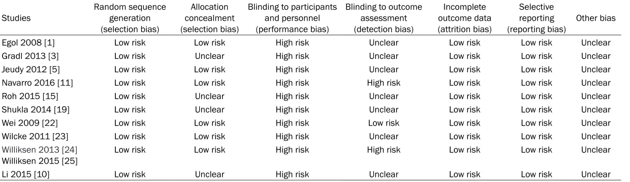
Table 2. Quality of observational studies
Studies Random sequence generation (selection bias)
Allocation concealment (selection bias)
Blinding to participants and personnel (performance bias)
Blinding to outcome assessment (detection bias)
Incomplete outcome data (attrition bias)
Selective reporting
(reporting bias) Other bias Egol 2008 [1] Low risk Low risk High risk Unclear Low risk Low risk Unclear Gradl 2013 [3] Low risk Unclear High risk Unclear Low risk Low risk Unclear Jeudy 2012 [5] Low risk Low risk High risk Unclear Low risk Low risk Unclear Navarro 2016 [11] Low risk Low risk High risk High risk Low risk Low risk Unclear Roh 2015 [15] Low risk Unclear High risk Unclear Low risk Low risk Unclear Shukla 2014 [19] Low risk Unclear High risk Unclear Low risk Low risk Unclear Wei 2009 [22] Low risk Low risk High risk Low risk Low risk Low risk Unclear Wilcke 2011 [23] Low risk Low risk High risk Unclear Low risk Low risk Unclear
Williksen 2013 [24]
[image:3.792.91.711.296.475.2]exclusions were removed, 10 RCTs (11 articles)
satisfied the inclusion criteria after screening
and assessing potentially relevant studies. Figure 1 shows the flow of studies through the
trial.
Characteristics of included trials and quality evaluation
Except for a study by Shukla [12], all RCTs
reported fractures using the AO/OTA classifica -tion. Ten RCTs with a total of 932 patients were included. Of these, 451 were in the VLP group
and 481 in the EF group. All studies focused on
function and complications, including 4 studies [13-17] that examined DASH scores and 6 [13, 18-22] that evaluated radiological measure-ments. One study [16, 17] had a follow-up peri-od of 60 months, with 12-28 months reported in a study by Li, 6 months in a study by Jeudy, and 12 months in 7 other studies [12-14, 16- 18, 21, 22] (Table 1).
The results of the quality evaluation are sh- own in Table 2. Random sequence generation, incomplete outcome data, and selective report-ing showed a low risk of bias in all studies. Allocation concealment showed a low risk of bias in 6 studies [13, 19, 21, 14-17], but was unclear in 4 studies. Blinding to participants and personnel showed a high risk of bias in all studies. Blinding to outcome assessment showed a high risk of bias in 2 studies [21, 16, 17] and low risk in 1 study [14], but bias was unclear in 7 studies [13, 18, 19, 20, 22, 12, 15]. Other sources of bias were unclear. Primary endpoints
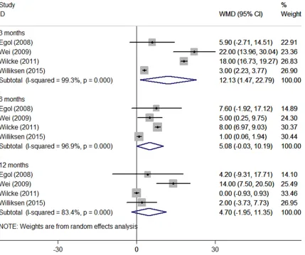
DASH scores
DASH scores for EF and VLPs were compared
[image:4.612.95.524.72.431.2]to the study design. I2 values for heterogeneity
at 3, 6, and 12 months were 99.3% (P<0.001), 96.9% (P<0.001), and 83.4% (P<0.001), re- spectively. The random-effects model was ap-
plied to these studies. DASH scores in the EF
group were superior to those in the VLP group at 3 months (WMD=12.13; 95% CI: 1.47-22.79;
P<0.001) and 6 months (WMD=5.08; 95% CI: -0.03-10.19; P<0.001). However, there was no
difference between the EF and VLP group at
12 months (WMD=4.70; 95% CI: -1.95-11.35;
P=0.166). Range of motion
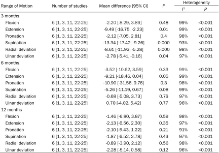
Range of motion was assessed in 6 studies (Table 3). At 3 months, wrist extension in the
EF group was less than in the VLP group
(MD=-9.49; 95% CI: -16.75--2.23; P=0.01). Supination, radial deviation, and ulnar
devia-tion in the EF group were also less than in the
VLP group at 3 months (MD=-13.34; 95% CI: -17.42--9.26; P<0.001, MD=-8.61; 95% CI: -11.93--5.28; P<0.001, and MD=-2.78; 95% CI: -5.41--0.16; P=0.04, respectively). Other items showed no differences. Range of motion also
showed no difference between EF and VLP
groups at 6 and 12 months.
Grip strength
Two studies examined grip strength. The results
of comparisons between the EF and VLP groups
are shown in Figure 3. Grip strength in the EF group at 3 months was significantly lower than
that in the VLP group (WMD=-13.60; 95% CI: -23.61--3.59; P<0.001), but the differences
were not significantly different at 6 (WMD=
-2.77; 95% CI: -11.32-5.78; P=0.526) and 12 months (WMD=-1.98; 95% CI: -7.84-3.88; P=0.507).
Secondary endpoints
We also compared VAS scores, complication rates, and radiological measurements. The VAS scores (Figure 4) showed no significant differ
-ences between the EF and VLP groups after
adopting a random-effects model.
We compared the radiological outcomes in the- se studies 12 months after surgery and
[image:5.612.91.523.87.388.2]exam-ined whether EF was associated with a higher Table 3. Range of motion at 3, 6, and 12 months postoperatively
Range of Motion Number of studies Mean difference [95% CI] P Heterogeneity
I2 P 3 months
Flexion 6 [1, 3, 11, 22-25] -2.20 [-8.29, 3.89] 0.48 99% <0.001
Extension 6 [1, 3, 11, 22-25] -9.49 [-16.75, -2.23] 0.01 99% <0.001
Pronation 6 [1, 3, 11, 22-25] -2.12 [-7.05, 2.81] 0.4 98% <0.001
Supination 6 [1, 3, 11, 22-25] -13.34 [-17.42, -9.26] 0.000 93% <0.001
Radial deviation 6 [1, 3, 11, 22-25] -8.61 [-11.93, -5.28] 0.000 98% <0.001
Ulnar deviation 6 [1, 3, 11, 22-25] -2.78 [-5.41, -0.16] 0.04 97% <0.001 6 months
Flexion 6 [1, 3, 11, 22-25] -3.52 [-10.62, 3.59] 0.33 99% <0.001
Extension 6 [1, 3, 11, 22-25] -9.21 [-18.46, 0.04] 0.05 99% <0.001
Pronation 6 [1, 3, 11, 22-25] -10.90 [-31.56, 9.76] 0.3 98% <0.001
Supination 6 [1, 3, 11, 22-25] -5.26 [-11.19, 0.67] 0.08 99% <0.001
Radial deviation 6 [1, 3, 11, 22-25] -0.68 [-5.08, 3.73] 0.76 97% <0.001
Ulnar deviation 6 [1, 3, 11, 22-25] 0.70 [-4.02, 5.42] 0.77 96% <0.001 12 months
Flexion 6 [1, 3, 11, 22-25] -1.46 [-6.80, 3.87] 0.59 98% <0.001
Extension 6 [1, 3, 11, 22-25] -2.13 [-6.56, 2.30] 0.35 97% <0.001
Pronation 6 [1, 3, 11, 22-25] -2.10 [-5.43, 1.22] 0.21 91% <0.001
Supination 6 [1, 3, 11, 22-25] -1.87 [-6.52, 2.78] 0.43 97% <0.001
Radial deviation 6 [1, 3, 11, 22-25] -0.89 [-3.90, 2.12] 0.56 98% <0.001
ulnar deviation (MD=0.94; 95% CI: 0.8-1.70;
P<0.001). The results are shown in Table 4. Complications included wound and pin-track infections, tendon rupture, carpal tunnel syn-drome, complex regional pain synsyn-drome, and need for reoperation. We found that patients
undergoing EF were much more susceptible
to wound and pin-track infection than those undergoing VLP (RR=7.15; 95% CI: 2.36-21.64;
P=0.0005). However, the reoperation rate was
higher for VLP than for EF (RR=0.32; 95% CI:
0.16-0.64; P=0.001; Table 5).
Publication bias
We also assessed publication bias using grip strength at 12 months. The results showed no publication bias. The Begg test (z=0.62,
P=0.563; Figure 5) and Egger test (t=0.28,
P=0.788) did not indicate bias.
Discussion
In this meta-analysis, we observed better early postoperative functional outcomes in the VLP
group than in the EF group, but no difference
between the groups was seen at 1 year.
Patients undergoing EF were more likely to
develop wound and pin-track infections. Pat-
ients in the VLP group had significantly higher grip strength than those in the EF group. How-ever, there was a significantly higher rate of
reoperation in the VLP group.
Differences in DASH scores were statistically
significant during follow-up. However, the mini -mal clinically important difference for the DASH score ranged between 10 and 15 points [11]. Therefore, the difference in DASH scores at 3 months after surgery indicated substantial
[image:6.612.96.523.76.437.2]functional improvement [20]. Significantly bet -ter results in DASH scores are consistent with Figure 3. Forest plot comparing grip strength. The EF group showed lower grip strength than the VLP group at 3
Figure 4. Forest plot comparing VAS scores. The results showed no difference at 3, 6, or 12 months.
Table 4. Radiological measurements
Radiological outcomes Number of studies Mean difference [95% CI] P Heterogeneity
I2 P Radial length 3 [1, 10, 22] 0.40 [-0.51, 1.30] 0.39 53% 0.12 Radial inclination 5 [1, 10, 11, 15, 22] 0.19 [-1.18, 1.55] 0.79 77% 0.002 Ulnar variance 4 [1, 5, 11, 22] 0.94 [0.80, 1.07] 0.000 0% 0.61 Volar tilt 7 [1, 3, 5, 10, 11, 15, 22] 2.40 [-0.37, 5.17] 0.09 100% <0.001
Table 5. Complications
Complications Number of studies RR [95% CI] P Heterogeneity
[image:7.612.92.525.617.707.2]results reported by Walenkamp et al. [11]. As
the external fixators were removed after bone
healing, the DASH scores were likely to change. The lower DASH score after using VLPs may be associated with worse wrist range of motion exercise [20].
Comparison of functional outcomes between
EF and VLPs showed that extension, supina -tion, radial devia-tion, and ulnar deviation were
significantly better in the VLP group than in the EF group at the 3-month follow-up. This may be
attributable to functional exercise during the early postoperative period. The change in dy- namic grip strength with VLP was larger than
with EF in the early postoperative stage and
may be helpful in functional exercise. Patients found functional exercises to be inconvenient
because of wrist joint limitations caused by EF,
which could lead to unsatisfactory outcomes for grip strength and range of motion.
The VAS score is an important indicator but
showed no difference between the EF and VLP
groups during follow-up. Radiological analysis
revealed that EF did not maintain ulnar vari -ance to the same extent as VLPs, similar to reports by other authors [19].
There were more complications in the EF group than in the IF group, mainly wound and
pin-track infections. A pin-site infection rate of up to 43% has been reported, but no further
treat-gested that use of VLPs led to faster recovery. No difference was observed in functional
out-come, radiological findings, and complication
rates at 1 year. The need for reoperation was more common with VLP use. Compared with
the findings reported by Li [20], other studies
showed similar outcomes. Moreover, Li’s study included 6 articles, while our study included 10 RCTs.
Our meta-analysis has several potential
limita-tions. First, statistical heterogeneity was large. This may be the result of differences in EF tech
-nique, as some studies reported temporary fix -ation with Kirschner wires. Second, external
fixators were removed at different time points,
which could have contributed to heterogeneity.
External fixators were removed at a minimum
of 5 weeks to a maximum of 6 weeks [14].
Third, some patients underwent “pure” EF, while some underwent EF with supplementary
K wires. These are two largely different
opera-tions. With the use of K wires, EF acts as a neu
-tralization plate whereas, “pure” EF relies on
ligamentotaxis. Thus, these results should be cautiously interpreted.
VLP use may improve functional recovery early after surgery in patients with unstable distal
radial fractures. However, VLP use and EF
[image:8.612.93.373.73.260.2]sh-owed comparable functional scores 12 mon- ths postoperatively. Nonetheless, the results need to be interpreted with caution due to Figure 5. Funnel plot comparing fusion rates for EF and VLP. The y-axis rep
-resents WMD, and the x-axis standard error of log (WMD). The dashed line in the middle is the log (WMD) value calculated from the fusion rate. Sloped lines represent the 95% CI boundaries and circles indicate the 7 studies evaluated.
ment was required [23-25]. Quality of life decreased once infections appeared. Except for a higher rate of reopera-tion in the VLP group, other complications showed no dif-ference. However, other stud-ies [26, 15, 16] were inconclu-sive. The need for reoperation was mainly due to tendon rup-ture and carpal tunnel synd- rome.
Distal radial fractures have been treated by surgeons
using bridging EF in prefer -ence to using VLPs, as many
sug-study heterogeneity and various sources of bias. High-quality, large-sample clinical rese- arch is needed for further validation.
Acknowledgements
This study was supported by the Social De-
velopment Foundation of Shaanxi Province (grant numbers 2017ZDXM-SF-009,
2017SF-050). We thank all colleagues working in the Department of Orthopedic Trauma, Honghui Hospital, Xi’an Jiaotong University.
Disclosure of conflict of interest
None.
Address correspondence to: Kun Zhang, Depart- ment of Orthopedic Trauma, Honghui Hospital, Xi’an Jiaotong University, Beilin District, No. 555 Youyi East Road, Xi’an 710054, Shaanxi Province, China. Tel: +8615829208357; E-mail: hhzhangkun@163. com
References
[1] Zhang B, Chang H, Yu K, Bai J, Tian D, Zhang G, Shao X and Zhang Y. Intramedullary nail versus
volar locking plate fixation for the treatment of
extra-articular or simple intra-articular distal radius fractures: systematic review and meta-analysis. Int Orthop 2017; 41: 2161-2169. [2] Rozental TD, Blazar PE, Franko OI, Chacko AT,
Earp BE and Day CS. Functional outcomes for
unstable distal radial fractures treated with
open reduction and internal fixation or closed reduction and percutaneous fixation: a pro-spective randomized trial. J Bone Joint Surg Am 2009; 91: 1837-1846.
[3] Handoll HH, Huntley JS and Madhok R.
Differ-ent methods of external fixation for treating
distal radial fractures in adults. Cochrane Da-tabase Syst Rev 2008: CD006522.
[4] Rader CP, Rauber C, Rehm KE and Koebke J.
Internal fixation of the distal radius: a compar -ative, experimental study. Arch Orthop Trauma Surg 1995; 114: 340-343.
[5] Orbay JL and Fernandez DL. Volar fixation for
dorsally displaced fractures of the distal radi-us: a preliminary report. J Hand Surg Am 2002; 27: 205-215.
[6] Johnson NA, Cutler L, Dias JJ, Ullah AS, Wildin CJ and Bhowal B. Complications after volar
locking plate fixation of distal radius fractures.
Injury 2014; 45: 528-533.
[7] Lattmann T, Meier C, Dietrich M, Forberger J
and Platz A. Results of volar locking plate os-teosynthesis for distal radial fractures. J Trau-ma 2011; 70: 1510-1518.
[8] Phadnis J, Trompeter A, Gallagher K, Bradshaw L, Elliott DS and Newman KJ. Mid-term
func-tional outcome after the internal fixation of dis -tal radius fractures. J Orthop Surg Res 2012; 7: 4.
[9] Sahu A, Charalambous CP, Mills SP, Batra S and Ravenscroft MJ. Reoperation for metal-work complications following the use of volar locking plates for distal radius fractures: a united kingdom experience. Hand Surg 2011; 16: 113-118.
[10] Wolf JC, Weil WM, Hanel DP and Trumble TE. A biomechanic comparison of an internal radio-carpal-spanning 2.4-mm locking plate and
ex-ternal fixation in a model of distal radius frac -tures. Hand Surg 2006; 31: 1578-1586. [11] Walenkamp MM, Bentohami A, Beerekamp
MS, Peters RW, van der Heiden R, Goslings JC
and Schep NW. Functional outcome in patients
with unstable distal radius fractures, volar
locking plate versus external fixation: a
meta-analysis. Strategies Trauma Limb Reconstr 2013; 8: 67-75.
[12] Shukla R, Jain RK, Sharma NK and Kumar R.
External fixation versus volar locking plate for
displaced intra-articular distal radius frac-tures: a prospective randomized comparative study of the functional outcomes. Ital J Orthop Traumatol 2014; 15: 265-270.
[13] Egol K, Walsh M, Tejwani N, McLaurin T, Wynn
C and Paksima N. Bridging external fixation and supplementary kirschner-wire fixation ver -sus volar locked plating for unstable fractures of the distal radius: a randomised, prospective trial. J Bone Joint Surg Br 2008; 90: 1214-1221.
[14] Wei DH, Raizman NM, Bottino CJ, Jobin CM, Strauch RJ and Rosenwasser MP. Unstable
dis-tal radial fractures treated with external fixa -tion, a radial column plate, or a volar plate. A prospective randomized trial. J Bone Joint Surg Am 2009; 91: 1568-1577.
[15] Wilcke MK, Abbaszadegan H and Adolphson PY. Wrist function recovers more rapidly after
volar locked plating than after external fixation
but the outcomes are similar after 1 year. Acta Orthop 2011; 82: 76-81.
[16] Williksen JH, Frihagen F, Hellund JC, Kvernmo
HD and Husby T. Volar locking plates versus
external fixation and adjuvant pin fixation in
unstable distal radius fractures: a randomized, controlled study. Hand Surg 2013; 38: 1469-1476.
[17] Williksen JH, Husby T, Hellund JC, Kvernmo
HD, Rosales C and Frihagen F. External fixation
and adjuvant pins versus volar locking plate
fixation in unstable distal radius fractures: a
[18] Gradl G, Wendt M, Mittlmeier T, Kundt G and
Jupiter JB. Non-bridging external fixation em -ploying multiplanar K-wires versus volar locked plating for dorsally displaced fractures of the distal radius. Arch Orthop Trauma Surg 2013; 133: 595-602.
[19] Jeudy J, Steiger V, Boyer P, Cronier P, Bizot P and Massin P. Treatment of complex fractures of the distal radius: a prospective randomised
comparison of external fixation ‘versus’ locked
volar plating. Injury 2012; 43: 174-179. [20] Zhang LH, Wang YN, Mao Z, Zhang LC, Li HD,
Yan H, Liu XX and Tang PF. Volar locking plate versus external fixation for the treatment of un -stable distal radial fractures: a meta-analysis of randomized controlled trials. J Surg Res 2015; 193: 324-333.
[21] Mellstrand Navarro C, Ahrengart L, Törnqvist H and Ponzer S. Volar locking plate or external
fixation with optional addition of k-wires for
dorsally displaced distal radius fractures. J Or-thop Trauma 2016; 30: 217-224.
[22] Roh YH, Lee BK, Baek JR, Noh JH, Gong HS and Baek GH. A randomized comparison of
vo-lar plate and external fixation for intra-articuvo-lar
distal radius fractures. J Hand Surg Am 2015; 40: 34-41.
[23] Flinkkila T, Ristiniemi J, Hyvonen P and Ham
-alainen M. Nonbridging external fixation in the
treatment of unstable fractures of the distal forearm. Arch Orthop Trauma Surg 2003; 123: 349-352.
[24] Hove LM, Krukhaug Y, Revheim K, Helland P
and Finsen V. Dynamic compared with static external fixation of unstable fractures of the
distal part of the radius: a prospective, ran-domized multicenter study. J Bone Joint Surg Am 2010; 92: 1687-1696.
[25] Egol KA, Paksima N, Puopolo S, Klugman J, Hiebert R and Koval KJ. Treatment of external
fixation pins about the wrist: a prospective,
randomized trial. J Bone Joint Surg Am 2006; 88: 349-354.
[26] Leung F, Tu YK, Chew WY and Chow SP. Com
-parison of external and percutaneous pin fixa
-tion with plate fixa-tion for intra-articular distal
radial fractures. A randomized study. J Bone Joint Surg Am 2010; 90: 16-22.
[27] Payandeh JB and McKee MD. External fixation