Original Article
MicroRNA-152-3p negatively
regulates ischemia-reperfusion induced
cardiomyocyte apoptosis by inhibiting PTEN
Peng Liu1,2,4, Yumei Xie3, Mingyang Qian3, Shushui Wang3, Junjie Li3, Xu Zhang3, Shibian Xi3, Gang Pan4,
Zhiwei Zhang1,2,3
1Southern Medical University, NO. 1023, South Shatai Road, Baiyun District, Guangzhou 510515, Guangdong, People’s Republic of China; 2Affiliated South China Hospital, Guangdong Provincial People’s Hospital, Southern Medical University, Guangdong, People’s Republic of China; 3Department of Heart Disease Institute, Guangdong General Hospital, Guangdong Academy of Medical Sciences, Guangzhou 510080, People’s Republic of China; 4Department of Critical Care Medicine, Yueyang First People’s Hospital, Yueyang 414000, People’s Republic of China
Received October 8, 2018; Accepted November 8, 2018; Epub January 15, 2019; Published January 30, 2019
Abstract: microRNAs (miRNAs) play a vital role in the treatment of many cardiovascular diseases. Myocardial isch-emia/reperfusion (I/R) injury has been associated with adverse cardiovascular outcomes. However, whether
152-3p is involved in I/R induced cardiomyocyte injury remains ill-defined. In this study, it was found that
miR-152-3p expression was markedly down-regulated in H9c2 cells after hypoxia/reoxygenation (H/R). H/R treatment
significantly elevated the levels of LDH and MDA, and enhanced cell apoptosis in H9c2 cells. Ectopic expression of miR-152-3p markedly blocked LDH & MDA production and apoptosis in H9c2 cells after H/R, while depletion
of miR-152-3p in H9c2 cells induced cell apoptosis. Mechanistic analyses indicated that phosphatase and tensin
homolog (PTEN) was regulated by miR-152-3p directly. Additionally, subsequent investigations identified that the
anti-apoptotic effect of miR-152-3p was partially reversed when H9c2 cells were co-transfected with PTEN mimic.
In addition, up-regulation of miR-152-3p significantly elevated the levels of p-AKT and p53 under H/R. Furthermore, therapeutic promotion of miR-152-3p decreased LDH content, cardiac hypertrophy, and apoptosis after I/R in vivo.
These findings show that miR-152-3p could suppress I/R injury induced cardiomyocyte apoptosis by targeting PTEN.
Therefore, miR-152-3p and PTEN could be regarded as potential therapeutic targets for the clinical study of I/R injury.
Keywords: Cardiomyocyte, miR-152-3p, ischemia-reperfusion, PTEN, apoptosis
Introduction
Ischemic heart disease is one of the leading causes of mortality and morbidity all over the world. The current standard treatment for myo-cardial ischemic incidents is the rapid reperfu-sion [1, 2]. However, the acute restoration of blood flow can cause the cardiac injury by it- self, a process termed myocardial I/R injury. Myocardial I/R induce cardiac injury in the pro-cess of myocardial ischemia, cardiac arrest, or cardiac surgery, which contributes to irrevers-ible damage to the myocardium [3-5]. It has been elucidated that the molecular mecha-nisms underlying myocardial I/R injury are com-plex and multifaceted, including oxygen free
mation, and mitochondrial permeability [6, 7]. However, the complete profile of molecular pathways underlying biologic processes in- volved in reperfusion injury remains mostly unknown. Recently, apoptosis was suggested to be the major mechanism leading to the myo-cardial I/R injury [8-10]. Therefore, further stud-ies on the apoptotic mechanisms underlying the development of myocardial I/R injury, may lead to the identification of effective targeted therapy.
evidence has indicated that miRNAs are involved in the regulation of various biological processes that are essential for normal devel-opment and cellular homeostasis [13, 14]. Re- cent studies have reported that several miR-NAs are involved in the regulation of pathologi-cal and physiologipathologi-cal process of cardiac diseas-es [15, 16]. As an example, miRNA microarray analysis shows that miR-320 is up-regulated in cardiomyocytes after I/R treatment, and that silencing of endogenous miR-320 inhibits I/R-induced cardiomyocyte apoptosis by target- ing HSP-20 [17]. Similarly, miR-320 has been shown to promote cardiomyocyte apoptosis and myocardial I/R injury, and its downregula-tion reduces cardiomyocyte apoptosis and death in mice [18]. Conversely, miR-17 is sensi-tive to I/R injury and attenuated I/R-induced apoptosome formation by inhibiting apoptotic protease activation factor 1 expression [19]. Among many miRNAs, miR-152-3p has been identified to play a key role in a number of essential biological processes, including tumor-igenesis, cell proliferation, differentiation, and apoptosis [20, 21]. However, the role of miR-152-3p in cardiac I/R injury remains largely unknown. Therefore, the purpose of this study was to investigate the effects of miR-152-3p on myocardial I/R injury, and to identify mecha-nisms by which miR-152-3p may ameliorate the cardiomyocyte apoptosis and apoptosis related genes. In this study, miR-152-3p was found to have anti-apoptotic effects on cardiomyocyte and protective effects against myocardial I/R injury. These results provide novel mechanistic insights into the role of miR-152-3p in myocar-dial I/R injury.
Materials and methods
Cell culture and reagents
H9c2 cells was purchased from the Cell Bank of Chinese Academy of Science and cultured in a medium of Dulbecco’s Modified Eagle Medium (Invitrogen, CA, USA) supplemented 10% FBS. Cells were cultured with 1% penicil-lin/streptomycin and 1% glutamine in a humidi-fied incubator with 5% CO2 at 37°C. Lactate dehydrogenase (LDH) and malondialdehyde (MDA) commercial kits were obtained from Beyotime Institute of Biotechnology (Shanghai, China). In situ cell death detection kit was pur-chased from Roche (Basel, Switzerland). The antibodies against Bax, Bcl-2 and caspase-3
were obtained from Santa Cruz Biotechnology (Santa Cruz, St Louis, Mo, USA). The antibodies against PTEN, phosopho-AKT, AKT, p53, HIF-α and β-actin were purchased from Abcam Biotechnology (Abcam, Cambridge, MA, USA).
In vitro I/R injury model
A simulated H/R cell model was performed as previously description [14, 15]. Briefly, H9c2 cells were firstly perfused in normal Hank’s solution with 95% oxygen and 5% CO2. To simu-late ischemia, nitrogen gas was flushed into the incubator to decrease oxygen to 1%. To simu-late reperfusion, cardiomyocytes were subject-ed to 6 hours of hypoxia followsubject-ed by re-oxygen-ation by incubre-oxygen-ation in normal Hank’s solution with a gas mixture of 95% oxygen and 5% CO2 for 1 hour. Cells under normoxia throughout the experiments were set as control group.
Cell transfection
miR-152-3p mimic and its negative control (mimic control), as well as miR-152-3p inhibitor and its negative control (anti-control) were pur-chased from QIAGEN (Duesseldorf, Germany). The plasmid for PTEN over-expression (PTEN) was constructed by Sesh-biotech (Shanghai, China). Cells were seeded in 6-well or 96-well plates and transfected using Lipofectamine 2000 (Invitrogen, CA, USA) for 24 hours, accord-ing to the manufacturer’s protocol. Transfected cells were used for further analysis.
Quantitative real-time PCR
Total RNAs from either cultured cells or tissue samples were isolated using Trizol Reagent (QIAGEN, Duesseldorf, Germany). First-strand cDNA was synthesized using a TaqMan Reverse Transcription Reagents (Life Technologies). The levels of mRNAs were detected using Taqman MicroRNA Assay Kits (Applied Biosystems, Foster City, CA) specific for miR-152-3p. The universal reverse quantitative PCR primer was provided in the miScript SYBR Green PCR Kit. The qRT-PCR results were analyzed using the comparative cycle threshold method (2-∆∆CT). Each sample was performed in triplicate. Detection of LDH and MDA
Apoptotic cell determination
Apoptotic cells were determined by TUNEL method as per manufacturer’s instructions. The percentage of TUNEL-positive cardiomyo-cytes per high powered field was observed under a fluorescence microscope (Olympus, Tokyo, Japan) by two different pathologists. For quantification, the apoptotic index was eval-uated by counting the number of TUNEL-posi- tive apoptotic cells per 100 cells in three ran-domly selected myocardial areas with 400 × magnification.
Western blot analysis
Cells or tissues were lysed in RIPA buffer (Beyotime, Jiangsu, China) containing protease inhibitors (Beyotime), and the protein concen-trations were measured using a BCA Kit (Sigma, St. Louis, MO, USA). A 25 µg sample of protein was separated by 10% SDS-PAGE and then transferred to PVDF membranes. The mem-branes were blocked with 5% non-fat powder- ed milk in PBS and then probed with primary antibodies. After primary antibody incubation, membranes were probed with peroxidase-labeled secondary antibodies. The results were normalized to β-actin as the relative density.
Dual luciferase reporter assay
The wild-type PTEN 3’UTR containing the pre-dicted miR-152-3p binding sites was amplified by specific primers using genomic DNA as a template and then inserted into the down-stream of the luciferase gene sequence in the psiCHECK-2 vector (Promega, WI, USA). The mutant-PTEN 3’UTR (deletion of AAAAUGU) was also accomplished by PCR. All vectors were identified by DNA sequencing. Cells were plated into 96-well plates in 5 replicates. For transfec-tion, 2 µl of 20 µM of either miR-152-3p mimic or miR-152-3p inhibitor and 150 ng reporter plasmids (either wt-PTEN or mut-PTEN) were mixed with 2 µl Lipofectamine 2000 (Invitrogen, CA, USA). Firefly luciferase activity was mea-sured using a Dual-Luciferase Assay System (Promega, WI, USA) and was normalized to Renilla activity.
In vivo gene transfection myocardial I/R model
A total of 60 female Sprague-Dawley rats (weight, 250 g-350 g; age, 10-14 weeks) were
purchased from the animal experimental cen-ter of the Central South University. Animals were housed at a temperature of 25±2°C and the humidity was set at 60%. Photoperiod was maintained a 12:12 hour light-dark cycle with standard forage and drinking water ad libitum. This study’s protocols were approved by the animal ethics committee of the Xiangya Ho- spital of Central South University. The 60 rats were randomly divided into three groups (n = 20). 1) Sham-operated group: the heart was exposed after left thoracotomy without ligat- ing the left anterior descending artery (LAD). 2) I/R group: The LAD was ligated reversibly using silk suture. Following 30 min of ischemia, the ligature was loosened, allowing the ischemic myocardium to re-perfuse for 2 h. 3) I/R + Ad-miR-152-3p group: Four days after the rats received intramyocardial injection of Ad-miR-152-3p recombinant adenovirus, myocardial I/R treatment was induced as above. Rats were sacrificed at the time of coronary artery flow of 120 minutes to obtain the hearts and blood samples for subsequent tests. MiR-152-3p mimic lentivirus and empty viral vectors were purchased from QIAGEN (Duesseldorf, Germany).
Histology
Heart samples were cut transversely into blocks and fixed in 4% paraformaldehyde. Tese were then embedded in OCT compound (BHD, UK) and then transversely cut into 4 μm sec-tions. Next, the slides were incubated with Masson’s trichrome for assessment of infarct size. Results were analyzed as the sum of the epicardial and endocardial scar length, divided by the sum of LV epicardial and endocardial cir-cumferences. Cardiomyocyte hypertrophy was assessed by calculating the average cardio-myocyte cross-sectional areas in five random fields.
Statistical analysis
All statistical results were analyzed using SPSS 18.0 statistical software. Experimental data from 3 independent experiments are present- ed as the mean ± SD. Quantitative variables between groups were analyzed using either
Results
miR-152-3p is down-regulated in H9c2 after H/R
We first detected miR-152-3p expression in H9c2 cells after 10 hours of hypoxia and 2.5 hours of reoxygenation by qRT-PCR. The results demonstrate that the expression of miR-152-3p is reduced markedly in H9c2 cells following
H/R treatment (Figure 1A). This finding indi-cates that miR-152-3p may play a vital role in the process of H/R injury.
miR-152-3p regulates H/R-induced cardiomyo
-cyte injury in vitro
The effects of miR-152-3p modification on the H/R injury of H9c2 cells were then explored. In
vitro gain and loss-of-function assays were per-Figure 1. Effects of miR-152-3p on H9c2 cells following H/R injury. (A) The expression of miR-152-3p was decreased
significantly after H/R injury. (B) H9c2 cells were transfected with 3p mimic or mimic-control, and
miR-152-3p expression was detected by qRT-PCR. (C) H9c2 cells were transfected with anti-miR-152-miR-152-3p or anti-control, and
expression of miR-152-3p was detected by qRT-PCR. The levels of LDH (D) and MDA (E) were measured in H9c2
[image:4.612.88.520.73.526.2]formed by the transfection of miR-152-3p mimic or inhibitor in H9c2 cells. As a result, transfection of miR-152-3p mimic increased expression of miR-152-3p dramatically (Figure 1B). Moreover, the relative level of miR-152-3p was reduced significantly after transfection with miR-152-3p inhibitor (Figure 1C). LDH release is an indicator of cellular injury. Our results demonstrate that the level of LDH sig-nificantly increases in response to H/R treat-ment in the culture media. Expression of LDH was repressed significantly by miR-152-3p over-expression. In contrast, the miR-93 inhibi-tor promoted LDH release after H/R (Figure
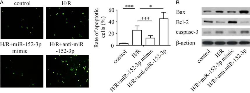
tosis, the miR-152-3p inhibitor was transfected into H9c2 cells to inhibit the expression of miR-152-3p and the responses to H/R were investi-gated. The results show that the ratio of apop-totic cardiomyocyte was promoted notably after H/R treatment compared with that in the con-trol group. However, transfection with miR-152-3p mimic significantly inhibited H/Rinduced cell apoptosis, and silencing of miR-152-3p promot-ed H/R-inducpromot-ed apoptosis in H9c2 cells (Figure 2A).
[image:5.612.92.521.73.234.2]Consistent with morphologic changes, the rep-resentative Western blot analysis showed that Figure 2. Effects of miR-152-3p on H/R-induced cardiomyocyte apoptosis. A: Ectopic expression of miR-152-3p attenuates cardiomyocyte apoptosis during H/R injury and its down-regulation promotes myocyte apoptosis. B: Western blot analysis of Bax, Bcl-2 and caspase-3 staining in H9c2 cells. β-action was used as endogenous control. ***P < 0.001, *P < 0.05.
Figure 3. miR-152-3p down-regulated PTEN by interacting with its 3’UTR. A: miR-152-3p and its putative binding sequences in the PTEN 3’UTR. B: H9c2 cells were co-transfection of PTEN-3’UTR-wt or PTEN-3’UTR-mut with
miR-152-3p mimic, anti-miR-miR-152-3p or appropriate controls and the relative lucif-erase activities were measured. **P < 0.01.
[image:5.612.93.375.305.477.2]1D). MDA is regarded as a bio-marker for cardiomyocyte oxi-dative damage. As shown in Figure 1E, the H/R-induced MDA release was reduced in H9c2 cells with over-expres-sion of miR-152-3p, whereas the depletion of miR-152-3p in H9c2 cells increased MDA expression (Figure 1E).
miR-152-3p down-regulation
is required for H/R-induced
cardiomyocyte apoptosis
apop-significant down-regulation in Bcl-2 expression dramatically increased Bax & caspase-3 ex- pression in H9c2 cells after H/R treatment. However, expression of Bcl-2 was reduced sig-nificantly, Bax was decreased & caspase-3 increased in the H/R + miR-152-3p inhibitor group compared with the H/R group, whereas ectopic expression of miR-152-3p markedly rescued the effect of H/R on expression of Bax, Bcl-2, and caspase-3 (Figure 2B). These results demonstrate that up-regulation of miR-152-3p reverses H/R-induced apoptosis ability.
PTEN is a direct target of miR-152-3p
To determine which gene was affected by miR-152-3p in cardiomyocytes, a bioinformatic analysis was performed on TargetScan to obtain putative target gene of miR-152-3p. The results indicate that human PTEN mRNA con-tains a binding site for miR-152-3p (Figure 3A). In addition, dual-luciferase reporter assay was employed to reveal whether miR-152-3p regu-lates PTEN directly or indirectly. When the wt-PTEN plasmid was co-transfected with the miR-152-3p inhibitor, the luciferase activity of H9c2
cells was decreased significantly, whereas this effect was almost abolished when the PTEN-3’UTR binding site was mutated. In contrast, the promotional effect of miR-152-3p on lucif-erase activity was obviously abrogated by the co-transfection of PTEN-3’UTR-mut in H9c2 cells (Figure 3B). These data support the assumption that miR-152-3p directly targets PTEN.
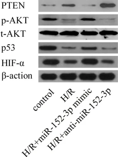
To further validate whether miR-152-3p regu-lates endogenous PTEN, the effects of miR-152-3p on PTEN were then explored in H9c2 cells by Western blot. As shown in Figure 4, the level of endogenous PTEN protein was signifi-cantly repressed in miR-152-3p-ectopic H9c2 cells and markedly enhanced in miR-152-3p-depleted H9c2 cells.
[image:6.612.91.288.73.335.2]The PI3K/Akt signaling pathway has been reported as a main regulator of H/R induced myocardial cell apoptosis [7]. As depicted in Figure 4, compared with the control group, the levels of p53 and phosphorylation of Akt were inhibited by H/R treatment, and this inhibition was ameliorated by over-expression of miR-152-3p. Furthermore, miR-152-3p knockdown reduced the levels of p53 and phosphorylated-Akt in H9c2 cells. These observations indicate that miR-152-3p may protect against H/R-induced myocardial apoptosis via up-regulation of PI3K/Akt signaling.
PTEN restoration partially reverses the effect
of miR-152-3p on H/R-induced cardiomyocyte injury
To assess the role of PTEN in H/R-induced car-diomyocyte injury, expression of PTEN was up-regulated in H9c2 cells after transfection with PTEN mimic. The effects of PTEN restoration on cardiomyocyte injury following the H/R treat-ment were determined. As shown in Figure 5A and 5B, PTEN up-regulation reversed the inhibi-tion of LDH and MBA induced by miR-152-3p mimic.
Our results demonstrate that up-regulation of miR-152-3p markedly inhibited cardiomyocyte apoptosis induced by H/R. However, the effects of miR-152-3p mimic were reversed by co-transfection with PTEN mimic (Figure 5C). These data present that the effects of miR-152-3p on H/R-induced cardiomyocyte injury were achieved by direct targeting of PTEN. Figure 4. Western blot analysis of PTEN,
miR-152-3p protects against myocardial I/R injury in vivo
As the miR-152-3p plays a vital role in sup-pressing cardiomyocyte injury in vitro, the next step was to explore the effects of miR-152-3p on cardiomyocyte injury in vivo. As shown in Figure 6A, the expression of miR-152-3p was significantly decreased in rat hearts after I/R treatment. Elevated levels of miR-152-3p in myocardium were observed after transfection of Ad-miR-152-3p by qRT-PCR. Furthermore, consistent with the results obtained from the in
vitro assays, the activity of serum LDH was notably up-regulated in the I/R group in com-parison with the sham group, whereas eleva-tion in LDH level was significantly down-regulat-ed in the I/R + Ad-miR-152-3p group than the I/R group (Figure 6B). Then, the effect of miR-152-3p on I/R-induced myocardial infarct was
(Figure 6E). Taken together, such outcomes indicate that miR-152-3p can inhibit myocardial I/R injury in vivo effectively.
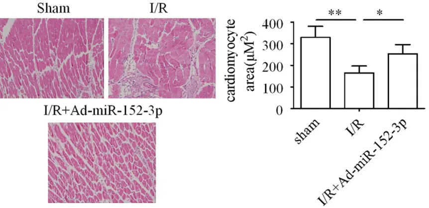
Cardiomyocyte hypertrophy is a pathological response associated with heart failure and sudden death [19, 20]. This study attempted to investigate the effect of miR-152-3p upon car-diomyocyte hypertrophy in rats. The results dis-close that the cell size of cardiomyocytes was significantly decreased in the myocardium by I/R treatment, whereas transfection with Ad-miR-152-3p markedly increased the cell size of cardiomyocytes following I/R treatment (Figure 7).
Discussion
[image:7.612.88.376.71.390.2]Myocardial I/R injury after coronary reperfusion is a complex biologic process which ultimately Figure 5. PTEN restoration partially reversed the protective effect of
miR-152-3p on cardiomyocyte. The inhibition of LDH (A) and MBA (B) induced by
miR-152-3p over-expression was almost completely abrogated by the raised expression of PTEN. (C) The anti-apoptotic effect of miR-152-3p was mark-edly rescued by the up-regulation of PTEN in H9c2 cells. ***P < 0.001, **P < 0.01, *P < 0.05.
examined. Our results show that the myocardial infarct area size is reduced to ~63% by the Ad-miR-152-3p trans-fection (Figure 6C).
Figure 6. miR-152-3p exerts protective effect against myocardial I/R injury in vivo. (A) Expression of miR-152-3p was
determined after myocardial I/R injury. Effects of miR-152-3p on the release of LDH (B) and myocardial infarct size (C) following I/R injury. (D) Representative TUNEL stained images of apoptotic cardiomyocytes. (E) Effects of
miR-152-3p on the levels of apoptosis-related factors after myocardial I/R injury. ***P < 0.001, *P < 0.05.
[image:8.612.93.524.480.689.2]causes further damage to ischemic myocardi-um [1-4]. It has been well established that apoptosis plays a vital role in cardiac I/R injury [8-10]. Hence, investigations of underlying apoptotic mechanisms will help develop new strategies for the treatment of I/R injury. Recently, several miRNAs have been shown to correlate with the regulation of pathological and physiological processes of heart disease owing to their effects on targeted genes associ-ated with apoptosis [15, 16]. Here, the role of miR-152-3p was explored during the cardiac I/R injury. In vitro experiments, RT-PCR showed that the level of miR-152-3p was reduced after H/R treatment as compared with controls. Therefore, miR-152-3p may act as a valuable I/R-related miRNA in cardiomyocyte. However, the potential role of miR-152-3p in the cardio-myocyte apoptosis of I/R injury remains unclear. In the present study, it is demonstrated that up-regulation of miR-152-3p inhibits H/R-induced apoptotic cell death and further study shows that the silencing of miR-152-3p increases the cardiomyocyte apoptosis in response to H/R in
vitro. It is known that the Bax/Bcl-2 ratio is rel-evant to the regulation of myocardial apoptosis during reperfusion [23]. This study reveals that miR-152-3p down-regulated the ratio of Bax/ Bcl-2 by decreasing Bax as well as increasing Bcl-2 expression, indicating that miR-152-3p participates in H/R injury process by regulat- ing the Bax/Bcl-2 ratio. Our prior studies dem-onstrated that free radicals and myocardial enzymes are generated excessively during isch-emic conditions [3-5]. These results confirm that the release of LDH and MDA increased sig-nificantly owing to H/R injury, while overexpres-sion of miR-152-3p could reduce LDH and MDA contents. These findings elucidate that miR-152-3p serves as a protector of myocardium against I/R injury in vitro.
Further experiments were conducted to verify the hypothesis that the protective effect of miR-152-3p on cardiomyocyte are sustained in vivo. In vivo studies have shown that the inhibition of apoptosis by the ectopic expression of miR-152-3p in rats is associated with decreased LDH release, infarct area and the ratio of Bax/ Bcl-2.
A growing body of evidence points that I/R inju-ry induces cardiac hypertrophy, the remarkable reduce in myocyte hypertrophy may be a
con-tributor to the noted improvement in cardiac function [23, 24]. Several studies have demon-strated that miRNAs protect against cardiac hypertrophy [25]. In the present study, upon histological examination, a cell size decrease was observed in the non-infarcted area of the LV following I/R injury, whereas miR-152-3p suppressed myocyte hypertrophy that were induced by the I/R injury. On this ground, miR-152-3p may be useful in alleviating I/R injury. These in vivo findings are coincide with the changes observed in the cultured cardiomyo-cyte. These data also could strongly support the idea that miR-152-3p plays a vital role in the protection of I/R injury both in vitro and in
vivo.
The PI3K/Akt signaling pathway is a classical protective pathway, and it has been involved in the maintenance of cell proliferation, apoptosis and inflammatory responses in most tissues [33, 34]. A study conducted by Chen et al. indi-cated that the PI3K/Akt signaling pathway can act as an endogenous negative feedback regu-lator to exert anti-apoptotic and proliferative effects via the phosphorylation of anti-apoptot-ic and pro-apoptotanti-apoptot-ic substrates, such as Bax, Bcl-2, p70s6k, and caspase9 [35]. The PI3K/ Akt signaling pathway can be critical for the development of myocardial infarction and car-diac dysfunction under the condition of isch-emia [36]. Increased PI3K/Akt activity has been reported to correlate with attenuation of myocardial I/R injury [37]. Moreover, PTEN was demonstrated to be a major negative regulator of PI3K/Akt pathway [38]. Hence, we hypothe-sized that activation of PI3K/Akt signaling path-way could play a role in cardioprotection against I/R injury by miR-152-3p. In this study, the lev-els of phosphorylated PI3K/Akt were down-reg-ulated in cardiomyocyte following I/R injury, while up-regulation of miR-152-3p in cardio-myocyte significantly increased the PI3K/Akt activity both in vivo and in vitro. Thus, these data indicate that cardiomyocyte protection properties of miR-152-3p are likely to be medi-ated by activation of the PI3K/Akt pathway. Our results only open a window and more inves-tigations are needed to further decode the mechanism for the anti-apoptotic property of miR-152-3p.
Acknowledgements
This trial was supported by State Key Project of Research and Development (No. 2016YFC11- 0300) and Guangdong Science and Technology Project China (No. 2015B070701008).
Disclosure of conflict of interest
None.
Abbreviations
miRNAs, microRNAs; I/R, ischemia/reperfu-sion; H/R, hypoxia/reoxygenation; PTEN, Kir-sten rat sarcoma viral oncogene homolog; UTRs, untranslated regions; LDH, lactate dehy-drogenase; LDA, malonic dialdehyde; TUNEL, TdT-mediated dUTP Nick-End Labeling; LAD, left anterior descending; qRT-PCR, quantitative
real-time polymerase chain reaction; PI3K, phosphoinositide 3-kinase; wt, wild-type; mut, mutant.
Address correspondence to: Zhiwei Zhang, Sou-
thern Medical University, NO. 1023, South Shatai Road, Baiyun District, Guangzhou 510515, People’s Republic of China; Affiliated South China Hospital, Guangdong Provincial People’s Hospital, Southern Medical University, Guangdong, People’s Republic of China; Department of Heart Disease Institute,
Guangdong General Hospital, Guangdong Academy
of Medical Sciences, Guangzhou 510080, People’s
Republic of China. Tel: 13575090873; E-mail: m18273080291@163.com
References
[1] Giustino G, Dangas GD. Ischemia-reperfusion injury and ischemic post-conditioning in acute myocardial infarction: lost in translation. Cath-eter Cardiovasc Interv 2017; 90: 1068-1069. [2] Zhou X, Shrestha SS, Luman E, Wang G, Zhang
P. Medical expenditures associated with dia-betes in myocardial infarction and ischemic stroke patients. Am J Prev Med 2017; 53: S190-S196.
[3] Wang Y, Song X, Yue X, Su H, Gu Y, Bowman L,
Ding M, Zou B, Zhao J, Lin X. Protection by ni-trite against the ischemic effects induced by acute myocardial infarction in mice. Anatol J Cardiol 2017; 18: 315-320.
[4] Elbadawi A, Awad O, Raymond R, Badran H, Mostafa AE, Saad M. Impact of remote isch-emic postconditioning during primary percuta-neous coronary intervention on left ventricular remodeling after anterior wall ST-segment ele-vation myocardial infarction: a single-center experience. Int J Angiol 2017; 26: 241-248. [5] Joliat GR, Halkic N, Pantet O, Ben-Hamouda N.
Ischemic stroke and ST-elevation myocardial infarction revealing infective endocarditis. Eur Rev Med Pharmacol Sci 2017; 21: 4640-4641. [6] Zhou FZ, Song W, Yin LH, Song ZF, Yang S, Yang
FB, Liu JF, Song YG, Zhang HY, Zhang ZM. Ef-fects of remote ischemic preconditioning on myocardial injury and endothelial function and prognosis after percutaneous coronary inter-vention in patients with acute coronary syn-drome. Eur Rev Med Pharmacol Sci 2017; 21: 4642-4648.
[7] Mandel’ IA, Podoksenov AY, Sukhodolo IV, Po
-doksenov YK, Svirko YS, Kamenshchikov NO,
Mikheev SL, Sementsov AS, Rogovskaya YV, An
[8] Wei Y, Meng T, Sun C. Protective effect of diltia-zem on myocardial ischemic rats induced by isoproterenol. Mol Med Rep 2018; 17: 495-501.
[9] Alqahtani F, Aljohani S, Tarabishy A, Busu T, Ad -cock A, Alkhouli M. Incidence and outcomes of myocardial infarction in patients admitted with acute ischemic stroke. Stroke 2017; 48: 2931-2938.
[10] Kernan WN, Viscoli CM, Dearborn JL, Kent DM, Conwit R, Fayad P, Furie KL, Gorman M, Guari
-no PD, Inzucchi SE, Stuart A, Young LH;
Insu-lin Resistance Intervention After Stroke (IRIS) Trial Investigators. Targeting pioglitazone hy-drochloride therapy after stroke or transient ischemic attack according to pretreatment risk for stroke or myocardial infarction. JAMA Neu-rol 2017; 74: 1319-1327.
[11] Moraes LN, Fernandez GJ, Vechetti-Júnior IJ, Freire PP, Souza RWA, Villacis RAR, Rogatto SR, Reis PP, Dal-Pai-Silva M, Carvalho RF. Inte-gration of miRNA and mRNA expression
profiles reveals microRNA-regulated networks during muscle wasting in cardiac cachexia. Sci Rep 2017; 7: 6998.
[12] Song YS, Joo HW, Park IH, Shen GY, Lee Y, Shin
JH, Kim H, Kim KS. Bone marrow mesenchy-mal stem cell-derived vascular endothelial growth factor attenuates cardiac apoptosis via regulation of cardiac 23a and miRNA-92a in a rat model of myocardial infarction. PLoS One 2017; 12: e0179972.
[13] Tu Y, Wan L, Bu L, Shen B. Retraction of: abs-tract P71, in vitro and in vivo direct monitoring of miRNA-22 expression in isoproterenol-in-duced cardiac hypertrophy by biolumines-cence imaging. Cardiovasc Res 2017; 113: 248.
[14] Zhu K, Liu D, Lai H, Li J, Wang C. Developing
miRNA therapeutics for cardiac repair in ischemic heart disease. J Thorac Dis 2016; 8: E918-E927.
[15] Bernardo BC, Ooi JY, Matsumoto A, Tham YK, Singla S, Kiriazis H, Patterson NL, Sadoshima
J, Obad S, Lin RC, McMullen JR. Sex differenc-Sex differenc-es in rdifferenc-esponse to miRNA-34a therapy in mouse
models of cardiac disease: identification of
sex-, disease- and treatment-regulated miR-NAs. J Physiol 2016; 594: 5959-5974. [16] Pereira BL, Arruda FC, Reis PP, Felix TF, Santos
PP, Rafacho BP, Gonçalves AF, Claro RT, Aze
-vedo PS, Polegato BF, Okoshi K, Fernandes AA, Paiva SA, Zornoff LA, Minicucci MF. Tomato (Ly-copersicon esculentum) supplementation in-duces changes in cardiac miRNA expression, reduces oxidative stress and left ventricular mass, and improves diastolic function. Nutri-ents 2015; 7: 9640-9.
[17] Song CL, Liu B, Diao HY, Shi YF, Zhang JC, Li YX,
Liu N, Yu YP, Wang G, Wang JP, Li Q. Down-reg
-myocyte apoptosis and protects against myocardial ischemia and reperfusion injury by
targeting IGF-1. Oncotarget 2016; 7: 39740-39757.
[18] Song CL, Liu B, Diao HY, Shi YF, Li YX, Zhang JC,
Lu Y, Wang G, Liu J, Yu YP, Guo ZY, Wang JP,
Zhao Z, Liu JG, Liu YH, Liu ZX, Cai D, Li Q. The protective effect of microRNA-320 on left ven-tricular remodeling after myocardial ischemia-reperfusion injury in the rat model. Int J Mol Sci 2014; 15: 17442-17456.
[19] Shi J, Bei Y, Kong X, Liu X, Lei Z, Xu T, Wang H,
Xuan Q, Chen P, Xu J, Che L, Liu H, Zhong J, Sluijter JP, Li X, Rosenzweig A, Xiao J. miR-17-3p contributes to exercise-induced cardiac growth and protects against myocardial isch-emia-reperfusion injury. Theranostics 2017; 7: 664-676.
[20] Luan W, Li R, Liu L, Ni X, Shi Y, Xia Y, Wang J, Lu
F, Xu B. Long non-coding RNA HOTAIR acts as a competing endogenous RNA to promote malig-nant melanoma progression by sponging miR-152-3p. Oncotarget 2017; 8: 85401-85414. [21] Sun J, Tian X, Zhang J, Huang Y, Lin X, Chen L,
Zhang S. Regulation of human glioma cell apoptosis and invasion by miR-152-3p through
targeting DNMT1 and regulating NF2: miR-152-3p regulate glioma cell apoptosis and invasion. J Exp Clin Cancer Res 2017; 36: 100. [22] Al-Rasheed N, Faddah LM, Hasan IH, Ali HM,
Al-Rasheed N, El Oraby NF, Mohamed MH.
Amelioration of panadol-induced nephrotoxici-ty via down-regulation of Bax/Bcl2 ratio with some antioxidants. Pharmacol Rep 2017; 69: 1088-1093.
[23] Fang L, Ellims AH, Beale AL, Taylor AJ, Murphy A, Dart AM. Systemic inflammation is associ
-ated with myocardial fibrosis, diastolic dysfunc -tion, and cardiac hypertrophy in patients with hypertrophic cardiomyopathy. Am J Transl Res 2017; 9: 5063-5073.
[24] Jin L, Piao ZH, Sun S, Liu B, Kim GR, Seok YM, Lin MQ, Ryu Y, Choi SY, Kee HJ, Jeong MH. Gal-lic acid reduces blood pressure and attenu-ates oxidative stress and cardiac hypertrophy in spontaneously hypertensive rats. Sci Rep 2017; 7: 15607.
[25] Ooi JY, Bernardo BC, McMullen JR. The thera-peutic potential of miRNAs regulated in settings of physiological cardiac hypertrophy.
Future Med Chem 2014; 6: 205-222.
[26] Moretti F, D’Antona P, Finardi E, Barbetta M, Dominioni L, Poli A, Gini E, Noonan DM, Impe -ratori A, Rotolo N, Cattoni M, Campomenosi P. Systematic review and critique of circulating miRNAs as biomarkers of stage I-II non-small cell lung cancer. Oncotarget 2017; 8: 94980-94996.
radiation in greenhouse through high-throu- ghput sequencing. BMC Genomics 2017; 18: 938.
[28] Song DD, Zhang Q, Li JH, Hao RM, Ma Y, Wang
PY, Xie SY. Single nucleotide polymorphisms rs701848 and rs2735343 in PTEN increases cancer risks in an Asian population. Oncotar-get 2017; 8: 96290-96300.
[29] Gupta A, Anjomani-Virmouni S, Koundouros N,
Poulogiannis G. PARK2 loss promotes cancer
progression via redox-mediated inactivation of PTEN. Mol Cell Oncol 2017; 4: e1329692. [30] Chen XΕ, Chen P, Chen SS, Ma T, Shi G, Zhou Y,
Li J, Sheng L. miR106b5p promotes cell cycle progression of malignant melanoma by target-ing PTEN. Oncol Rep 2018; 39: 331-337. [31] Cen M, Hu P, Cai Z, Fang T, Zhang J, Lu M.
TIEG1 deficiency confers enhanced myocardial
protection in the infarcted heart by mediating the Pten/Akt signalling pathway. Int J Mol Med 2017; 39: 569-578.
[32] Wang X, Ha T, Hu Y, Lu C, Liu L, Zhang X, Kao R, Kalbfleisch J, Williams D, Li C. MicroRNA-214 protects against hypoxia/reoxygenation in-duced cell damage and myocardial ischemia/ reperfusion injury via suppression of PTEN and Bim1 expression. Oncotarget 2016; 7: 86926-86936.
[33] Chang H, Li C, Wang Q, Lu L, Zhang Q, Zhang Y,
Zhang N, Wang Y, Wang W. QSKL protects
against myocardial apoptosis on heart failure
via PI3K/Akt-p53 signaling pathway. Sci Rep
2017; 7: 16986.
[34] Zhao X, Xiang Y, Cai C, Zhou A, Zhu N, Zeng C. Schisandrin B protects against myocardial
ischemia/reperfusion injury via the PI3K/Akt
pathway in rats. Mol Med Rep 2018; 17: 556-561.
[35] Chen L, Liu P, Feng X, Ma C. Salidroside sup -pressing LPS-induced myocardial injury by
in-hibiting ROS-mediated PI3K/Akt/mTOR path -way in vitro and in vivo. J Cell Mol Med 2017; 21: 3178-3189.
[36] Zhou Q, Meng G, Teng F, Sun Q, Zhang Y. Ef- Ef-fects of astragalus polysaccharide on apopto-sis of myocardial microvascular endothelial cells in rats undergoing hypoxia/reoxygenation
by mediation of the PI3K/Akt/eNOS signaling
pathway. J Cell Biochem 2018; 119: 806-816. [37] Tang L, Mo Y, Li Y, Zhong Y, He S, Zhang Y, Tang
Y, Fu S, Wang X, Chen A. Urolithin A alleviates
myocardial ischemia/reperfusion injury via
PI3K/Akt pathway. Biochem Biophys Res Com -mun 2017; 486: 774-780.
[38] Wang J, Chen H, Zhou Y, Su Q, Liu T, Wang XT, Li L. Atorvastatin inhibits myocardial apoptosis in a swine model of coronary