Case Report
Root canal therapy of a supernumerary tooth
fused with maxillary second molar: a case report
Linrui Wu1, Bohong Shi2, Ru Zhang3, Jiarong Liu1
1Department of Stomatology, Union Hospital, Tongji Medical College, Huazhong University of Science and
Technology, Wuhan, Hubei, P. R. China; 2Department of Stomatology, People’s Hospital of Rizhao, Rizhao,
Shandong, P. R. China; 3Department of Stomatology, Central Hospital of Rizhao, Rizhao, Shandong, P. R. China
Received December 7, 2017; Accepted October 9, 2018; Epub April 15, 2019; Published April 30, 2019
Abstract: Presented in this report is a referral case of maxillary second molar with developmental anomaly. A small crown of a supernumerary tooth fused to the second molar on its buccal side. The unusual morphology and
compli-cated root canal system rendered diagnosis and treatment difficult. Periapical radiography and cone-beam comput -ed tomography were employ-ed for the evaluation, diagnosis and treatment planning in this case. Micro-ultrasound instrument and operating microscope helped us handle unusual complexities of the root canal system. The radio-logical examinations, in combination with other state-of-art clinical instruments clearly revealed the anatomical anomalies and successfully assisted the treatment.
Keywords: CBCT, fused teeth, root canal therapy
Introduction
A fused tooth represents a developmental anomaly characterized by uncommon morpho-logical features and a complicated root canal system [1]. Pindborg [2] defined fusion as a union between dentin and/or enamel of two or more separate developing teeth. The incidence stands somewhere between 0.1%-1% in per- manent dentition and 0.5%-2.5% in primary dentition [3-5]. Fused teeth are relatively less common in the molar area than in the anterior area [6]. The developmental stage of union dictates whether the pulp chambers and root canals are merged or separated [7, 8]. Dia- gnosis and endodontical treatment of such cases are extremely difficult due to their atypi-cal root anatomy and unpredictable root canal system. We successfully treated a maxillary second molar with developmental anomaly by non-surgicalroot canal therapy and the thera-peutic process is reported as follows.
Case report Clinical information
A 23-year-old female presented with a chief complaint of spontaneous and radiating pain in the right maxillary posterior region, and the pain prevented her from falling asleep during
the night. The patient was referred to the De- partment of Endodontics, Union Hospital, Hua- zhong University of Science and Technology, Wuhan, China, and was diagnosed with irre-versible pulpitis by the first attending dentist on the basis of immediate, excruciating and painful sensation on cold pulp test. Then, the caries was removed, the pulp chamber was opened and the tooth was mediated with antip-ulp by the dentist.
Clinical examinations revealed a temporary restoration in tooth #17 and an abnormal mor-phology of the crown. A supernumerary tooth was detected on the buccal aspect of the maxil-lary right second molar, suggesting probable fusion with an adjacent supernumerary tooth. No discomfort was perceived upon palpation, percussion or bite testing after the treatment by the first dentist. The mobility was within physiologically normal limits. Periodontal prob-ing exhibited no periodontal pocket around the tooth.
Figure 1. Plane of coronal CBCT section of maxillary right second molar. A and B. Showed different sections on coronal plane of maxillary right second molar, and the supernumerary tooth is on the buccal aspect, the periodontal ligament space was intact around teeth and there existed no periapical pathosis.
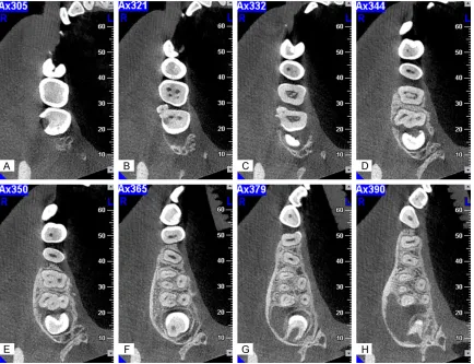
Figure 2. Plane of axial CBCT sections of maxillary right second molar. A-H. Showed the sections from crown to root apex, fused maxillary second molar and concrescent supernumerary tooth can be seen. The main tooth has three canals, including mesiobuccal canal (MB), distobuccal canal (DB), and palatal canal (P). The supernumerary tooth’s canal was separated from mesiobuccal canal and they join together in the apical area.
paramolar and maxillary right second molar.
[image:2.612.91.525.309.642.2]Treatment plan
The treatment plan was to complete the root canal therapy. The patient was informed of imaging examination results and the diagnosis, then treatment was administered with a written informed consert obtained from her.
Therapeutic process
The pulp chamber was exposed upon removal of the temporary restoration (Figure 3). The canal orifices were mesiobuccal (MB), distobuc-cal (DB), palatal (P), mesial (M), and
mesiobuc-sealer (DentsplyDeTrey, Konstanz, Germany). After removal of the excess gutta percha in the chamber, the crown was restored with glass ionomer cement (3M ESPE, USA). The whole therapeutic process was performed under a dental operating microscope (DOM). The postoperative radiography revealed that all canals were properly and densely filled (Figure 4D).
[image:3.612.90.376.73.191.2]During the follow-up period, the patient did not report any signs and symptoms related to the treated region. A porcelain crown was prepared in a subsequent session.
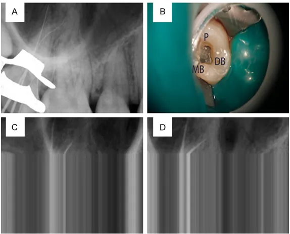
Figure 3. Pre-operation images. (A) (Clinical occlusal) and (B) (buccal) showed the view of maxillary second molar and supernumerary tooth after the removal of the temporary restoration. Caries on buccal and disto-side were all removed.
Figure 4. Operation images. A. Measurement image: K-file (Size#10) was used into root canal, and the working length of MB, ML, D, M were 23, 21, 20.5, 16 mm, B. Image of root canal orifice under a microscope; C. The trail
point film: all points were reaching the apical area. D. The postoperative radiograph after obturation: all canals were properly filled.
[image:3.612.89.376.258.489.2]Discussion
Dental fusion is defined as the merging of two or more teeth at enamel and/or dentinal level [2]. The fusion may be either complete or incomplete, depending upon the developmen-tal stage of the teeth at the time of union [9]. Such developmental anomaly is often confused with gemination. Gemination is a rare morph-anatomic anomaly that develops when the bud of a single tooth attempts to divide, which often causes crowding [10, 11]. Fusion occurs when two teeth buds join, while gemination results from the attempted division of one tooth bud into two [12]. In fact, gemination and fusion are anomalies with similar clinical presentations and unclear aetiology [13]. The fusion could be differentiated from germination by counting the number of teeth in the arch, with the anoma-lous crown being counted in. A full complement of teeth is indicative of gemination, whilst one tooth less than normal is suggestive of fusion [14]. However, this rule does not apply if a nor-mal tooth fuses with a supernumerary tooth. In this case, all teeth erupt normally including the wisdom tooth, and clinically, a small abnor-mal fused crown of a supernumerary tooth makes it can be classified as either dental fusion of the second maxillary molar with a supernumerary tooth or gemination of the sec-ond molar. Considering that the CBCT images revealed independent pulp cavities in the coro-nal area of the paramolar, we tended to use the term “fused tooth”.
Both fusion and gemination are more prevalent in primary dentition than in deciduous denti-tion. There is a subtle difference in the predilec-tion site of the fused teeth among areas in dental arch [15-17]. Shapira & Kuftinec [18] identified the order of decreasing frequency as: upper central incisors, molars (especially upper molars), premolars, followed by lateral incisors and canines. The present case had an involved molar in the right upper area and reports on such scenario have been very scanty.
Those fused teeth are more vulnerable to car-ies and/or periodontal conditions [19, 20]. Due to their unusual shape, it is difficult to make a definite diagnosis and to give right treatment, since the conventional intraoral periapical imaging produces only a 2-dimensional image. As to this case, the first referral dentist advised the patient to take a CBCT when she found the fused tooth. The 3-dimensional images, plus its
high resolution, offered us more valuable infor-mation about the root system, which substan-tially facilitated the treatment [21-24].
Fused teeth were usually treated by extraction in the past. Recently, several novel treatment alternatives has being proposed in the litera-ture, including non-surgical root canal treat-ment (NSRCT) or sectioning of the adjacent teeth to retain the teeth [25, 26]. Reports on fused maxillary molars or germination in molar area are rare. Song et al. [27] reported a case who had the maxillary right molar involved, with a large extra cusp on the buccal aspect. The canal of the supernumerary tooth and disto-buccal root canal of the first molar fused 2 mm below the cement-enamel junction. Root canal treatment was administered only on the super-numerary tooth, given that the maxillary first molar responded normally to stimuli. After 1 year of follow-up, he had no clinical symptoms, and the maxillary right first molar remained vital. Foran et al. [28] reported a concrescence of maxillary second and third molars that necessitated NSRCT, and a 15-month follow-up showed the prognosis was excellent. Saeed Asgary [29] reported a maxillary right second molar fused with a supernumerary tooth, which was diagnosed as chronic apical abscess, and treated with NSRCT. Radiological examination 1 year after the treatment showed healthy peri-odontal condition and complete bone regenera-tion. In this case report, we presented a case of fused right second molar with a paramolar on the buccal side. The CBCT images helped us to have a three-dimensional view of the root sys-tem and the auxiliary equipment made the treatment easier. For instance, rubber dam iso-lated the involved tooth from other parts and saliva, and created a clean operative field. Micro-ultrasound instrument and operating microscope helped us precisely locate the root canal.
Conclusion
Acknowledgements
This work was supported by grants from the Hubei Natural Science Foundation (No 2017- CFB747, the grant holder is Jiarong Liu). Disclosure of conflict of interest
None.
Address correspondence to: Dr. Jiarong Liu, Depart- ment of Stomatology, Union Hospital, Tongji Medi- cal College, Huazhong University of Science and Technology, 1277# Jiefang Avenue, Wuhan, Hubei, P. R. China. Tel: +86-27-85726480; +86-1533726- 3509; E-mail: [email protected]
References
[1] Duncan WK, Helpin ML. Bilateral fusion and gemination: a literature analysis and case re-port. Oral Surg Oral Med Oral Pathol 1987; 64: 82-7.
[2] Pindborg JJ. Pathology of the dental hard tis-sues. Philadelphia: WB Saunders; 1970; 58-64.
[3] Aryanpour S, Bercy P, Van Nieuwenhuysen JP. Endodontic and periodontal treatments of
a geminated mandibular first premolar. Int
Endod J 2002; 35: 209-14.
[4] Liang RZ, Wu JT, Wu YN, Smales RJ, Hu M, Yu JH, Zhang GD. Bilateral maxillary fused second and third molars: a rare occurrence. Int J Oral Sci 2012; 4: 231-4.
[5] Salem Milani A. Endodontic management of a fused mandibular second molar and paramo-lar: a case report. Iran Endod J 2010; 5: 131-4. [6] Rajab LD, Hamdan MA. Supernumerary teeth: review of the literature and a survey of 152 cases. Int J Paediatr Dent 2002; 12: 244-54. [7] Croll TP, Rains NJ, Chen E. Fusion and
gemina-tion in one dental arch: report of case. ASDC J Dent Child 1981; 48: 297-9.
[8] Brook AH, Winter GB. Double teeth. A retro-spective study of ‘geminated’ and ‘fused’ teeth in children. Br Dent J 1970; 129: 123-30.
[9] Ballal S, Sachdeva GS, Kandaswamy D. Endo- dontic management of a fused mandibular second molar and paramolar with the aid of spiral computed tomography: a case report. J Endod 2007; 33: 1247-51.
[10] Gupta S, Singla S, Marwah N, Dutta S, Goel M. Synodontia between permanent maxillary lat-eral incisor and a supernumerary tooth: treat-ment prespective. J Oral Health Comm Dent 2007; 1: 52-5.
[11] Cho KM, Jang JH, Park SH. Clinical manage-ment of a fused upper premolar with
supernu-merary tooth: a case report. Restor Dent Endod 2014; 39: 319-23.
[12] Gadimli C, Sari Z. Interdisciplinary treatment of a fused lower premolar with supernumerary tooth. Eur J Dent 2011; 5: 349-53.
[13] Tsesis I, Steinbock N, Rosenberg E, Kaufman AY. Endodontic treatment of developmental anomalies in posterior teeth: treatment of ge-minated/fused teeth-report of two cases. Int Endod J 2003; 36: 372-9.
[14] Humerfelt D, Hurlen B, Humerfelt S. Hyper- dontia in children below four years of age: a radiographic study. ASDC J Dent Child 1985; 52: 121-4.
[15] al Shalabi RM, Omer OE, Glennon J, Jennings M, Claffey NM. Root canal anatomy of
maxilla-ry first and second permanent molars. Int Endod J 2000; 33: 405-14.
[16] Rwenyonyi CM, Kutesa AM, Muwazi LM, Buwembo W. Root and canal morphology of
maxillary first and second permanent molar
teeth in a Ugandan population. Int Endod J 2007; 40: 679-83.
[17] Neelakantan P, Subbarao C, Ahuja R, Subbarao CV, Gutmann JL. Cone-beam computed tomog-raphy study of root and canal morphology of
maxillary first and second molars in an Indian
population. J Endod 2010; 36: 1622-7. [18] Shapira Y, Kuftinec MM. Multiple
supernumer-ary teeth. Report of two cases. Am J Dent 1989; 2: 28-30.
[19] Nunes E, de Moraes IG, de Novaes PM, de Sousa SM. Bilateral fusion of mandibular sec-ond molars with supernumerary teeth: case report. Braz Dent J 2002; 13: 137-41.
[20] Ghoddusi J, Zarei M, Jafarzadeh H. Endodontic treatment of a supernumerary tooth fused to a mandibular second molar: a case report. J Oral Sci 2006; 48: 39-41.
[21] Peters OA, Laib A, Rüegsegger P, Barbakow F. Three-dimensional analysis of root canal geo-metry by high-resolution computed tomogra-phy. J Dent Res 2000; 79: 1405-9.
[22] Yin X, Cheung GS, Zhang C, Masuda YM, Kimura Y, Matsumoto K. Micro-computed to-mographic comparison of nickel-titanium rota-ry versus traditional instruments in C-shaped root canal system. J Endod 2010; 36: 708-12. [23] Paqué F, Ganahl D, Peters OA. Effects of root canal preparation on apical geometry assess- ed by micro-computed tomography. J Endod 2009; 35: 1056-9.
[24] Fan B, Cheung GS, Fan M, Gutmann JL, Bian Z. C-shaped canal system in mandibular second molars: part I-anatomical features. J Endod 2004; 30: 899-903.
[28] Foran D, Komabayashi T, Lin LM. Concrescence of permanent maxillary second and third mo-lars: case report of non-surgical root canal treatment. J Oral Sci 2012; 54: 133-6.
[29] Asgary S. Endodontic treatment of a maxillary second molar with developmental anomaly: a case report. Iran Endod J 2007; 2: 73-6. [26] de Siqueira VC, Braga TL, Martins MA, Raitz R,
Martins MD. Dental fusion and dens evagina-tus in the permanent dentition: literature re-view and clinical case report with conservative treatment. J Dent Child (Chic) 2004; 71: 69-72.
[27] Song CK, Chang HS, Min KS. Endodontic man-agement of supernumerary tooth fused with