Original Article
Value of joint detection of serum TK-1 and tumor
markers in gastric cancer screening
Shuanping Zhen1, Ziying Gao2
1Department of Clinical Laboratory, Traditional Chinese Medicine Hospital of Baoji, Baoji, Shaanxi Province, China; 2Department of Clinical Laboratory, Baoji Central Hospital, Baoji, Shaanxi Province, China
Received March 30, 2019; Accepted July 9, 2019; Epub August 15, 2019; Published August 30, 2019
Abstract: Objective: To explore the value of joint detection of serum thymidine kinase-1 (TK-1) and tumor markers in gastric cancer screening. Methods: We included 45 patients with gastric cancer as the cancer group and 160 patients with benign gastric cancer as the benign group from May 2017 to May 2018. In addition, 35 healthy vol-unteers were recruited as the control group. Levels of carcino-embryonic antigen (CEA), carbohydrate antigen 72-4 (CA72-4) and carbohydrate antigen 19-9 (CA19-9) were determined by electrochemiluminescence immunoassay,
and TK-1 level was determined by enzyme-linked immuno sorbent assay (ELISA) for confirming the sensitivity and specificity of joint detection of TK-1 and those tumor markers in gastric cancer diagnosis. Then TK-1 expression lev -els of the gastric cancer patients with different pathological characteristics as well as the correlation between TK-1 and the three biomarkers were analyzed. Results: The levels of TK-1, CEA, CA72-4 and CA19-9 in patients of the
cancer group were significantly higher than those in patients of the benign group and the control group (all P<0.05).
The areas under the curve (AUC) of TK-1, CEA, CA72-4 and CA19-9 were all greater than 0.7 at respective and joint determination. TK-1 levels in patients with lymph node metastasis, low degree of differentiation or no
differentia-tion, tumor diameter of ≥4 cm, or large depth of invasion were significantly higher than those in patients without the pathological indices (all P<0.05). The sensitivity and specificity of CEA, CA19-9, CA72-4 and TK-1 detection alone were significantly lower than joint detection of those four indices (P<0.05). TK-1 had obvious correlation with CEA, CA19-9 and CA72-4 (r=0.685, 0.659, 0.685, respectively; all P<0.05). Conclusion: Serum TK-1, CEA, CA72-4 and
CA19-9 can be used as markers for diagnosis of gastric cancer. The joint detection of those four tumor markers can effectively improve the sensitivity and accuracy of gastric cancer, which is conducive to early diagnosis and interven-tion, presenting a relatively high value in clinical application.
Keywords: TK-1, tumor markers, gastric cancer, joint detection
Introduction
Gastric cancer is one of the most common malignant tumors in clinic, with poor prognosis and adverse affects on health of the patients. Early detection and treatment are at present the keys to improving the recovery rate, quality of life and survival time of patients with gastric cancer among the medical community [1-3]. In 2012, approximately 950,000 new gastric can-cer cases and 720,000 deaths were present worldwide, which ranked respectively the 5th in the incidence rate and the 3rd in the mortality rate of malignant tumors, and more than 1/3 of the new cases occurred in China [4]. Patients in the early stage of gastric cancer have no obvi-ous symptoms, and most of them are in the
middle or advanced stages when receiving inspection in a hospital, thus missing the best opportunity for treatment [5, 6]. Gastroscopy combined with pathological diagnosis is the gold standard for diagnosis of gastric cancer, but poor tolerance to gastroscopy, high eco-nomic costs and low acceptability among patients make it unsuitable for basic screening [7, 8].
Metabolites directly produced by tumor cells
and released into body fluids or tissues, or pro
-duced by normal cells when fighting against
Their presence or changes in quantity can
reflect the occurrence and progression of
tumors, with advantages of convenient detec-tion, strong acceptability and low costs, which improves the detection rate of primary tumors, monitor tumor recurrence or metastasis, and evaluate the prognosis effect [10]. However,
tumor markers are not specific antigens of
tumors. Wide range of concentration of a single tumor marker in serum lacks strong sensitivity,
specificity and accuracy, which is easy to cause
misdiagnosis or missed diagnosis, and reduces the clinical application value [11]. Therefore, joint detection of different kinds of malignant tumor markers has high application value for early diagnosis and treatment of gastric cancer [12].
Materials and methods
Patients
Forty-five patients with gastric cancer were
included as the cancer group and one hundred and sixty patients with benign gastric cancer as the benign group from May 2017 to May 2018.
In addition, thirty-five healthy volunteers in the
same period were recruited as the control group. Inclusion criteria: Gastric cancers or
benign gastric tumors were confirmed by patho -logical diagnosis by gastroscopic biopsy or gas-tric samples after surgery, and the judgement for the pathological diagnosis accorded to 2017 consensus for diagnostic pathology in biopsies of chronic gastritis and epithelial neo-plasms released by the Group of Digestive Diseases of Chinese Society of Pathology [13]. All the included patients received neither radio-therapy nor chemoradio-therapy. In addition, gastric cancer in the included patients was pathologi-cally proved to be adenocarcinoma. Exclusion criteria: (1) Patients without complete examina-tion or testing items; (2) Patients with coagu- lation disorder or endocrine disorders; (3) Patients who were in poor basic condition, com-plicated with other serious diseases; (4) Patients who were in pregnancy or lactation, or who were blood donors. The study was approved by the Medical Ethics Committee of Baoji Central Hospital, and informed consents were obtained from all the patients or their families.
Methods
A total of 5 mL fasting venous blood was col-lected from each subject and centrifuged at
3,000 rpm per 10 min for isolating the serum. The levels of carcinoembryonic antigen (CEA; Amyjet, Wuhan, China), carbohydrate antigen 72-4 (CA72-4; Savant, Beijing, China) and car-bohydrate antigen 19-9 (CA19-9; Linc-Bio, Shanghai, China) of all subjects were deter-mined by fully automated chemiluminescent immunoassay analyzer (Roche, Switzerland). The level of thymidine kinase-1 (TK-1; Cusabio, Wuhan, China) was determined by enzyme-linked immuno sorbent assay using a Thermo
Scientific™ Multiskan™ FC microplate reader.
All the procedures were carried out in strict accordance with the instructions of the kits. The pathological data of the patients were obtained from the pathology laboratory of gas-tric cancer of Baoji Central Hospital. The condi-tions including lymph node metastasis, tumor size, degree of differentiation, and depth of invasion of the patients were evaluated by pro-fessional pathologists for the postoperative samples [14].
Outcome measures
Primary indicators: The expression levels of CEA, CA19-9, CA72-4 and TK-1 in the patients of the three groups were recorded. The correla-tions between TK-1 and various pathological indices were analyzed. The detection
sensitivi-ty, specificity and accuracy of each tumor
maker as well as joint detection were calculat-ed. According to the kit instructions, CEA >4.3 ng/mL, CA19-9 >27 U/mL, CA72-4 >6.9 U/mL, TK-1 >2 pmol/L were in the positive range [15]. Single index was positive when its testing value was greater than the critical value, or negative when its testing value was less than or equal to the critical value. While joint detection showed a positive result if at least one index was test-ing positive, otherwise the detection result was negative.
Secondary indicators: The receiver operating characteristic (ROC) curves of TK-1, CEA, CA19-9 and CA72-4 in diagnosis of gastric cancer were statistically analyzed. Relationships be- tween TK-1 and age, CEA, CA19-9, and CA72-4 were respectively analyzed.
Statistical analysis
data conformed to normal distribution were expressed as mean ± standard deviation (_x ± sd). Measurement data between two groups were compared using t tests. Comparison of independent samples of multiple groups was carried out by one-way ANOVA. The post hoc SNK method was used for pair-wise compari-sons. Enumeration data were expressed as the number of cases/percentage (n/%), for
which a χ2 test was performed. The correlation analysis was conducted by the Spearman Rank
Correlation method. For all analyses, P<0.05 was considered statistically significant.
Results
Baseline characteristics
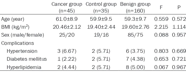
There were no differences in age, sex, body mass index (BMI) and the number of patients with complications among the three groups,
presenting no statistical significances (all
P>0.05). See Table 1.
Expression levels of tumor markers
The levels of CEA, CA19-9, CA72-4 and TK-1 in
the cancer group were significantly higher than
those in the benign group and the control group, and the differences were statistically
significant (all P<0.05). However, there was no
difference between the benign group and the control group (P>0.05). See Figure 1.
Expression level of TK-1 and different patho-logical indices
TK-1 levels in patients with lymph node metas-tasis, low degree of differentiation or no
differ-entiation, tumor diameter of ≥4 cm, or large depth of invasion (T3-T4) were significantly
cancer
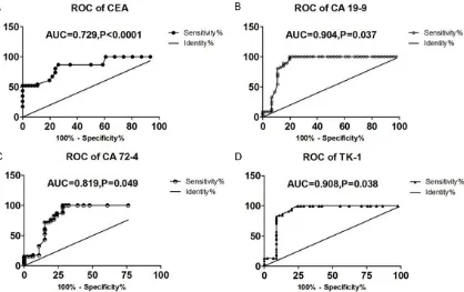
The ROC curves of the four included tumor markers were respectively made to obtain the areas under the curve by taking gastric cancer patients as the disease group and healthy patients as the control group. According to the areas under the curve, CEA had moderate diag-nostic value for gastric cancer, while CA19-9, CA72-4 and TK-1 presented high diagnostic value. See Figure 3.
Sensitivity, specificity and accuracy of TK-1 and tumor markers
The sensitivity and specificity of CEA, CA19-9, CA72-4 and TK-1 detection alone were signifi -cantly lower than joint detection of those four
indices (P<0.05). See Table 2.
Correlations between tumor markers and TK-1
TK-1 had no correlation with the patient’s age (P>0.05). However, TK-1 presented obvious cor-relations with CEA, CA19-9, and CA72-4, and
the differences were statistically significant (all P<0.05). See Figure 4.
Discussion
Lesions of gastric cancer originate from gastric mucosa epithelial tissue and spread through lymphatics and blood vessels under serosa in the early stage. Early stage gastric cancer often
[image:3.612.91.396.89.207.2]has no specific clinical manifestations, which poses difficulties in distinguishing other diseas -es like gastritis, gastric ulcer from gastric can-cer. The detection rate of early gastric cancer is very low in China. Lack of obvious discomfort symptoms in the early stage with late inspec-tion in a hospital, and imperfecinspec-tion of the early Table 1. Baseline characteristics of the three groups (_x± sd) n (%)
Cancer group
(n=45) Control group (n=35) Benign group (n=160) F P
Age (year) 61.0±8.9 59.9±9.5 59.3±9.7 0.559 0.572
BMI (kg/m2) 20.46±2.12 19.40±2.44 19.60±2.76 2.215 1.114
Sex (male/female) 25/20 19/16 85/75 0.088 0.957
Complications
Hypertension 3 (6.67) 2 (5.71) 6 (3.75) 0.803 0.669
Diabetes mellitus 1 (2.22) 2 (5.71) 7 (4.38) 0.653 0.721 Hyperlipidemia 2 (4.44) 2 (5.71) 8 (5.00) 0.067 0.967
Note: BMI, body mass index.
higher than those in patients with lymph no- de metastasis, interme-diate or high degree of differentiation, tumor
diameter of <4 cm, or
shallow depth of inva-sion (T1-T2). The differ-ence was statistically
significant (all P<0.05).
See Figure 2.
Figure 1.Expression levels of the tumor markers in the three groups (_x ± sd). Comparison between the cancer group and the control group, ***P<0.001; comparison between the cancer group and the benign group, ###P<0.001. A: CEA level of the three groups; B: CA19-9 level of the three groups; C: CA72-4 level of three groups; D: TK-1 level of three groups. The higher the level, the higher the risk of cancer; TK-1, thymidine kinase-1; CEA, carcinoembryonic antigen; CA72-4, carbohydrate antigen 72-4; CA19-9, carbohydrate antigen19-9.
Figure 2.Expression level of TK-1 in gastric cancer patients with different pathological indices. A: TK-1 level in gas-tric cancer patients with lymph node metastasis or otherwise, the presence or absence of lymph node metastasis is related to the prognosis of the patients; B: Differentiation of tumors, the lower the degree of differentiation, the
worse the prognosis of the patients; C: Tumor diameter, the larger the tumor diameter, the more difficulty the surgery
[image:4.612.91.516.406.634.2]detection system for high-risk groups mainly account for the low detection rate [16]. At pres-ent, gastroscopy, cytologic examination of the gastric juice, barium meal and CT are common-ly used for gastric cancer detection in clinic, but relatively high economic costs and certain damages to the body lead to low acceptability
among patients, with difficulty in clinical popu -larization [17]. Moreover, sample collection of routine serum tumor markers are convenient with quick detection, but the detection
pres-ents low sensitivity, specificity and accuracy. As a result, confirmed cases with gastric cancer
often appear in locally progressive or advanced
indicators for this study. CEA is a heavily glyco-sylated protein that belongs to the CEA-related cell adhesion molecule (CEACAM) family, which
can reflect invasion and metastasis of a tumor
and thus can be used as an indicator to evalu-ate the patients’ prognosis. Current studies
have confirmed that CEA presents an expres -sion rate of 20%-80% in gastrointestinal tumors
but with poor sensitivity and specificity, and it is
[image:5.612.100.518.75.338.2]no longer used as a biomarker for single detec-tion in cancer screening, and other markers are required for joint detection [19]. CA72-4 is a mucin-like carbohydrate antigen and exists in various malignant tumor tissues such as gas-Figure 3.Diagnostic value of the tumor markers for gastric cancer. A: ROC curve of CEA; B: ROC curve of CA19-9; C: ROC curve of CA72-4; D: ROC curve of TK-1; ROC curve, receiver operating characteristic curve; TK-1, thymidine kinase-1; CEA, carcinoembryonic antigen; CA72-4, carbohydrate antigen 72-4; CA19-9, carbohydrate antigen19-9.
Table 2. Comparison between TK-1 and tumor markers in
sensi-tivity, specificity and accuracy
Tumor marker Sensitivity (%) Specificity (%) Accuracy (%)
CEA 29.41 87.59 72.20
CA19-9 31.75 82.39 66.83
CA72-4 35.29 82.47 70.73
TK-1 34.89 81.48 71.71
CEA+CA19-9+CA72-4+TK-1 46.51 99.10 75.12
Note: TK-1, thymidine kinase-1; CEA, carcinoembryonic antigen; CA72-4, carbohy-drate antigen 72-4; CA19-9, carbohycarbohy-drate antigen19-9.
stage, which significantly
[image:5.612.91.370.429.507.2]tric cancer. However, CA72-4 does not express in non-epithelial malignant tumors. Studies
have proved that the specificity of CA72-4 in
diagnosing gastric cancer is over 95%, and the expression level of CA72-4 has obvious correla-tion with tumor differentiacorrela-tion, clinical stage and degree of invasion. Though CA72-4 serves
at present as one of the efficient indicators for
diagnosis of gastric cancer, its low sensitivity makes it less helpful for meeting the clinical needs [20]. CA19-9 is a mucin-like
glycopro-tein. It is specific to various gastrointestinal
tumors, and it has the highest sensitivity to pancreatic cancer. TK-1, a soluble protein, can catalyze the conversion of thymidine to thymi-dine monophosphate, and it is positively
corre-lated with DNA synthesis and can reflect the
proliferation of tumor cells. TK-1 presents low content in a healthy body, but its activity and content will be elevated with the rapid prolifera-tion of tumor cells when the body is under can-cerization. In recent years, a large number of
studies have confirmed that TK-1 is closely
related to the occurrence of tumors. Nisman et al. found that TK-1 content was closely related to the tumor stage and grades of lung cancer [21]. Moreover, TK-1 can serve as an indepen-dent prognostic factor for leukemia, lymphoma,
lung cancer and kidney cancer, but the signifi -cance of TK-1 in diagnosis of gastric -cancer has not been systematically reported [22].
This study found that the levels of CEA, CA19-9, CA72-4 and TK-1 in the cancer group were
sig-nificantly higher than those in the benign group
and the control group, which is consistent with
the findings of Li et al. [23]. Some scholars
believe that CA19-9, CA72-4 and CEA have low sensitivity and cannot be used as adjuvant diagnostic indicators for gastric cancer alone, and joint detection of CEA, CA19-9 and CA72-4 is a preferred combination for gastric cancer
diagnosis [24]. Felix et al. has confirmed that
serum TK-1 has become a new biomarker for prognosis of pancreatic cancer [25]. Besides,
[image:6.612.94.519.77.377.2]Wang et al. confirmed that TK1 expression in
patients with ovarian serous adenocarcinoma was correlated with MDACC grading, pathologi-cal stage, lymph node metastasis, the recur-rence rates and overall survival rates [26]. Therefore, TK-1 alone for gastric cancer
detec-tion presents relatively low specificity. While
this study proved that TK-1 levels in patients with lymph node metastasis, low degree of dif-ferentiation or no difdif-ferentiation, tumor
diame-ter of ≥4 cm, or large depth of invasion (T3-T4) were significantly higher than those in patients
without the above pathological features, indi-cating that TK-1 can be used as a relatively potent biomarker for prognosis of gastric can-cer. Ning et al. found that joint detection of TK-1 with CEA and CA19-9 that commonly used in
clinic could significantly improve the specificity
of detection of gastrointestinal tumors [27]. Moreover, TK-1 has obvious correlations with CEA, CA19-9 and CA72-4, and their combina-tions can further improve the accuracy of gas-tric cancer detection. Therefore, serum TK-1 has the forewarning function for the diagnosis of precancerous lesions and early-stage tumors, and it can be used as an indicator for tumor screening in physical examination. Plus, TK-1 can obviously improve the sensitivity and
specificity of diagnosis of early-stage tumor
when included in combination detection with other tumor markers, which is worthy of clini-cally further promotion [27].
Although joint detection is of significance in
guiding clinical diagnosis of gastric cancer, its diagnostic sensitivity is still not high, which accounts for its application in preliminary tumor screening in clinical practice. As a result, patients with positive serum markers require conventional imaging and pathological exami-nation, and patients with negative results need to be diagnosed as soon as possible when dis-comfort symptoms or abnormal indicators
appear. Besides, we also expect to find more
ideal biomarkers for gastric cancer detection as well as more economical diagnostic meth-ods with less invasive damages. However, this study has limited sample size, and the contents of tumor markers of the included patients after treatment were not recorded and taken into consideration. In future studies, we will cooper-ate with several departments to explore the relationship between TK-1 and prognosis and survival time of gastric cancer patients, so as to provide more data to support its application value for large-scale clinical promotion.
In conclusion, serum TK-1, CEA, CA72-4 and CA19-9 can be used as adjuvant biomarkers for diagnosis of gastric cancer. The joint detec-tion of those four tumor markers can effectively improve the sensitivity and accuracy of gastric cancer, which is conducive to early diagnosis and intervention, presenting a relatively high value in clinical application.
Disclosure of conflict of interest
None.
Address correspondence to: Ziying Gao, Depart- ment ofClinical Laboratory, Baoji Central Hospital, No. 8 Jiangtan Road, Weibin District, Baoji 721008, Shaanxi Province, China. Tel: +86-0917-3391664; E-mail: gaoziying7u2@163.com
References
[1] Van Cutsem E, Sagaert X, Topal B, Haustermans K and Prenen H. Gastric cancer. Lancet 2016; 388: 2654-2664.
[2] Szász AM, Lánczky A, Nagy Ã, Förster S, Hark K, Green JE, Boussioutas A, Busuttil R,
Szabó A and Győrffy B. Cross-validation of sur -vival associated biomarkers in gastric cancer using transcriptomic data of 1,065 patients. Oncotarget 2016; 7: 49322-49333.
[3] Li K and Li J. Current molecular targeted thera-py in advanced gastric cancer: a comprehen-sive review of therapeutic mechanism, clinical trials, and practical application. Gastroenterol Res Pract 2016; 2016: 4105615.
[4] Yang L, Zheng R, Wang N, Yuan Y, Liu S, Li H, Zhang S, Zeng H and Chen W. Incidence and mortality of stomach cancer in China, 2014. Chin J Cancer Res 2018; 30: 291-298. [5] Ono H, Yao K, Fujishiro M, Oda I, Nimura S,
Yahagi N, Iishi H, Oka M, Ajioka Y, Ichinose M and Matsui T. Guidelines for endoscopic sub-mucosal dissection and endoscopic sub-mucosal resection for early gastric cancer. Dig Endosc 2016; 28: 3-15.
[6] Men F, Wei L, Liu B, Wu F, Liu J, Guo N and Niu Q. Comparison of the safety of the application of painless gastroscopy and ordinary gastros-copy in chronic hypertension patients com-bined with early gastric cancer. Oncol Lett 2018; 15: 3558-3561.
[7] Ji ZH, Peng KW and Li Y. Intraperitoneal free cancer cells in gastric cancer: pathology of peritoneal carcinomatosis and rationale for traperitoneal chemotherapy/hyperthermic in-traperitoneal chemotherapy in gastric cancer. Transl Gastroenterol Hepatol 2016; 1: 69. [8] Cappello F, Logozzi M, Campanella C, Bavisotto
levels in human body fluids: a tumor marker by
themselves? Eur J Pharm Sci 2017; 96: 93-98. [9] Sembiring J, Sarumpaet K, Ganie RA. Diag-
nostic test pepsinogen I and combination with tumor marker CEA in gastric cancer. Iop Conference Series: Earth & Environmental Science 2018.
[10] Zhang Q, Qu H, Sun G, Li Z, Ma S, Shi Z, Zhao E, Zhang H and He Q. Early postoperative tu-mor marker responses provide a robust prog-nostic indicator for N3 stage gastric cancer. Medicine (Baltimore) 2017; 96: e7560. [11] Virgilio E, Proietti A, D’Urso R, Cardelli P,
Giarnieri E, Montagnini M, Giovagnoli MR, Mercantini P, Balducci G and Cavallini M. Measuring intragastric tumor markers in gas-tric cancer patients: a systematic literature
re-view on significance and reliability. Anticancer
Res 2017; 37: 2817-2821.
[12] Huo YR, Huang Y, Liauw W, Zhao J and Morris DL. Prognostic value of carcinoembryonic antigen (CEA), AFP, CA19-9 and CA125 for pa-tients with colorectal cancer with peritoneal carcinomatosis treated by cytoreductive sur-gery and intraperitoneal chemotherapy. Anti- cancer Research 2016; 36: 1041.
[13] The Group of Digestive Diseases of Chinese Society of Pathology. Consnesus for diagnostic pathology in biopsies of chronic gastritis and epithelial neoplasms. Chinese Journal of Pathology 2017; 46: 289.
[14] Research Group of “Molecular Typing and Individualized Diagnosis and Treatment of Gastric Cancer” of National High-tech R&D Program (863 Program). Suggestions on
path-ological classification and diagnostic criteria of
gastric cancer. Chinese Journal of Pathology 2010; 39: 266-269.
[15] Hu ZS and Xu YH. Diagnostic value of joint detection of serum gastrin-17, thymidine ki-nase-1, carcinoembryonic antigen, carbohy-drate antigen 19-9, 12-5, and 72-4 in gastric cancer. Chinese Journal of Gerontology 2015; 8: 2107-2108.
[16] Bibi F, Alvi SA, Sawan SA, Yasir M, Sawan A, Jiman-Fatani AA and Azhar EI. Detection and genotyping of helicobacter pylori among gas-tric ulcer and cancer patients from saudi ara-bia. Pak J Med Sci 2017; 33: 320-324. [17] Wang Y, Li Z, Shan F, Miao R, Xue K, Li Z, Gao
C, Chen N, Gao X, Li S and Ji J. Current status of diagnosis and treatment of early gastric cancer in China--Data from China Gastroin- testinal Cancer Surgery Union. Chinese Journal of Gastrointestinal Surgery 2018; 21: 168-174.
[18] Zou WB, Yang F and Li ZS. How to improve the diagnosis rate of early gastric cancer in China. Journal of Zhejiang University (Medical Sciences) 2015; 44: 9-14.
[19] Feng F, Tian Y, Xu G, Liu Z, Liu S, Zheng G, Guo M, Lian X, Fan D and Zhang H. Diagnostic and prognostic value of CEA, CA19-9, AFP and CA125 for early gastric cancer. BMC Cancer 2017; 17: 737.
[20] Yang AP, Liu J, Lei HY, Zhang QW, Zhao L and Yang GH. CA72-4 combined with CEA, CA125 and CAl9-9 improves the sensitivity for the ear-ly diagnosis of gastric cancer. Clin Chim Acta 2014; 437: 183-186.
[21] Nisman B, Nechushtan H, Biran H, Gantz-Sorotsky H, Peled N, Gronowitz S and Peretz T. Serum thymidine kinase 1 activity in the prog-nosis and monitoring of chemotherapy in lung cancer patients: a brief report. J Thorac Oncol 2014; 9: 1568-1572.
[22] Li Y, Wei R and Song S. Diagnostic and prognostic value of serum thymidine kinase 1 in cancer patients. Indian J Hematol Blood Transfus 2018; 34: 168-170.
[23] Wei HE, Zhang LN and Room E. Assessment of prognosis and trauma extent in endoscopic re-section and traditional open radical rere-section treatment of early gastric cancer. Journal of Hainan Medical University 2016; 22.
[24] Zhao LS, Yun K and Dong XH. The correlation between CA72-4, CEA and CA19-9 expression in patients with gastric carcinoma and patho-logical features. Journal of China Medical University 2014; 43: 259-262.
[25] Felix K, Hinz U, Dobiasch S, Hackert T, Bergmann F, Neumuller M, Gronowitz S, Bergqvist M and Strobel O. Preoperative serum thymidine kinase activity as novel monitoring, prognostic, and predictive biomarker in pan-creatic cancer. Pancreas 2018; 47: 72-79. [26] Wang J, Liu Q, Zhou X, He Y, Guo Q, Shi
Q, Eriksson S, Zhou J, He E and Skog S. Thymidine kinase 1 expression in ovarian se-rous adenocarcinoma is superior to Ki-67: a new prognostic biomarker. Tumour Biology the Journal of the International Society for Oncodevelopmental Biology & Medicine 2017; 39: 1010428317706479.
[27] Ning S, Wei W, Li J, Hou B, Zhong J, Xie Y, Liu H,
Mo X, Chen J and Zhang L. Clinical significance