©IJRASET: All Rights are Reserved
2023
Detection of Sugarcane Disease and Classification
using Image Processing
Arpan Kumar1, Anamika Tiwari 2
1, 2Deptt. Of CSE, Bhabha Institute of Technology, Kanpur
Abstract: Image Processing has been an effective tool in solving a number of real life problems related to medical, science, weather etc. Analysis of the color degradation in diseased leaf or plant is an important step in manually identifying the disease and the type of disease. The work done in this thesis will automate this manual process of identifying the disease and inculcate the methods used by humans to distinguish between diseased and healthy plants. The current work has taken sugarcane plant as test dataset but the method can be generalized for other plant species as well. The soft computing techniques are helpful in developing the knowledge based systems, may be effectively utilized to develop the expert system. This system will be helpful for farmers to find the solutions to their farming problems. This research is carried out to study effectiveness of Image Processing and computer vision techniques for detection of disease in sugarcane plants by observing the leaves. Few major diseases in sugarcane plant like red rot, mosaic and leaf scald have been studied and detection algorithm for the same has been implemented in this research work.
Keywords: Sugarcane, leaf disease detection, computer vision, segmentation, image processing.
I. INTRODUCTION
Computer vision techniques are used for agricultural applications, such as detection of weeds in a field, sorting of fruit on a conveyer belt in fruit processing industry, etc. The underlying approach for all of these techniques is the same. First, digital images are acquired from environment around the sensor using a digital camera.
Then image-processing techniques are applied to extract useful features that are necessary for further analysis of these images. After that, several analytical discriminant techniques, such as statistical, bayesian or neural networks will be used to classify the images according to the specific problem at hand.
This constitutes the overall concept that is the framework for any vision related algorithm. Plants are often affected by various diseases and some of these disease can destroy the whole crop, if not diagnosed and treated in time. This may cause huge loss to the farmer and would also result in less yield of staple crops leading to price rise and burden on the economy[1]. Therefore, continuous and accurate disease detection is in great need and as a prerequisite to provide essential disease information, by which effective plant protection such as optimal decision of fungicide spraying [2, 3], detailed analysis of plant pathology [4], efficacy evaluation of fungicide application [5] can be facilitated. In most practices, the naked eye observation method is generally used to estimate disease that influence both quality and crop yield. Commonly pesticides are used to manage diseases in plant production. Pesticides are sometimes overused in production, may contaminate ground water and pose serious risk to producers, consumers and overall health or well-being of the environment. According to FAO, pesticides are very dangerous to people who are applying the pesticides and to those who are living or playing near the fields where pesticides are used. Pesticide misuse has adverse effects or hazards on the environment and human health and also causes economic loss to the grower.
Traditionally, the disease level is assessed by naked eye visual observation method. This method of disease detection is done by the experts who have the ability to detect subtle changes in leaf color. This method is very laborious, time consuming, expensive, impractical for large fields and different experts can detect the same part as different diseases. Using technological support, automated measurement of disease severity on crops is very important to estimate the extent of the disease and the amount of pesticides needed for use. This method is fast and accurate approach to measure the extent of disease in leaf. Moreover, this method has been the base to develop a new tool to identify the disease well in advance to enhance the cultivation. Digital image processing is used as a tool for early identification of leaf disease.
©IJRASET: All Rights are Reserved
2024
fertilizers and pesticides. Sugarcane crop accounts for almost 75% of the global sugar production and India is the largest consumer and second largest producer of sugar in the world.
But, being long durational crop, sugarcane is prone to the number of disease caused by pathogens viz. fungi, bacteria, viruses and phytoplasmas like organisms. Amongst all the diseases, red rot and smut are causing the major out-breaks in the recent years causing 30-100% yield loss in commercial sugarcane cultivars throughout India [12]. In this paper, an image processing approach has been presented to detect sugarcane plant disease.
II. LITERATURE REVIEW
Sachin Kharade et.al profounded that the identification of the plant diseases is the key to prevent the losses in the yield and quantity of the agricultural product. The studies of the plant diseases mean the studies of visually observable patterns seen on the plant. According to the authors, health monitoring and disease detection on plant is very critical for sustainable agriculture, though it is very difficult to monitor the plant diseases manually.
It requires tremendous amount of work, expertize in the plant diseases, and also require the excessive processing time. Hence, image processing is used for the detection of plant diseases. Disease detection involves the steps like image acquisition, image pre-processing, image segmentation, feature extraction and classification. The author discussed the methods used for the detection of plant diseases using their leaves images. This paper also discussed some segmentation and feature extraction algorithm used in the plant disease detection[1].
Wenjiang Huang et al developed the new spectral indices for identifying the winter wheat disease. They consider three different pests (Powdery mildew, yellow rust and aphids) in winter wheat for their study. The most and the least relevant wavelengths for different diseases were extracted using RELIEF-F algorithm. The classification accuracies of these new indices for healthy and infected leaves with powdery mildew, yellow rust and aphids were 86.5%, 85.2%, 91.6% and 93.5% respectively [2]. Enhanced images have high quality and clarity than the original image according to Thangadurai et.al.[3] Color images have primary colors red, green and blue. It is difficult to implement the applications using RGB because of their range i.e. 0 to 255. Hence they convert the RGB images into the grey images.
Then the histogram equalization which distributes the intensities of the images is applied on the image to enhance the plant disease images. Monica Jhuria et al uses image processing for detection of disease and the fruit grading in [4]. They have used artificial neural network for detection of disease. They have created two separate databases, one for the training of already stored disease images and other for the implementation of the query images. Back propagation is used for the weight adjustment of training databases. They consider three feature vectors, namely, color, textures and morphology [5]. They have found that the morphological feature gives better result than the other two features. Zulkifli Bin Husin et al, in their paper [4], they captured the chilli plant leaf image and processed to determine the health status of the chilli plant. Their technique is ensuring that the chemicals should apply to the diseased chilli plant only.
They used the MATLAB for the feature extraction and image recognition. In this paper pre-processing is done using the Fourier filtering, edge detection and morphological operations. Computer vision extends the image processing paradigm for object classification. Here digital camera is used for the image capturing and LABVIEW software tool to build the GUI. The segmentation of leaf image is important while extracting the feature from that image. Mrunalini R. Badnakhe, et.al. compare the Otsu threshold and the k-means clustering algorithm used for infected leaf analysis in [6]. They have concluded that the extracted values of the features are less for k-means clustering.
©IJRASET: All Rights are Reserved
2025
III.PROPOSED WORK
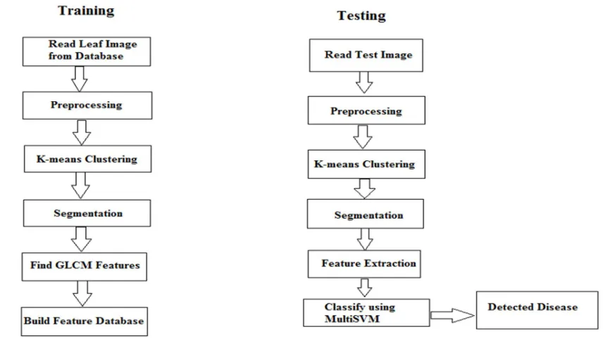
[image:3.595.99.532.242.488.2]This section discusses the implementation details of the proposed work. The proposed methodology can be divided into two phases Training and Testing. During the training phase, various types of leaf images containing diseased leaves are read. Pre-processing is applied to the images to improve the perceptual qualities and improve brightness of the images. Clustering is performed using k-means cluster algorithm. Based on this, the image is segmented into various regions containing diseased part and non-diseased part. Gray level Concurrence matrix(GLCM) algorithm is applied on the images to obtain features to obtain a database of the features. During the testing phase, the test images are input to the system and same process of pre-processing, k-means clustering based segmentation is performed and the GLCM feature for current image are evaluated. The Multi-SVM classifier is then used to classify images into the type of disease.
Figure 3: Flowchart of the Leaf Disease Detection Algorithm
IV.SIMULATION RESULTS
The above algorithm has been developed in MATLAB 2013a and both the training and testing phase are implemented. The input images consist of sugarcane leaves obtained from sugarcane disease dataset.
The idea in this thesis is to come up with a simple and effective method to detect the type of disease a sugarcane plant is affected, by analyzing the colour of the leaf. The major disease affecting the sugarcane plant are mosaic disease, eye spot disease and the red rot disease. Various images of these images were taken to be the training database. In all, there were 2 images with red rot disease, 3 with eye spot disease and 4 images taken for training were affected with the mosaic disease. The below figure 4 shows the dataset images.
[image:3.595.142.455.616.729.2]©IJRASET: All Rights are Reserved
2026
[image:4.595.62.495.112.347.2]Figure 5 shows the leaf dataset for the mosaic disease. It consists of four images.
Figure 5: Mosaic Disease Dataset
[image:4.595.96.500.422.730.2]
Figure 6: Red Rot Disease Datset Figure 6 shows the disease dataset for the red rot disease.
To detect the disease we first the take the image of the leaf and then enhance the image and then make the color transform and detect the color that is not visible by the human eyes, after that our next process is to apply which is k means clustering whose work is to make the cluster of images of same type of color.
The below images shows the various steps of the training part of the algorithm.
©IJRASET: All Rights are Reserved
2027
Figure 8: Selected Leaf Image
[image:5.595.216.380.381.547.2]The leaf image selected in one of the runs as shown in figure 8 is a sugarcane leaf having mosaic disease.
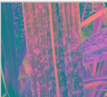
Figure 9: Image after intensity adjustment
[image:5.595.205.389.570.738.2]©IJRASET: All Rights are Reserved
2028
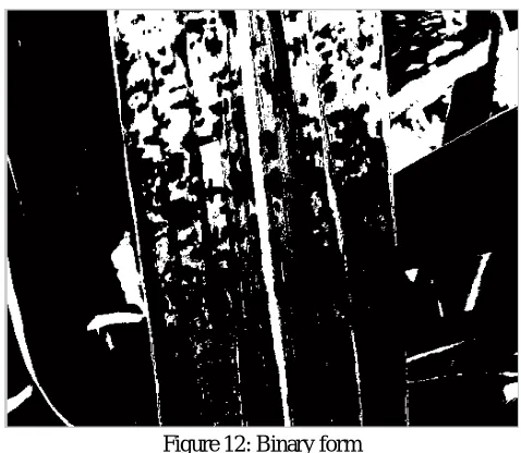
[image:6.595.111.489.112.239.2]Figure 11: Image after applying segmentation
Figure 12: Binary form
Figure 11 shows the binary converted image. The next step is to extract the GLCM features from the images. The GLCM feature matrix of the final trained dataset consists of 13 rows and 9 columns. The 13 rows depict the 13 GLCM features while the number of columns depict the number of training dataset images. Figure 13 below shows the trained feature matrix.
Figure 13: Training Features
[image:6.595.178.417.261.469.2]©IJRASET: All Rights are Reserved
2029
Table I : Training Labels
1 1 1 2 2 2 2 3 3
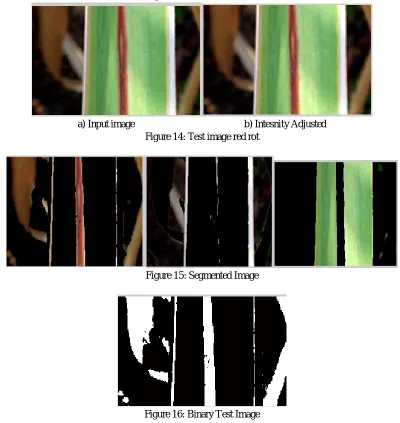
The testing part consists of similar steps to preprocess and extract features from the test image. The steps for the test image with the test image for a red rot disease is as shown in below figures.
a) Input image b) Intesnity Adjusted Figure 14: Test image red rot
[image:7.595.96.496.173.596.2]Figure 15: Segmented Image
Figure 16: Binary Test Image
A. Disease Detected
Red rot
The designed algorithm performs well with almost 97% accuracy.
Table II: Test results
©IJRASET: All Rights are Reserved
2030
V. CONCLUSION
In this paper a computer vision based technique to detect leaf disease in sugarcane plant has been implemented. Image processing is nowadays for a number of applications in agriculture. Combination of feature extractions like color, size and shape with different classifiers has added accuracy to these applications. Red Rot disease, eye spot disease and mosaic disease are three most common diseases occurring in sugarcane plant in India. A through study into the causes and symptoms of these diseases is presented in this research paper. Image processing techniques along with k-means classification has been used to classify the leaf dataset, according to the disease. The leaf image is compared with this database to detect the type of disease existing in the plant.
REFERENCES
[1] Sachin. D Kharade, A.B.Patil, “Plant Disease Detection Using Image Processing”, 2015 International Conference on Computing Communication Control and Automation.
[2] Wenjiang Huang, Qingsong Guan, Juhua Luo, Jingcheng Zhang, Jinling Zhao, Dong Liang, Linsheng Huang, and Dongyan Zhang, “New Optimized Spectral Indices for Identifying and Monitoring Winter Wheat Diseases”, IEEE journal of selected topics in applied earth observation and remote sensing,Vol. 7, No. 6, June 2014
[3] Dr.K.Thangadurai, K.Padmavathi, “Computer Vision image Enhancement For Plant Leaves Disease Detection”, 2014 World Congress on Computing and Communication Technologies.
[4] Monica Jhuria, Ashwani Kumar, and Rushikesh Borse, “Image Processing For Smart Farming: Detection Of Disease And Fruit Grading”, Proceedings of the 2013 IEEE Second International Conference on Image Information Processing (ICIIP-2013).
[5] Zulkifli Bin Husin, Abdul Hallis Bin Abdul Aziz, Ali Yeon Bin Md Shakaff Rohani Binti S Mohamed Farook, “Feasibility Study on Plant Chili Disease Detection Using Image Processing Techniques”, 2012 Third International Conference on Intelligent Systems Modelling and Simulation.
[6] Mrunalini R. Badnakhe, Prashant R. Deshmukh, “Infected Leaf Analysis and Comparison by Otsu Threshold and k-Means Clustering”, International Journal of Advanced Research in Computer Science and Software Engineering, Volume 2, Issue 3, March 2012.
[7] H. Al-Hiary, S. Bani-Ahmad, M. Reyalat, M. Braik and Z.AL Rahamneh, “Fast and Accurate Detection and Classification of Plant Diseases”, International Journal of Computer Applications (0975 – 8887)Volume 17– No.1, March 2011.
[8] Chunxia Zhang, Xiuqing Wang, Xudong Li, “Design of Monitoring and Control Plant Disease System Based on DSP&FPGA”, 2010 Second International Conference on Networks Security, Wireless Communications and Trusted Computing.
[9] Santanu Phadikar and Jaya Sil, “Rice Disease Identification using Pattern Recognition”, Proceedings of 11th International Conference on Computer and Information Technology (ICCIT 2008) 25-27 December, 2008, Khulna, Bangladesh.
[10] Anand.H.Kulkarni, Ashwin Patil R. K.” Applying image processing technique to detect plant diseases”, International Journal of Modern Engineering Research, Vol.2, Issue.5, Sep-Oct. 2012 pp-3661-3664
[11] Sabah Bashir, Navdeep Sharma “Remote Area Plant Disease Detection Using Image Processing”, IOSR Journal of Electronics and Communication Engineering, ISSN: 2278-2834 Volume 2, Issue 6 2012, PP 31-34
[12] Piyush Chaudhary et al. “Color Transform Based Approach for Disease Spot Detection on Plant Leaf”, International Journal of Computer Science and Telecommunications, Volume 3, Issue 6, June 2012
[13] Shitala P., Krishna M. K., Tripathi R. C., Relative sub-image based features for leaf recognition using support vector machine. In Proceedings of the 2011,International Conference on Communication, Computing & Security (ICCCS '11), 2011, 343-346.
[14] Cope J. S., Remagnino P., Barman S., Wilkin P., Plant texture classification using gabor co-occurrences,” in Proceedings of the 6th international conference on Advances in visual computing, 2, 2010, 669– 677.
[15] Amlekar M., Manza R.R., Yannawar P., Gaiwad A.T., Leaf Features Based Plant Classification Using Artificial Neural Network, IBMRD's Journal of Management and Research, Vol 3, No 1 2014,224-232.
[16] https://www.vsisugar.com/india/agriculture_divisions/plantpathology/mosaicdisease-sugarcane.htm
[17] VISHWANATHAN, R., MALATHI, P., AND PADMANABHAN, P. (2003). ‘Variation in Sugarcane red rot pathogen, Colletotrichum falcatum Went.’ SBI.
Coimbatore. Frontiers of Fungal Diversity in India (Eds., G.P. Rao, C. Manoharachari, D.J. Bhat, R.C. Rajak and T.N. Lakhanpal), International Book Distribution Co., Lucknow, pp. 639-667.