Original Article
Vitexin prevents myocardial infarction in rats via
inhibiting oxidative stress and myocardial apoptosis
Feifei Tian1,2, Yongjun Mao3, Xiaofei Sun2
1Qingdao University, Qingdao 266003, China; 2Department of Cardiology, Jining No.1 People’s Hospital, Jining 272000, China; 3Department of Geriatrics, The Affiliated Hospital of Qingdao University, Qingdao 266003, China Received February 18, 2019; Accepted May 10, 2019; Epub July 15, 2019; Published July 30, 2019
Abstract: Background: Myocardial infarction is a disease which seriously threatens people’s health and life. Vitexin is one of the main active components of the plant hawthorn, and it has a wide range of pharmacological effects. This study aimed to investigate the protective effects of vitexin on myocardial infarction in rats. Methods: Sixty SD rats were randomly divided into sham-operated, model, low-, middle- and high-dose vitexin and glyceryl trinitrate groups. The later 4 groups were intraperitoneally injected with 10, 20 and 40 mg/kg vitexin and 10 ml/kg glyceryl trinitrate, respectively. The treatment was performed for 7 days. After 1 h from the last administration, the rats in the model group, low-, middle- and high-dose vitexin groups and glyceryl trinitrate group received the coronary artery ligation. After 24 h, the hemodynamic indexes, serum biochemical indexes and the myocardial B-cell lymphoma-2 (Bcl-2) and Bcl-2 associated X (Bax) protein expressions were determined. Results: Compared with model group, in the high-dose vitexin group the ST segment and T wave elevations were significantly decreased (P < 0.05), the heart rate and left ventricular end diastolic pressure were significantly decreased (P < 0.05), the left ventricular systolic pressure, +dp/dtmax and -dp/dtmax were significantly increased (P < 0.05), the serum aspartate aminotransferase, creatine phosphokinase and lactate dehydrogenase levels were significantly decreased (P < 0.05), the serum superoxide dismutase, catalase and glutathione peroxidase levels were significantly increased (P < 0.05), the serum malondial -dehyde level was significantly decreased (P < 0.05), the myocardial Bcl-2 protein level was significantly increased (P < 0.05), and the Bax protein level was significantly decreased (P < 0.05). Conclusion: Vitexin has protective effects on myocardial infarction in rats. The mechanism may be related to its resistance of oxidative stress, and regulation of Bcl-2 and Bax protein expression in myocardial tissue.
Keywords: Vitexin, myocardial infarction, oxidative stress, Bcl-2, Bax
Introduction
Myocardial infarction is a myocardial ischemic injury and myocardial cell necrosis caused by insufficient supply of coronary blood flow. The formation of myocardial infarction is a complex biochemical, hemodynamic and histopathologi-cal process, accompanied by changes in arte-rial pressure parameters, preload, heart rate, as well as the release of myocardial injury marker enzymes and lipid peroxidation. These changes can cause the increase of reactive oxygen species, such as superoxide anion and hydroxyl radical, which leads to oxidative stress damage, thus damaging the lipid, protein and DNA of myocardial cell membranes [1, 2]. It is found that, the oxidative stress induced by increased reactive oxygen species plays an important role in the process of permanent
plate-This study investigated the protective effects of vitexin on myocardial infarction in rats and explored the related mechanisms. The objec-tive was to provide a basis for further develop-ment of vitexin related drugs for prevention of myocardial infarction.
Materials and methods
Animal grouping and treatment
Sixty Wistar rats (230 ± 30 g; Medical Labo- ratory Shandong Laboratory Animal Center, Jinan, China) were randomly divided into 6 groups: sham-operated group, model group, low-, middle- and high-dose vitexin groups and glyceryl trinitrate group, 10 rats in each group. The rats in the low-, middle- and high-dose vitexin groups were intraperitoneally injected with vitexin (HPLC purity ≥ 98%; Chengdu Man-siter Biotechnology Co., Ltd., Chengdu, China) with dose of 10, 20 and 40 mg/kg, respective-ly. The rats in glyceryl trinitrate group were intraperitoneally injected with glyceryl trinitrate (Shanxi Kangbao Biological Products Co., Ltd., Changzhi, China) with dose of 10 ml/kg. The rats in the sham-operated and model groups were intraperitoneally injected with normal saline. The administration was performed once per day and was continued for 7 days. After 1 h from the last administration, the rats in the model group, low-, middle- and high-dose vitex-in groups and glyceryl trvitex-initrate group received coronary artery ligation. The rats were fixed to the operating table under anesthesia. The tho-racic cavity between the left third fourth ribs was opened, and the heart was exposed. The left anterior descending branch of coronary artery was exposed. Except in the sham-operat-ed group, the coronary artery in the other groups was immediately ligated using 0# suture. Then, the heart was sent back to the thoracic cavity. The fluid and gas were squeezed out, and the thoracic cavity was quickly closed. The operation time of thoracic cavity opening was less than 30 s. After the model was successful, the rats were routinely cared for. In the sham-operated group, the rats only received the threading, without coronary artery ligation. During the whole experiment, no rats died. Test of hemodynamic indexes
After 24 h from coronary artery ligation, the rats were anaesthetized by intraperitoneal
injection of 10% chloral hydrate, with a dose of 30 mg/kg. The electrocardiograph electrode needle was subcutaneously embedded in the limbs of the rats, and the normal lead II electro-cardiogram was recorded. The changes of ST segment and T wave were observed. The right carotid artery was separated. A polyethylene plastic pipe with 1 mm diameter was inserted into the left ventricle through the left carotid artery to perform the cardiac catheterization and was connected with the biological signal acquisition system. The heart rate (HR), left ventricular systolic pressure (LVSP), left ven-tricular end diastolic pressure (LVEDP) and maximum left ventricular systolic/diastolic rate (± dp/dtmax) were measured.
Determination of serum biochemical indexes
After hemodynamic test, the abdominal cavity of the rats was opened. The blood was taken from the abdominal aorta. After centrifuging at 2000 r/min for 15 min, the serum was obtained. The serum aspartate aminotransferase (AST), creatine phosphokinas (CPK), lactate dehydro-genase (LDH), superoxide dismutase (SOD) catalase (CAT), glutathione peroxidase (GSH-Px) and malondialdehyde (MDA) levels were determined. The procedures were in accor-dance to the instructions of kits (Shanghai Sangon Biological Engineering Technology and Service Co., Ltd., Shanghai, China).
Determination of myocardial B-cell lympho-ma-2 and Bcl-2 associated X protein expres-sions
The hearts of the rats were taken. After rinsing with normal saline, about 100 mg heart tissue was taken, and homogenized. The protein was extracted. The expressions levels of B-cell lym-phoma-2 (Bcl-2) and Bcl-2 associated X (Bax) protein were determined using western blot assays. β-actin was used as the internal refer -ence. The relative expression level of the target protein was presented by the ratio of integral optical density of target protein to β-actin. The procedures were in accordance to the instruc-tions of kits (Beijing Zhongshan Golden Bridge Biotechnology Co., Ltd., Beijing, China).
Statistical analysis
data were presented as mean ± SD. The differ-ence between the two groups was analyzed using one-way analysis of variance with SNK-q test. P < 0.05 was considered as statistically significant.
Results
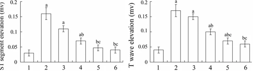
Vitexin decreased the ST segment and T wave elevations in electrocardiogram in myocardial infarction rats
After 24 h from myocardial infarction modeling, the ST segment and T wave in the electrocar-diogram in each group were elevated. Compared with sham-operated group, the ST segment and T wave elevations in the model group were sig-nificantly increased (P < 0.05). Compared with model group, the ST segment and T wave eleva-tions in the middle- and high-dose vitexin groups and glyceryl trinitrate group were signifi -cantly decreased (P < 0.05) (Figure 1).
Vitexin improved the hemodynamic indexes in myocardial infarction rats
As shown in Table 1, compared with sham-operated group, in the model group the HR and LVEDP were significantly increased (P < 0.05),
and the LVSP, +dp/dtmax and -dp/dtmax were sig-nificantly decreased (P < 0.05). Compared with model group, in the high-dose vitexin group and glyceryl trinitrate group the HR and LVEDP were significantly decreased (P < 0.05), and the LVSP, +dp/dtmax and -dp/dtmax were significantly increased (P < 0.05).
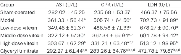
Vitexin decreased the serum AST, CPK and LDH levels in myocardial infarction rats
Table 2 showed that, the serum AST, CPK and LDH levels in the model group were significantly higher than those in sham-operated group (P < 0.05). Compared with the model group, the serum AST, CPK and LDH levels in the high-dose vitexin group and glyceryl trinitrate group were significantly decreased (P < 0.05).
Vitexin increased the serum SOD, CAT and GSH-Px levels and decreased the serum MDA level in myocardial infarction rats
[image:3.612.94.522.74.196.2]The serum SOD, CAT and GSH-Px levels in the model group were significantly lower than those in sham-operated group (P < 0.05), and the serum MDA level in the model group was signifi -cantly higher than that in sham-operated group (P < 0.05). Compared with the model group, the
Figure 1. ST segment and T wave elevations in different groups. 1: Sham-operated; 2: Model; 3: Low-dose vitexin; 4: Middle-dose vitexin; 5: High-dose vitexin; 6; Glyceryl trinitrate. aP < 0.05 compared with sham-operated group; bP <
[image:3.612.90.524.268.350.2]0.05 compared with model group; cP < 0.05 compared with low-dose vitexin group.
Table 1. Hemodynamic indexes in different groups
Group HR (beats/min) LVSP (mmHg) LVEDP (mmHg) +dp/dtmax (mmHg/s) -dp/dtmax (mmHg/s)
Sham-operated 392.45 ± 54.77 143.28 ± 16.22 8.76 ± 2.57 3501.96 ± 502.78 3446.36 ± 567.67 Model 412.72 ± 56.58a 111.47 ± 21.63a 18.34 ± 3.27a 2988.35 ± 345.27a 3117.26 ± 356.83a
Low-dose vitexin 412.36 ± 67.56a 114.27 ± 16.16a 16.17 ± 3.38a 3034.26 ± 482.36a 3134.63 ± 412.84a
Middle-dose vitexin 404.26 ± 73.26b 119.73 ± 23.62a 14.32 ± 2.41a 3205.65 ± 409.49b 3204.36 ± 444.52a
High-dose vitexin 399.63 ± 45.51b 129.26 ± 22.37b 12.45 ± 3.42b 3232.94 ± 501.63b 3302.52 ± 513.67b
Glyceryl trinitrate 398.36 ± 67.37b 133.58 ± 17.82b,c 10.21 ± 2.74b 3345.04 ± 412.22b,c 3349.27 ± 467.33b,c aP < 0.05 compared with sham-operated group; bP < 0.05 compared with model group; cP < 0.05 compared with low-dose vitexin group. HR, hear rate; LVSP, left ventricu
serum SOD, CAT and GSH-Px levels in the high-dose vitexin group and glyceryl trinitrate group were significantly increased (P < 0.05), and the serum MDA levels in high-dose vitexin group and glyceryl trinitrate group were significantly decreased (P < 0.05) (Table 3).
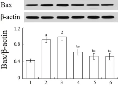
Vitexin increased the myocardial Bcl-2 protein level and decreased the myocardial Bax pro-tein level in myocardial infarction rats
Compared with the sham-operated group, the myocardial Bcl-2 protein expression levels in rats in the model group and low- and middle-dose vitexin groups were significantly decreased (P < 0.05), and the myocardial Bax protein expression levels in the model group and low-dose vitexin group were significantly increased (P < 0.05). Compared with the model group, the myocardial Bcl-2 protein levels in the high-dose vitexin group and glyceryl trinitrate group were significantly increased (P < 0.05), and the myo -cardial Bax protein levels and middle- and high-dose vitexin groups and glyceryl trinitrate group were significantly decreased (P < 0.05) (Figures 2 and 3).
Discussion
The flavonoids extracted from hawthorn fruits and leaves have the effect of lowering blood lipid, lowering blood pressure, increasing coro-nary artery blood flow, protecting myocardial ischemia and antioxidation [10-12]. Vitexin is one of the flavonoids extracted from the haw -thorn fruits and leaves. In this study, rats were pre-treated by intraperitoneal injection with vitexin, and then a myocardial infarction mo- del was established by coronary artery liga- tion. The protective effects of vitexin on myo-cardial infarction were investigated. Results sh-
0.05). This indicates that, the myocardial infarc-tion model has been successfully constructed. Compared with the model group, in the high-dose vitexin group, the ST segment and T wave elevations were significantly decreased (P < 0.05), the HR and LVEDP were significantly decreased (P < 0.05), and the LVSP, +dp/dtmax and -dp/dtmax were significantly increased (P < 0.05). This indicates that, vitexin has obvious protection effects on myocardial infarction model.
[image:4.612.92.398.84.178.2]Studies have shown that, under normal condi-tions, the production and clearance of oxygen free radicals is balanced in the body. When myocardial ischemia occurs, it causes the depletion of high-energy phosphate com-pounds, leading to damage of energy-depen-dent ion pumps. This causes the mitochondrial calcium overload and a large number of toxic oxygen radicals are produced through different signal transduction pathways. Therefore, the integrity of the granular membrane and myo-cardial cell membrane is destroyed, which increases the permeability of the myocardial cell membrane and leads to the release of myo-cardial enzymes [13, 14]. The release of CPK is considered to be one of the most sensitive indi-cators of myocardial injury. The depletion of CPK in the myocardium can reflect the extent of myocardial ischemia, and the amount of CPK in the blood is positively correlated with the degree of myocardial necrosis [15]. AST widely exists in many organs such as heart, liver and skeletal muscle, and its content is high in car-diac myocytes. The increased AST level in the blood is one of the indexes of myocardial injury [16]. It is found that, the activity of serum LHD is increased in the patients with myocardial infarction, and this is the result of the release of LHD from the damaged cardiac myocytes
Table 2. Serum myocardial enzyme levels in different groups
Group AST (U/L) CPK (U/L) LDH (U/L)
Sham-operated 282.02 ± 45.25 235.68 ± 53.37 466.37 ± 75.56 Model 361.33 ± 56.44a 505.74 ± 64.56a 702.73 ± 91.89a
Low-dose vitexin 349.46 ± 61.37a 486.58 ± 71.33a 678.27 ± 90.70a
Middle-dose vitexin 322.12 ± 57.30a 367.34 ± 65.94a,b 604.78 ± 94.42a
High-dose vitexin 303.67 ± 62.29b 331.21 ± 63.48a,b,c 513.12 ± 98.95b
Glyceryl trinitrate 292.27 ± 61.44b,c 283.26 ± 64.76a,b,c,d 471.78 ± 79.87b,c,d
aP < 0.05 compared with sham-operated group; bP < 0.05 compared with model group; cP < 0.05 compared with low-dose vitexin group; dP < 0.05 compared with middle-dose vi
-texin group. AST, aspartate aminotransferase; CPK, creatine phosphokinase; LDH, lactate dehydrogenase.
into the blood [17]. Results of this study showed that, compared with sham-operated group, the serum AST, CPK and LDH levels in the model group were significantly increased (P < 0.05). Compared with the model group, the serum AST, CPK and LDH levels in the high-dose vitex-in group were significantly decreased (P < 0.05). This suggests that, the pretreatment with vitexin can prevent the leakage of intracel-lular AST, CPK and LDH into blood, thus exert-ing myocardial protection function.
[image:5.612.91.524.85.178.2]In the process of myocardial infarction, with the production of a large number of free radi-cals, the activities of endogenous antioxidant enzymes are reduced. This oxidation-anti-oxi-dation imbalance causes the oxidative stress, leading to the damage of myocardial cells [18]. SOD, CAT and GSH-Px are the common antioxi-dant enzymes, which are the first line of defense against reactive oxygen species [19]. MDA is the product of lipid peroxidation. Excessive MDA indicates oxidative damage of cell mem-brane structure. Studies have shown that, there is a significant increase in MDA level during myocardial infarction, suggesting that oxidative damage occurs during the myocardial infarc-tion [20]. In the present study, the serum SOD, CAT and GSH-Px levels in the model group were significantly lower than those in sham-operated group (P < 0.05), and the serum MDA level in model group was significantly higher than that in sham-operated group (P < 0.05). Compared with model group, in high-dose vitexin group the serum SOD, CAT and GSH-Px levels were significantly increased (P < 0.05), and the serum MDA level was significantly decreased (P < 0.05). This indicates that, vitexin can prevent oxidative stress, thus protecting the myocardial injury.
Table 3. Serum oxidative stress index in different groups
Group SOD (U/ml) CAT (U/ml) GSH-Px (U/ml) MDA (mmol/ml)
Sham-operated 244.45 ± 45.68 2.34 ± 0.44 316.65 ± 67.45 5.23 ± 1.46
Model 163.23 ± 36.14a 1.05 ± 0.26a 217.33 ± 45.62a 9.45 ± 2.33a
Low-dose vitexin 182.11 ± 33.58a 1.34 ± 0.35a 223.12 ± 46.83a 8.13 ± 2.82a
Middle-dose vitexin 195.28 ± 31.12a 1.62 ± 0.36a 276.34 ± 53.56a 7.45 ± 3.17a
High-dose vitexin 211.05 ± 45.33a,b 2.11 ± 0.46b,c 298.89 ± 57.12b,c 5.92 ± 3.34b,c
Glyceryl trinitrate 226.46 ± 52.67b,c 2.25 ± 0.51b,c,d 302.56 ± 59.36b,c 6.02 ± 2.73b,c
aP < 0.05 compared with sham-operated group; bP < 0.05 compared with model group; cP < 0.05 compared with low-dose
vitexin group; dP < 0.05 compared with middle-dose vitexin group. SOD, superoxide dismutase; CAT, catalase; GSH-Px, glutathi
[image:5.612.90.290.230.382.2]-one peroxidase; MDA, malondialdehyde.
Figure 2. Myocardial Bcl-2 protein expressions in dif-ferent groups. 1: Sham-operated; 2: Model; 3: Low-dose vitexin; 4: Middle-Low-dose vitexin; 5: High-Low-dose vitexin; 6; Glyceryl trinitrate. aP < 0.05 compared
with sham-operated group; bP < 0.05 compared with
model group; cP < 0.05 compared with low-dose vi
-texin group; dP < 0.05 compared with middle-dose
vitexin group. Bcl-2, B-cell lymphoma-2.
Figure 3. Myocardial Bcl-2 protein expressions in dif-ferent groups. 1: Sham-operated; 2: Model; 3: Low-dose vitexin; 4: Middle-Low-dose vitexin; 5: High-Low-dose vi-texin; 6; Glyceryl trinitrate. aP < 0.05 compared with
sham-operated group; bP < 0.05 compared with mod
-el group; cP < 0.05 compared with low-dose vitexin
[image:5.612.91.288.491.635.2]Apoptosis is regulated by many related genes and proteins, among which Bcl-2 and Bax are the most concerned apoptosis-related genes. Bc1-2 can regulate the permeability of mito-chondrial membrane and inhibit cell apoptosis by preventing cytochrome C from releasing into the cytoplasm [21]. Bax can directly bind to the mitochondrial membrane, change the permea-bility of the membrane, cause cytochrome C to release into the cytoplasm, activate the Caspases family, and eventually cause the apoptosis [22]. Results of this study showed that, compared with the sham-operated gr- oup, the myocardial Bcl-2 protein expression level in the model group was significantly decreased (P < 0.05), and the Bax protein expression level was significantly increased (P < 0.05). Compared with the model group, in the high-dose vitexin group the myocardial Bcl-2 protein level was significantly increased (P < 0.05), and the Bax protein level was signifi -cantly decreased (P < 0.05). It is suggested that, vitexin can up-regulate the expression of Bax protein and down-regulate the expression of Bcl-2 protein in myocardial tissue, thus pre-venting myocardial infarction.
In conclusion, vitexin has protective effects on myocardial infarction in rats. The mechanism may be related to its resistance to oxidative stress, and regulation of Bcl-2 and Bax protein expression in myocardial tissue. This study has provided a theoretical basis for the clinical application of vitexin to prevention of myocar-dial infarction.
Disclosure of conflict of interest
None.
Address correspondence to: Yongjun Mao, Depart-
ment of Geriatrics, The Affiliated Hospital of Qing-dao University, 16 Jiangsu Road, QingQing-dao 266003, China. Tel: +86-532-82911847; E-mail: maoyong- junqd@126.com
References
[1] Malasky BR, Alpert JS. Diagnosis of myocardial injury by biochemical markers: problems and promises. Cardiol Rev 2002; 10: 306-317. [2] Ganesan B, Buddhan S, Anandan R,
Sivaku-mar R, AnbinEzhilan R. Antioxidant defense of betaine against isoprenaline-induced myocar-dial infarction in rats. Mol Biol Rep 2010; 37: 1319-1327.
[3] Zhou T, Chuang CC, Zuo L. Molecular charac-terization of reactive oxygen species in myocar-dial ischemia-reperfusion injury. Biomed Res Int 2015; 2015: 864946.
[4] Flora SJ, Shrivastava R, Mittal M. Chemistry and pharmacological properties of some natu-ral and synthetic antioxidants for heavy metal toxicity. Curr Med Chem 2013; 20: 4540-4574. [5] Chen J, Song SJ, Song N. Simultaneous deter-mination of 2”-o-rhamnosyl vitexin and vitexin in chinese hawthorn leaf and its extract by RP-HPLC. Journal of Chinese Pharmaceutical Sci-ences 2006; 15: 51-54.
[6] Dong L, Fan Y, Shao X, Chen Z. Vitexin protects against myocardial ischemia/reperfusion inju-ry in Langendorff-perfused rat hearts by at-tenuating inflammatory response and apopto -sis. Food Chem Toxicol 2011; 49: 3211-3216. [7] Abbasi E, Nassiri-Asl M, Shafeei M, Sheikhi M.
Neuroprotective effects of vitexin, a flavonoid, on pentylenetetrazole-induced seizure in rats. Chem Biol Drug Des 2012; 80: 274-278. [8] Dong LY, Chen ZW, Guo Y, Cheng XP, Shao X.
Mechanisms of vitexin preconditioning effects on cultured neonatal rat cardiomyocytes with anoxia and reoxygenation. Am J Chin Med 2008; 36: 385-397.
[9] Xue HF, Ying ZM, Zhang WJ, Meng YH, Ying XX, Kang TG. Hepatic, gastric, and intestinal first-pass effects of vitexin in rats. Pharm Biol 2014; 52: 967-971.
[10] Li H, Song F, Xing J, Tsao R, Liu Z, Liu S. Screen-ing and structural characterization of alpha-glucosidase inhibitors from hawthorn leaf fla -vonoids extract by ultrafiltration LC-DAD-MS(n) and SORI-CID FTICR MS. J Am Soc Mass Spec-trom 2009; 20: 1496-1503.
[11] Fan C, Yan J, Qian Y, Wo X, Gao L. Regulation of lipoprotein lipase expression by effect of haw-thorn flavonoids on peroxisome proliferator re -sponse element pathway. J Pharmacol Sci 2006; 100: 51-58.
[12] Ma LY, Liu RH, Xu XD, Yu MQ, Zhang Q, Liu HL. The pharmacokinetics of C-glycosyl flavones of hawthorn leaf flavonoids in rat after single dose oral administration. Phytomedicine 2010; 17: 640-645.
[13] Li X, Arslan F, Ren Y, Adav SS, Poh KK, Sorokin V, Lee CN, de Kleijn D, Lim SK, Sze SK. Meta-bolic adaptation to a disruption in oxygen sup-ply during myocardial ischemia and reperfu-sion is underpinned by temporal and quanti-tative changes in the cardiac proteome. J Pro-teome Res 2012; 11: 2331-2346.
[15] Yıldız K, Ince AT, Sarbay Kemik A, Sert S, Isen HC, Tozlu M, Baysal B, Akyüz U, Kocaman O, Danalıoğlu A. Role of serum myeloperoxidase, CPK, CK-MB, and cTnI tests in early diagnosis of myocardial ischemia during ERCP. Turk J Gastroenterol 2014; 25: 291-7.
[16] Wang Z, Zhang J, Ren T, Dong Z. Targeted me-tabolomic profiling of cardioprotective effect of Ginkgo biloba L. extract on myocardial isch-emia in rats. Phytomedicine 2016; 23: 621-631.
[17] Wang N, Min X, Li D, He P, Zhao L. Geranylgera-nylacetone protects against myocardial isch-emia and reperfusion injury by inhibiting high-mobility group box 1 protein in rats. Mol Med Rep 2012; 5: 521-524.
[18] Bagatini MD, Martins CC, Battisti V, Gasparetto D, da Rosa CS, Spanevello RM, Ahmed M, Schmatz R, Schetinger MR, Morsch VM. Oxida-tive stress versus antioxidant defenses in pa-tients with acute myocardial infarction. Heart Vessels 2011; 26: 55-63.
[19] Aydemir T, Oztürk R, Bozkaya LA, Tarhan L. Ef-fects of antioxidant vitamins A, C, E and trace elements Cu, Se on CuZn SOD, GSH-Px, CAT and LPO levels in chicken erythrocytes. Cell Biochem Funct 2000; 18: 109-115.
[20] Rozario RJ, Akhter N, Bhuiyan NI, Alom MM. Antioxidant effect of valsartan in experimental model of myocardial infarction. Mymensingh Med J 2011; 20: 472-477.
[21] Tian Y, Zhang W, Xia D, Modi P, Liang D, Wei M. Postconditioning inhibits myocardial apoptosis during prolonged reperfusion via a JAK2-STAT3-Bcl-2 pathway. J Biomed Sci 2011; 18: 53.