GALECTIN-3 IMMUNOHISTOCHEMICAL EXPRESSION IN NEOPLASTIC AND NON
1
Ajay Kr Singh, *
,2Chumila Thinley Bhutia
1
1
Department of Pathology, KGMU, Lucknow, UP, India
2Department of Pathology, Sikkim Manipal Institute of Medical
ARTICLE INFO ABSTRACT
Background: of the beta
malignant transformation in various organs including thyroid. Aim: To evaluate immunohistochemical expression of galectin thyroid lesions and correlated with his
Materials and Methods: histopathological
follicular, 02 solid variant, 01 micropapillary), 10 FTC, 14 benign neoplasm (including follicular adenoma and hurthle cell adenoma) and 13 non neoplastic (includes hyperplasi
lesions) cases. To rule out subjective differences during IHC, a 3 used.
Statistical analysis: sciences) version 15.0 statistic Results:
benign neoplasms (p<0.0001).
years and majority of subjects were female
neoplastic, neoplastic lesions had higher scores (p=0.012). Conclusion:
Copyright©2017, Ajay Kr Singh et al. This is an open access article distributed under the Creative Commons Att distribution, and reproduction in any medium, provided the original work is properly cited.
INTRODUCTION
The differential diagnosis of the thyroid nodule includes non neoplastic and neoplastic (benign and malignant) (
Asa, 2008; Aiad et al., 2008; Baloch and Livolsi
et al., 2004).Thyroid cancer is the most common endocrine malignancy and more then 95% of thyroid carcinoma originates from follicular epithelial cells. (Hundahi
Worldwide, incidence rates of thyroid cancer may vary, possibly due to relation with inherent racial, ethnic differences, geographical, environmental differences (including iodine excess or deficiency), radiation exposure (Leonard Wartofsky 2010) and specific mutations. various subtypes is providing novel diagnostic markers as well as targets fo
therapies. (Kondo et al., 2006) In India, the top five common sites of cancer in females are mouth, cervix, uterus, breast thyroid and stomach. (Kalyani and Das, 2010
*Corresponding author: Chumila Thinley Bhutia,
Department of Pathology, Sikkim Manipal Institute of Medical sciences, Gangtok, Sikkim, India.
ISSN: 0975-833X
Article History:
Received 03rd April, 2017
Received in revised form 19th May, 2017
Accepted 28th June, 2017
Published online 26th July, 2017
Citation: Ajay Kr Singh, Chumila Thinley Bhutia, Raj M 3 immunohistochemical expression in neoplastic and non
Key words:
Galectin-3, Thyroid neoplasia, Immunohistochemistry.
RESEARCH ARTICLE
3 IMMUNOHISTOCHEMICAL EXPRESSION IN NEOPLASTIC AND NON
LESIONS OF THYROID
Chumila Thinley Bhutia,
1Raj Mehrotra,
1Madhu Mati Goel,
1Mala Sagar
and
1Madhu Kumar
Department of Pathology, KGMU, Lucknow, UP, India
Department of Pathology, Sikkim Manipal Institute of Medical Sciences, Gangtok, Sikkim, India
ABSTRACT
Background: Thyroid cancer is the most common endocrine malignancy. The galectin
of the beta-galectoside family that play an important role in cellular growth, differentiation and malignant transformation in various organs including thyroid.
To evaluate immunohistochemical expression of galectin-thyroid lesions and correlated with histopathology.
Materials and Methods: In this observational study we evaluated gal
histopathological diagnosed sample including as follows: 30 PTC (included 9 classical variant, 18 follicular, 02 solid variant, 01 micropapillary), 10 FTC, 14 benign neoplasm (including follicular adenoma and hurthle cell adenoma) and 13 non neoplastic (includes hyperplasi
lesions) cases. To rule out subjective differences during IHC, a 3
Statistical analysis: The statistical analysis was done using SPSS (statistical package for social sciences) version 15.0 statistical analysis software.
Results: Galectin-3 expression was significantly higher in malignant thyroid neoplasm as compared to benign neoplasms (p<0.0001). Maximum number of malignant cases were aged between 41 to 50 years and majority of subjects were females. For scoring, it was observed that as compared to non neoplastic, neoplastic lesions had higher scores (p=0.012).
Conclusion: The Gal-3 to be used as adjunct for aiding thyroid cancer diagnosis
access article distributed under the Creative Commons Attribution License, which distribution, and reproduction in any medium, provided the original work is properly cited.
The differential diagnosis of the thyroid nodule includes non-neoplastic and non-neoplastic (benign and malignant) (Fischer and
Livolsi, 2002; Weber Thyroid cancer is the most common endocrine malignancy and more then 95% of thyroid carcinoma originates Hundahi et al., 1998) Worldwide, incidence rates of thyroid cancer may vary, racial, ethnic differences, geographical, environmental differences (including iodine Leonard Wartofsky, ) and specific mutations. various subtypes is providing novel diagnostic markers as well as targets for individualized In India, the top five common e mouth, cervix, uterus, breast,
2010) Survey also
Department of Pathology, Sikkim Manipal Institute of Medical sciences,
detected a belt of thyroid cancer in women in coastal districts of Kerela, Karnataka and Goa. (
pathologists have challenging role i
of thyroid lesions through FNAC/ Core biopsy / lobectomy. (De Lellis and Williams, 2004
follicular-patterned thyroid lesion even with histological analysis the distinction between benign and malignant neoplasm can be difficult. (Fischer
al., 1998) For this reason, the approach to these challenging tumors should include ancillary techniques, immunohistochemistry and molecular profiling.
useful markers for DTC (differe
Galectin-3(Gal-3), cytokeratin 19 (CK
endothelial growth factor, androgen receptor, p16, aurora and HBME-1. (Connie et al., 2010
cancer diagnostic marker panel study reported to date, was found to be the most accurate standard alone marker for DTC diagnosis when compared with a panel of 56 other molecular markers. (Wiseman
classification performance of Gal
International Journal of Current Research
Vol. 9, Issue, 07, pp.54025-54030, July, 2017
, Raj Mehrotra, Madhu Mati Goel, Malti Kumari, Mala Sagarand Madhu Kumar
3 immunohistochemical expression in neoplastic and non-neoplastic lesions of thyroid”, International Journal of Current Research
3 IMMUNOHISTOCHEMICAL EXPRESSION IN NEOPLASTIC AND NON-NEOPLASTIC
Madhu Mati Goel,
1Malti Kumari,
, Gangtok, Sikkim, India
Thyroid cancer is the most common endocrine malignancy. The galectin-3 is a member an important role in cellular growth, differentiation and
-3 in neoplastic and non-neoplastic
In this observational study we evaluated gal-3 expression in spectrum of 67 diagnosed sample including as follows: 30 PTC (included 9 classical variant, 18 follicular, 02 solid variant, 01 micropapillary), 10 FTC, 14 benign neoplasm (including follicular adenoma and hurthle cell adenoma) and 13 non neoplastic (includes hyperplasia and inflammatory lesions) cases. To rule out subjective differences during IHC, a 3- observe evaluation method was
The statistical analysis was done using SPSS (statistical package for social
3 expression was significantly higher in malignant thyroid neoplasm as compared to Maximum number of malignant cases were aged between 41 to 50 For scoring, it was observed that as compared to
non-3 to be used as adjunct for aiding thyroid cancer diagnosis.
ribution License, which permits unrestricted use,
detected a belt of thyroid cancer in women in coastal districts of Kerela, Karnataka and Goa. (Ganpati Mudur, 2005) The pathologists have challenging role in preoperative assessment of thyroid lesions through FNAC/ Core biopsy / lobectomy. , 2004) In many cases specially in patterned thyroid lesion even with histological analysis the distinction between benign and malignant Fischer and Asa, 2008; Hundahi et
) For this reason, the approach to these challenging tumors should include ancillary techniques, immunohistochemistry and molecular profiling. The most differentiated thyroid cancer) were 3), cytokeratin 19 (CK-19), Vascular endothelial growth factor, androgen receptor, p16, aurora-A, ., 2010) In the largest thyroid cancer diagnostic marker panel study reported to date, Gal -3 was found to be the most accurate standard alone marker for DTC diagnosis when compared with a panel of 56 other
et al., 2008) Furthermore, the classification performance of Gal-3 alone (accuracy of 86.9%)
INTERNATIONAL JOURNAL OF CURRENT RESEARCH
and Madhu Kumar, 2017.
was almost as good as the best multimarker panel (accuracy of 91.0%). (Connie et al., 2010)
Aims and Objectives
To evaluate Galectin-3 expression in Thyroid Neoplasia in terms of intensity and proportion of tumour cell stained and correlated with histopathology.
MATERIALS AND METHODS
Histopathologically diagnosed non-neoplastic and neoplastic (benign and malignant) cases of thyroid lesions were taken from archives of department of pathology of King George,s Medical university, Lucknow. These surgical specimen were fixed in 10% formalin, routinely processed, paraffine-embedded and the sections were stained with hematoxylin and eosine stain. Study duration was two year ie one year retrospective and one year prospective. The histopathological evaluation were performed by three pathologists and final diagnosis of the tumour histotype and subtype was made in accordance with world health organization. (Paron et al., 2003) For statistical purpose the histopathological diagnosis was considered as gold standard. The diagnosis of the neoplastic lesion were distributed as follows : 30 PTC (included 9 classical variant, 18 follicular, 02 solid variant, 01 micropapillary), 10 FTC, 14 benign neoplasm (including follicular adenoma and hurthle cell adenoma) and 13 non neoplastic (includes hyperplasia and inflammatory lesions) cases (Table-1).
Study sample: (Table -1)
Thyroid lesions No. of cases
Nonneoplastic
Inflammatory lesions : Hashimoto’s Thyroiditis Lymphocytic thyroiditis Reidel’s Thyroiditis
Hyperplasia : Colloid Nodule /goiter Multinodular Goiter Adenomatous Colloid Goiter
2 2 1
5 2 1
Neoplastic Benign : Follicular adenoma Hurthle cell adenoma
12 2
Malignant : Papillary Thyroid Carcinoma
Classical type
Follicular variant
Solid variant
Micropapillary Carcinoma
Follicular Thyroid Carcinoma
9 18
2 1 10
Total 67
Immunohistochemistry (Dabbs J David, 2010) (Standard
protocols were used) For immunohistochemical evaluation using Streptavidin Biotin immunoperoxidase method was done. Staining and evaluation using specific monoclonal antibody to Gal-3 was done as per following protocol: 1. Deparaffinization 2. Unmasking of antigenic sites by microwave antigen retrieval method 3. Peroxidase Treatment 4. Primary Antibody (NovocastraTM Lyophilized Mouse Monoclonal Antibody Galectin-3, product code : NCL-GAL-3 ; 1:100 dilution)- Slides were wiped off excess buffer and incubated overnight with primary antibody (monoclonal
antibody for Gal -3) 5. Biotinylated link antibody 6. Enzyme Conjugate- Enzyme conjugate (Streptavidin horse radish peroxidase) was applied for 30 minutes. 7. Substrate Chromogen Solution- Concentrated diaminobenzidine solution (DAB) used. 8. Counterstain and Mounting- Presence of brown coloured end product at the site of target antigen was indicative of positive reactivity. Positive tissue control (Carcinoma Prostate) and negative tissue control (Normal thyroid tissue) were taken. Immunohistochemical scoring of Gal-3 : Staining pattern of Gal -3 protein detected in many tissues and cell types, localized in the cytoplasm and / the nucleus, on the cell surface, or in the extracellular environment.
Interpretation of Gal-3 staining and corelation with histopathology features
Interpretation was done according to the criteria defined by Fabio Orlandi et al. in 1998 (Orlandi et al., 1998) and Katie B Weber et al. in 2004 (Weber et al., 2004) in a research paper titled (Gal-3 is a presurgical marker in Human Thyroid Carcinoma and The use of a combination of Gal-3 and Thyroid peroxidase for the diagnosis and prognosis of Thyroid Cancer respectively.
Qualitative criteria
Score Pattern
0 No staining
1 Light / weak staining
2 Moderate staining
3 Intense staining
Quantitative criteria
Score Pattern
+++ >60% of neoplastic cells in cytoplasm / nucleus/ both
(cytoplasm and nucleus) /extracellular matrix
++ 30 - 60% of neoplastic cells in cytoplasm /nucleus/ both
(cytoplasm and nucleus) /extracellular matrix
+ <30% of neoplastic cells in cytoplasm /nucleus/ both
(cytoplasm and nucleus) /extracellular matrix
- No reactivity
In our study, only those cases have been taken who had scoring grade ++ and
+++
Statistical tools employed
The statistical analysis was done using SPSS (Statistical Package for Social Sciences) statistical Analysis Software. The values were represented in Number (%) and Mean±SD. The Statistical formulas were used ie. Chi square test and Interobserver differences (Kappa) used. P <0.05 was considered statistically Significant And =0.20-0.40 is considered as Fair agreement.
RESULTS
Maximum number of patients in non-neoplastic group were aged between 31-40 years whereas maximum number of malignant cases were aged between 41 to 50 years and maximum no. of benign cases were aged between 21-30 years. Overall as well as for all the three groups individually, majority of subjects were females. Majority of cases (n=54; 80.6%) were finally diagnosed as neoplastic. There were a total of 13 (19.4%) cases which were diagnosed as
neoplastic. Among cases diagnosed as neoplastic – a total of 40 (74.1%) were malignant and 14 (25.9%) were benign whereas among those 13 cases diagnosed as non-neoplastic, a total of 8 (61.5%) were hyperplastic and remaining 5 (38.5%) were inflammatory lesions (Table-2).
Table 2. Distribution of Specimen according to the Final Diagnosis (n=67)
S.No. Final Diagnosis No. of cases Percentage
1. Neoplastic 54 80.6
Malignant 40 59.7
Benign 14 20.9
2. Non-neoplastic 13 19.4
Multiple observer evaluation galectin-3 IHC
To rule out subjective differences during IHC evaluation, 3-observer evaluation method was used - Assessment of Level of Agreement between Observer 1 and Observer 2 -The reliability of assessment was done using Kappa-statistic as follows: On scoring of staining the % of agreement (53.7%), order of agreement remained to be fair only (=0.369). On intensity of staining, total % agreement between two observers was observed to be 55.2% which was of fair order (=0.391). Assessment of Level of Agreement between Observer 1 and Observer 3- On scoring of staining the % of agreement (52.2%), order of agreement was found to be fair (=0.353). On intensity of staining, total % agreement between two observers was observed to be 47.8%, fair agreement (=0.288). Assessment of Level of Agreement between observer 2 and Observer 3- On scoring of staining the % of agreement (67.2%), category of agreement was moderate (=0.557). On intensity of staining, total % agreement between two observers was observed to be 53.7%, fair agreement (=0.372). As level of agreement between observers ranged from fair to moderate, the observations of single observer were not reliable. Hence, wherever grading was done either agreement of at least two observers and in case there was no agreement in all the three observers then the median grade of three observers was considered to be applicable for final assessment.
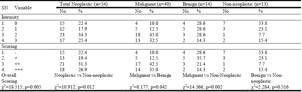
Intensity and Diagnosis (Table-3)
Scoring and Diagnosis
For scoring, it was observed that as compared to non-neoplastic, neoplastic lesions had higher scores (p=0.012).
[image:3.595.302.566.271.431.2]Similarly, more than three-fourth (77.5%) of malignant cases had scores ++ and +++ as compared to only 35.7% of benign and 23.1% of non-neoplastic cases, thus malignant cases showed a statistically significant difference from both benign as well as non-neoplastic lesions (p=0.042 and 0.002). With increasing intensity, a significant association with malignancy was evident. There were 31/40 (77.5%) of malignant cases, 5/14(35.7%) of benign cases and 3/13(23.1%) of non-neoplastic cases accorded with intensity 2 and 3. Gal-3 expression was identified in 76.7% (23/30) of PTC cases , and by histological subtypes of PTC Gal-3 expression was identified in classical, follicular variant, solid variant and micropapillary as 77.7%(7/9), 72.2% (13/18), 100%(2/2) and 100%(1/1) respectively (Figure-1,2) (Table-4). In FTC 80% (8/10) cases showed positivity for Gal-3 (Figure-3).
Table 4. Evaluation of IHC Gal-3 expression in thyroid neoplasm
Diagnosis by Histopathology
Total No.
No. +ve of Gal-3 (%)
“p” value
Sens %
Spec %
A. Malignant Neoplasm 40 31 (77.5%) <0.001 77.5 69.2
1. Papillary thyroid carcinoma
Classical 09 07 (77.7%) 0.201 77.8 43.1
Follicular variant 18 13 (72.2%) 0.159 72.2 44.9
Solid variant 02 02 (100%) 0.224 100.0 39.7
Micropapillary 01 01 (100%) 0.393 100 40.9
2. Follicular thyroid carcinoma
10 08 (80%) 0.130 80 43.9
B. Benign Neoplasm (Follicular adenoma and Hurthle Cell adenoma)
14 05 (35.7%) 0.055 35.7 34.0
C. Non-neoplastic (Hyperplasia and inflammatory lesions)
13 03 (23.1%) 0.004 23.1 31.5
*Only those cases have been taken as positive who had scoring grade ++ or +++.
Overall, Galectin-3 expression evaluated utilizing IHC techniques for thyroid malignancy shows sensitivity (77.5%). In benign and non-neoplastic lesion , Gal-3 expression was observed as 35.7% (5/14) and 23.1% (3/13) respectively (Figure-4).
DISCUSSION
Galectin-3 is a member of the beta-galactoside binding protein family which is normally expressed in macrophages, mast Table 3. Association of IHC Findings with Final Histopathological Diagnosis
SN Variable Total Neoplastic (n=54) Malignant (n=40) Benign (n=14) Non-neoplastic (n=13)
No. % No. % No. % No. %
Intensity
1. 0 15 22.4 4 10.0 4 28.6 7 53.8
2. 1 12 17.9 5 12.5 5 28.6 3 23.1
3. 2 23 34.3 18 45.0 3 28.6 1 7.7
4. 3 17 25.4 13 32.5 2 14.3 2 15.4
Scoring
1. - 15 22.4 4 10.0 4 28.6 7 53.8
2. + 13 19.4 5 12.5 5 35.7 3 23.1
3. ++ 21 31.3 17 42.5 3 21.4 1 7.7
4. +++ 18 26.9 14 35.0 2 14.3 2 15.4
Overall Scoring
Neoplastic vs Non-neoplastic Malignant vs Benign Malignant vs Non-Neoplastic Benign vs
Non-neoplastic
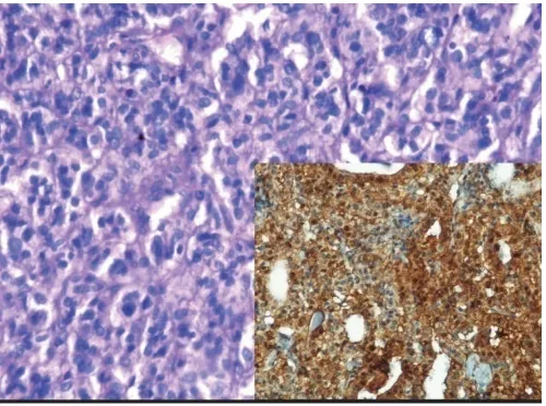
[image:3.595.59.545.582.730.2]Figure 1. (H&E 20X)- Photomicrographs showing classical papillary thyroid carcinoma, Inset- (IHC 20X)- showing gal-3 expression in classical papillary thyroid carcinoma. Score- ++, Intensity-2, Pattern- Cytoplasm
Figure 2. (H&E 40X)- Photomicrograph showing papillary thyroid carcinoma (solid variant), Inset- (IHC 40X)- showing gal-3 expression in papillary thyroid carcinoma (solid variant). Score- +++, Intensity-3, Pattern- Cytoplasm
Figure 3. (H&E 20X)- Photomicrograph showing Follicular thyroid carcinoma, Inset- (IHC 20X)- showing gal-3 expression follicular thyroid carcinoma. Score- +++, Intensity-3, Pattern- Cytoplasm
Figure 4. (H&E 20X)- Photomicrograph showing lymphocytic thyroiditis, Inset - (IHC 20X)- showing gal-3 expression in extracellular matrix in lymphocytic thyroiditis
cells, langerhans cells and various malignant cells including thyroid gland cells (Fernandez et al., 1997). Galectin-3 possesses affinity for beta galactoside containing glycoconjugates which is found mainly in intracellular, extracellular and cell-surface and they participates in cellular functions like cell proliferation, apoptosis, cellular transformation, tumour progression and metastasis of cancer cells (Cvejic et al., 2005; Nangia –Makkar et al., 2007). In our study to analyze this protein by immunohistochemistry (IHC) in67 histopathological samples, and for IHC interpretation utilized a multi-pathologist consensus scoring strategy. In which we have reported the 3 independent observer evalutaion of Gal-3 and which is more reliable determination and assess the interobserver variability. As level of agreement between 3 observers ranged from slight to moderate. Hence for each parameter (intensity and scoring), criteria of agreement of at least two observers was done. For scoring, it was observed that as compared to non-neoplastic, neoplastic lesions had higher scores (p=0.012). Similarly, more than three-fourth (77.5%) of malignant cases had scores ++ and +++ as compared to only 35.7% of benign and 23.1% of non-neoplastic cases. Thus with increasing intensity, a statistically significant association with malignancy was evident. The majority of studies reported Gal-3 positivity in 90% to 100% of PTC cases. [Connie et al., 2010] Very few studies have reported Gal-3 expression in PTC by histological subtypes. Similarly, in our study, Gal-3 expression was identified in 77% (23/30) of PTC cases , and by histological subtypes of PTC Gal-3 expression was identified in classical, follicular variant, solid variant and micropapillary as 77.7%(7/9), 72.2% (13/18), 100%(2/2) and 100%(1/1) respectively. Expression of Gal-3 has also ranged from 20% to 100% in reported cases of FTC. The largest series, reported by Bartolazzi et al, identified Gal-3 expression in 95% (54/57) of FTC cases. (Bartolazzi et al., 2008) Similarly, in our study, 80% (8/10) cases of FTC showed positivity for Gal-3. Hence, Gal-3 is highly expressed in thyroid malignancies. There is no significant difference found in Gal-3 expression in PTC histological subtypes and FTC. Although it appears likely that a small proportion of follicular carcinomas are negative for Gal-3, likewise in our study 20% (2/10) of FTC cases were negative for Gal-3, it remains elusive whether the negativity is true or false. Orlandi et al. failed to detect Gal-3 positivity in some FTCs, where they succeeded in showing focal positivity in the same cases when analyzed with differently prepared sections. (Bartolazzi et al., 2008) Immunohistochemical
[image:4.595.306.553.51.233.2] [image:4.595.37.287.54.243.2] [image:4.595.37.284.313.498.2] [image:4.595.37.287.558.744.2]analysis of FTCs with focal Gal-3 expression in this kind of tumor, we could also possibly hypothesize that a false negative result yielded when the areas negative for Gal-3 are sampled. Also (a well-known fact in the literature is that poorly-differentiated FTC subtypes show a weaker Gal-3 expression or have absolutely no expression compared with the well differentiated carcinomas). (Hidenori et al., 1999) According to Kawachi et al. (2000) Primary lesions of papillary carcinomas with metastasis contained significantly higher concentrations of Gal-3 than tumours of this type without metastases. In our single case of known metastatic PTC we found strong Gal-3 positivity.
Follicular thyroid lesions have been rightly referred to as ‘ the bane of the pathologist’ in a recent article by Baloch and LiVolsi (2002). The follicular variant of papillary thyroid carcinoma (PTCFV) often poses a diagnostic challenge in which the differential diagnosis includes other follicular patterned lesions such as follicular adenoma (FA) and follicular carcinoma. The distinction between these lesions is important because prognosis and management differ. Because FVPTC is diagnosed almost solely based on subjective cytologic criteria (nuclear grooves, nuclear clearing, nuclear overlap, and intranuclear pseudoinclusions) and no uniform minimal diagnostic criteria exist (Renshaw and Gould, 2002; Chan, 2002). It is not surprising that disagreements in the diagnosis of PTC have underscored the interobserver variability in the evaluation of these lesions. In our study, Gal-3 was expressed in 72.2%(13/18) of FVPTC, 80%(8/10) of FTC and 35.7%(5/14) of benign neoplasm (follicular and hurthle cell adenomas). In an attempt to resolve this common diagnostic difficulties, many immunohistochemical markers have been evaluated for their potential in distinguishing PTC from other follicular lesions, the main ones including Gal-3, cytokeratin (CK-19) and HBME-1. Gal-3 has recently generated much interest and has repeatedly been suggested as a suitable marker to fulfill this role (Boila et al., 2014; Cui et al., 2012). In several studies, Gal-3 expression was not identified in any thyroid goiter cases, with the exception of the reports from Beesely et al. (2002) and Prasad et al. (2005) found that Gal-3 positivity in 38% and 55% of goiter specimens respectively. Similarly in our study, Gal-3 was expressed in 23.1% (3/13) of non-neoplastic lesions. The most likely explanation for the Gal-3 expression of non-neoplastic follicular cells in an inflamed area are neosynthesis effected by cytokines secreted by inflammatory cells or the simple permeation of Gal-3 abundantly shed by lymphocytes into the neighboring follicular cells (LIoyd, 2001)
Conclusion
Use of strict criteria and guidelines for histological grading can result in acceptable levels of interobserver agreement and also identify areas that might benefit from refinement. Gal-3 is highly expressed in thyroid malignancies (specially in supporting the diagnosis of PTC and its variants.Immunopanel evaluation with Gal-3, HBME-1 and CK-19 have the most commonly studied adjunctive markers and may further improve sensitivity for thyroid malignancies . No single marker by itself is 100% sensitive for malignancy. Gal-3 to be used as adjunct for aiding thyroid cancer diagnosis.
Disclosure
The authors report no conflict of interest in this work.
REFERENCES
Aiad HA, Kandil MA, Assad NY, El-Kased AM. et al. 2008. Galectin-3 immunostaining in cytological and histopathological diagnosis of thyroid lesions. Journal of Egyptian nat. Cancer Inst., 20:30-46
Baloch ZW, Livolsi VA. 2002. Follicular–Patterned lesion of thyroid. The bane of the pathologist. Am J Clin Pathol., 117:143-50
Bartolazzi A, Oralandi F, Saggiorato E, Volante M, Arecco F.
et al. 2008. Galectin-3 expression analysis in the surgical selection of follicular thyroid nodules with indeterminate fine needle aspiration cytology: a prospective multicentre study. Lancet Oncol., 9:543-549.
Boila AN, Catana R, Loghin A et al. 2014. Diagnostic valu of HBME-1 CD-56, Galectin-3 and Cytokeratin-19 in papillary thyroid carcinomas and thyroid tumours of uncertain malignant potential. Rom J Morphol Embryol., 55(1):49-56.
Bsseley MF, KM Mc Laren. 2002. Cytokeratin-19 and galectin-3 immunohistochemistry in the differential diagnosis of solitary thyroid nodules. Histopathology, 41:236-243.
Chan JK. 2002. Strict criteria should be applied in the diagnosis of encapsulated follicular variant of papillary thyroid carcinoma. Am J Clin Pathol., 117:16-18.
Connie GC, Scott S. Strugnell, Obil. G, Steven JM et al. 2010. Diagnostic utility of gal-3 in thyroid cancer: The American Journal of Pathology, Vol. 176, No. 5.
Cui W, Sang W, zheng S, Ma Y. et al. 2012. Usefullness of cytokeratin-19, galectin-3 and Hector Battifora mesotheial-1 in the diagnosis of benign and malignant thyroid nodules.
Clin Lab., 58(7-8):673-80
Cvejic D, Savin S, Patrovic I, Selemetjev S. et al. 2005. Galectin-3 and proliferating cell nuclear antigen (pcna) expression in papillary thyroid carcinoma. Exp Oncol., 27(3):210-14.
Dabbs J David. 2010. Diagnostic immunohistochemistry: chapter 1:edition 2; Elsevier. 1-37.
De Lellis RA, Williams ED, 2004. Pathology of the thyroid and parathyroid in: DeLellis RA, Lloyd RV,Eng C(eds), Pathology and genetics of tumours of endocrine organs, world health organization classification of tumours, IARC press, Lyon, 57-66
Fernandez PL, Merino MJ, Gomez M, Campo E. 1997. Galectin-3 and laminin expression in neoplastic and non-neoplastic thyroid tissue. J Pathol., 181(1):80-86.
Fischer S, Asa SL. 2008. Application of immune histochemistry to thyroid neoplasms. Arch Pathol Lab Med., 132:359-72.
Ganpati Mudur, 2005. New Delhi, India has some of the highest cancer rates in world; BMJ, Vol: 330.
Hidenori I, Yoichiro H, Tadashi Y, Shiro A. 1999. Expression of galectin-3 in fine needle aspiration as a diagnostic marker differentiating benign from malignant thyroid neoplasms. Cancer, 85:2475-84.
Hundahi SA, Fleming ID, Flemgen AM. et al. 1998. A national cancer data base report on 53,856 cases of thyroid carcinoma treated in the US., 1985-1995. Cancer, 83:2638-48.
Kalyani R, Das S, ML Harendra Kumar. 2010. Pattern of Cancer in adolescent and young adults : 10 yrs study in India; Asian Pacific Journal of Cancer Prevention, vol 11. Kawachi K, Matsushita Y, Yonezawa S, Nakano S. 2000.
possible role in metastasis formation. Hum Pathol., 31:428-433.
Kondo T, Ezzats, Asa SL. 2006. Pathogenetic mechanisms in thyroid follicular cell neoplasia. Nat Rev Cancer, 6: 292-306
Leonard Wartofsky, 2010. Increasing world incidence of thyroid cancer: Increased detection Or higher radiation exposure ? Hormones, 9(2):103-108.
LIoyd RV. 2001. Distinguishing benign from malignant thyroid lesions; Gal-3 as the latest candidate. Endocrine Pathol., 3:255-257.
Nangia –Makkar P, Nakhara S, Hogan V, Raj A. 2007. Galectin -3 in apoptosis, a novel therapeutic target. J Bioenerg Biomembr., 39(1):79-84.
Orlandi F, Saggiorato E, Pivano G, Puligheddu B, Termine A, Cappia S, et al. 1998. Galectin-3 is a presurgical marker of human thyroid carcinoma. Cancer Res., 58:3015–20 Paron I, Scaloni A, Pines A, Bachi A. et al. 2003. Nuclear
localization of galectin-3 in transformed thyroid cells: a role in transcriptional regulation. Biochem Biophys Res Commun., 302:545-553.
Prasad ML, Pellegata NS, Huang Y, Nagaraja HN, et al. 2005. Galectin-3, fibronectin-1, CITED-1, HBME1 and cytokeratin-19 immunohistochemistry is useful for the differential diagnosis of thyroid tumours. Mod Pathol., 18:48–57.
Renshaw AA. and Gould EW. 2002. Why there is the tendency to “overdiagnose the follicular variant of papillary thyroid carcinoma. Am J Clin Pathol., 117:19-21.
Weber KB, Shroyer KR, Heinz DE et al. 2004. The use of a combination of galectin-3 and thyroid peroxidise for the diagnosis and prognosis of thyroid cancer. Am J Clin Pathol., 122:524-31.
Wiseman SM, Adrienne Melck, Hamid Masoudi et al. 2008. Molecular phenotyping of thyroid tumors identifies a marker panel for differentiated thyroid cancer diagnosis.
Ann Surg Oncol., 15:2811- 2826.