ISSN Online: 2164-005X ISSN Print: 2163-9914
DOI: 10.4236/ojra.2018.83010 Aug. 29, 2018 93 Open Journal of Rheumatology and Autoimmune Diseases
Impact of an Exercise Training Program in a
Patient with Relapsing Polychondritis:
A Case Report
Alexandre Moura dos Santos, Diego Sales de Oliveira, Rafael Giovani Misse,
Letícia Alves Perin, Jean Marcos de Souza, Fernanda Rodrigues Lima,
Rosa Maria Rodrigues Pereira, Samuel Katsuyuki Shinjo
*Division of Rheumatology, Hospital das Clinicas HCFMUSP, Faculdade de Medicina, Universidade de Sao Paulo, Sao Paulo, Brazil
Abstract
Background: Exercise training has demonstrating to be safe and promote benefits for several rheumatologic autoimmune diseases. However, no study has evaluated the safety and benefits of exercise in relapsing polychondritis. Aim: To evaluate the effectiveness of an exercise training program in a pa-tient with relapsing polychondritis. Case presentation: A 67-year-old female patient with relapsing polychondritis in remission was submitted to a 12-week, twice weekly, aerobic and resistance training program. Aerobic ca-pacity, muscle strength and function caca-pacity, as well as body composition, were evaluated at baseline and after 12-weeks. Conclusions: Exercise training program demonstrated to be effective for increasing aerobic capacity, muscle strength and function, and for improving body composition in the patient. Further studies are necessary to confirm these findings.
Keywords
Exercise Training, Relapsing Polychondritis, Aerobic Training, Resistance Training
1. Introduction
Relapsing polychondritis (RP) is a rare autoimmune systemic disease characte-rized by inflammation of proteoglycan-rich structures and cartilaginous tissues [1] [2]. Patients with RP have recurrent inflammation of auricular pinna, nasal cartilage, peripheral joints and vertebral disks [2] [3]. Moreover, these patients How to cite this paper: dos Santos, A.M.,
de Oliveira, D.S., Misse, R.G., Perin, L.A., de Souza, J.M., Lima, F.R., Pereira, R.M.R. and Shinjo, S.K. (2018) Impact of an Exer-cise Training Program in a Patient with Relapsing Polychondritis: A Case Report. Open Journal of Rheumatology and Au-toimmune Diseases, 8, 93-98.
https://doi.org/10.4236/ojra.2018.83010
Received: August 3, 2018 Accepted: August 26, 2018 Published: August 29, 2018
Copyright © 2018 by authors and Scientific Research Publishing Inc. This work is licensed under the Creative Commons Attribution International License (CC BY 4.0).
DOI: 10.4236/ojra.2018.83010 94 Open Journal of Rheumatology and Autoimmune Diseases may exhibit clinical manifestations, such as auricular chondritis, non-erosive polyarthritis, nasal cartilage chondritis, inflammation of ocular structures, res-piratory tract chondritis, as well as vestibular and cochlear damage [2] [3] [4].
Structural deformities can lead to life-threatening cardiopulmonary manife-stations, such as airway collapse and valvarcardiac regurgitation [4] [5]. Gener-ally, the primary cause of death is respiratory failure due to RP, followed by lung infections and cardiovascular events [1] [2] [3] [4] [5].
Exercise training has proven to be an important tool for mitigating clinically related symptoms in autoimmune rheumatic diseases, such as systemic lupus erythematosus, rheumatoid arthritis and systemic autoimmune myopathies [6] [7]. In addition, exercise training has been shown to promote an increase in aerobic capacity, muscle strength and function, and to improve quality of life in autoimmune rheumatic diseases [6] [7] [8] [9]. Importantly, these benefits of exercise training have been observed without exacerbating the inflammatory mi-lieu [9] [10], proving that this non-pharmacological strategy is effective and safe for the management of autoimmune rheumatic diseases. However, to the best of our knowledge, there are no studies evaluating the effectiveness and safety of ex-ercise training programs for patients with RP.
2. Case Study
A 67-year-old female with RP, according to criteria of McAdam et al. [2] was as-sessed in the present case study. She presented the first symptoms 10 years prior, with polyarthralgia, bilateral diffuse anterior scleritis, hoarseness and acute chondritis of the right auricle. There was no cardiac or interstitial lung involve-ment. At the time of the initial manifestations, she was an active attorney, non-smoker, with controlled diabetes mellitus and systemic arterial hyperten-sion. She was promptly treated with the combination of methotrexate (15 mg/week) and prednisone (0.5 mg/kg/day), with complete resolution of symp-toms. One year later, she presented hoarseness and ear chondritis again, during glucocorticoid withdrawal, and was successfully treated with a new prednisone dose (0.5 mg/kg/day again) and methotrexate adjustment (to 25 mg/week). Her disease has since remained stable, without related complaints and normal labor-atory tests. Prednisone was suspended 8 years ago and methotrexate 2 years lat-er, without further relapse.
DOI: 10.4236/ojra.2018.83010 95 Open Journal of Rheumatology and Autoimmune Diseases per session, including leg press, bench press, leg extension, pulley, leg flexion, seated row and abdominal exercises. The patient performed three sets of 12 re-petitions, with 1 min rest between sets. Stretching exercises were performed after the resistance training. Training intensity was adjusted according to a gradual increase in strength, such that the patient was able to perform no more than 12 repetition maximum (RM). At least two physical education professional super-vised and monitored all sessions. The patient was adsuper-vised to report any adverse events throughout the exercise training program.
Before and after the exercise training program, the patient completed a max-imal graded treadmill cardiopulmonary exercise test to determine peak oxygen uptake (peak VO2). Additionally, muscle strength tests (assessed by one-RM and handgrip test), functional performance (assessed by Timed Up and Go [TUG] and Timed-Stands Test [TST]), were evaluated as described elsewhere [9] [10]. Fat-free mass, fat mass and body fat were measured using dual X-ray absorpti-ometry (DXA).
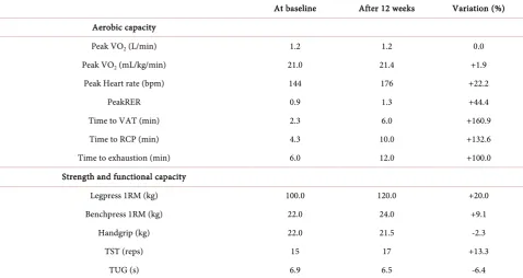
Data on aerobic capacity and muscle strength at baseline and after 12-week of exercise intervention are given in Table 1. After the 12-week exercise training program, the patient exhibited a substantial increase in aerobic capacity, dem-onstrated by increased time to ventilatory anaerobic threshold, time torespira-tory compensation point and time to exhaustion, with no changes in absolute (L/min) or relative (mL/kg/min) peak VO2. Moreover, increases in peak heart rate peak and respiratory exchange rate were observed. In addition, muscle strength was increased on the leg press and bench press exercises and improve-ment was seen in the TUG test, suggesting improved muscle function.
Body composition data are presented in Figure 1. The patient had a signifi-cant decrease in body fat and fat mass as well as an increase in fat-free mass, suggesting improved body composition after the 12-weeks exercise training program. The patient signed the consent form and consents with the publishing of this case report.
3. Discussion
This is the first study to evaluate the safety and benefits of exercise training in a patient with RP. The present study showed that a 12-week exercise training was able to increase aerobic capacity, as well as upper- and lower-limb muscle strength and functional capacity. In addition, a decrease in body fat and fat mass and an increase in fat-free mass were also observed. Notably, there was no dis-ease relapse or adverse events during the exercise training program.
DOI: 10.4236/ojra.2018.83010 96 Open Journal of Rheumatology and Autoimmune Diseases
Table 1. Aerobic capacity, muscle strength and functional capacity in the patient at baseline and after 12-weeks exercise training program.
At baseline After 12 weeks Variation (%) Aerobic capacity
Peak VO2 (L/min) 1.2 1.2 0.0
Peak VO2 (mL/kg/min) 21.0 21.4 +1.9
Peak Heart rate (bpm) 144 176 +22.2
PeakRER 0.9 1.3 +44.4
Time to VAT (min) 2.3 6.0 +160.9
Time to RCP (min) 4.3 10.0 +132.6
Time to exhaustion (min) 6.0 12.0 +100.0
Strength and functional capacity
Legpress 1RM (kg) 100.0 120.0 +20.0
Benchpress 1RM (kg) 22.0 24.0 +9.1
Handgrip (kg) 22.0 21.5 -2.3
TST (reps) 15 17 +13.3
TUG (s) 6.9 6.5 -6.4
RER: respiratory exchange rate; RCP: respiratory compensation point; RM: repetition maximum; TST: timed-stands test; TUG: timed-up-and-go. VAT: ventilatory anaerobic threshold; Peak VO2: peak oxygen uptake.
Figure 1. Body composition in the patient before and after 12-week exercise training program.
Impairmentsin aerobic capacity, muscle strength and function in autoimmune rheumatic disease have been associated with disease activity, impaired quality of life and increased cardiovascular risk in patients with autoimmune rheumatic diseases [11] [12]. In the present study, a 12-week exercise training program was shown to be effective for improving aerobic capacity and increasing muscle strength and function (i.e. TUG) in the RP patient, with no related adverse events reported, suggesting a potential therapeutic effect of exercise training in this disease.
[image:4.595.69.537.399.526.2]DOI: 10.4236/ojra.2018.83010 97 Open Journal of Rheumatology and Autoimmune Diseases strongly associated with worsening inflammation [14] [15]. Notably, recent stu-dies have demonstrated that exercise has a potential anti-inflammatory effect in inflammatory diseases [9] [10]. One mechanism that contributes to the an-ti-inflammatory effects of exercise training is a decrease in adipose tissue [14]. In the present study, substantial decreases in body fat and fat mass, besides an in-crease in fat-free mass, were observed. These results suggest that exercise train-ing can improve body composition and possibly help attenuate inflammatory markers in RP. Inflammatory markers were not evaluated in this case study and therefore further studies involving a larger number of patients are necessary to confirm this assumption.
4. Conclusion
In conclusion, the present case report suggests that a chronic exercise training program increased aerobic capacity, muscle strength and functional capacity in the patient. Additionally, a decrease in body fat and fat mass, and an increase in fat-free mass, suggests that exercise training can improve body composition in this disease. No adverse events were reported during the exercise training pro-gram, indicating that the exercise training was safe in this case study. However, studies assessing inflammatory markers, safety and the exercise training pro-gram in the disease are necessary to confirm these findings.
Conflicts of Interest
All authors declare no conflict of interest.
Fund
This work was supported by Fundação de Amparo à Pesquisa do Estado de São Paulo (FAPESP) [#2016/19771-5 to D.S.O, #2016/23574-0 to R.G.M., #2017/ 06794-0 to L.A.P]; Federico Foundation, Faculdade de Medicina da USP and Fundação Faculdade de Medicina to S.K.S.
References
[1] Trentham, D.E. and Le, C.H. (1998) Relapsing Polychondritis. Annals of Internal Medicine, 129, 114-122. https://doi.org/10.7326/0003-4819-129-2-199807150-00011
[2] McAdam, L.P., O’Hanlan, M.A., Bluestone, R. and Pearson, C.M. (1976) Relapsing Polychondritis: Prospective Study of 23 Patients and a Review of the Literature.
Medicine (Baltimore), 55, 193-215.
https://doi.org/10.1097/00005792-197605000-00001
[3] Pallo, P.A.O., Levy-Neto, M., Pereira, R.M.R. and Shinjo, S.K. (2017) Relapsing Po-lychondritis: Prevalence of Cardiovascular Diseases and Its Risk Factors, and Gen-eral Disease Features According to Gender. Revista Brasileira de Reumatologia Engl Ed, 57, 338-345. https://doi.org/10.1016/j.rbr.2017.01.002
[4] Schumacher, S. and Pieringer, H. (2017) Relapsing Polychondritis: A Chameleon among Orphan Diseases. Wiener Medizinische Wochenschrift, 167, 227-233.
DOI: 10.4236/ojra.2018.83010 98 Open Journal of Rheumatology and Autoimmune Diseases
[5] Michet Jr., C.J., McKenna, C.H., Luthra, H.S. and O’Fallon, W.M. (1986) Relapsing Polychondritis. Survival and Predictive Role of Early Disease Manifestations. An-nals of Internal Medicine, 104, 74-78. https://doi.org/10.7326/0003-4819-104-1-74
[6] Perandini, L.A., de Sá-Pinto, A.L., Roschel, H., Benatti, F.B., Lima, F.R., Bonfá, E. and Gualano, B. (2012) Exercise as a Therapeutic Tool to Counteract Inflammation and Clinical Symptoms in Autoimmune Rheumatic Diseases. Autoimmunity Re-views, 12, 218-224. https://doi.org/10.1016/j.autrev.2012.06.007
[7] Benatti, F.B. and Pedersen, B.K. (2015) Exercise as an Anti-Inflammatory Therapy for Rheumatic Diseases-Myokine Regulation. Nature Reviews Rheumatology, 11, 86-97. https://doi.org/10.1038/nrrheum.2014.193
[8] Stavropoulos-Kalinoglou, A., Metsios, G.S., Veldhuijzen van Zanten, J.J., Nightin-gale, P., Kitas, G.D. and Koutedakis, Y. (2013) Individualised Aerobic and Resis-tance Exercise Training Improves Cardiorespiratory Fitness and Reduces Cardi-ovascular Risk in Patients with Rheumatoid Arthritis. Annals of the Rheumatic Diseases, 72, 1819-1825. https://doi.org/10.1136/annrheumdis-2012-202075
[9] Perandini, L.A., Oliveira, D.S., Mello, S.B., Camara, N.O., Benatti, F.B., Lima, F.R., Borba, E., Bonfa, E., Sá-Pinto, A.L., Roschel, H. and Gualano, B. (2014) Exercise Training Can Attenuate the Inflammatory Milieu in Women with Systemic Lupus Erythematosus. Journal of Applied Physiology, 117, 639-647.
https://doi.org/10.1152/japplphysiol.00486.2014
[10] Oliveira, D.S., Shinjo, S.K., Silva, M.G., de Sá-Pinto, A.L., Lima, F.R., Roschel, H., Mello, S.B.V., Costa-Hong, V., Irigoyen, M.C.C., Pereira, R.M. and Gualano, B. (2017) Exercise in Takayasu Arteritis: Effects on Inflammatory and Angiogenic Factors and Disease-Related Symptoms. Arthritis Care & Research (Hoboken), 69, 892-902. https://doi.org/10.1002/acr.23011
[11] Khoja, S.S., Almeida, G.J., Chester Wasko, M., Terhorst, L. and Piva, S.R. (2016) Association of Light-Intensity Physical Activity with Lower Cardiovascular Disease Risk Burden in Rheumatoid Arthritis. Arthritis Care & Research (Hoboken), 68, 424-431. https://doi.org/10.1002/acr.22711
[12] Poulsen, K.B., Alexanderson, H., Dalgard, C., Jacobsen, S., Weile, L. and Diederich-sen, L.P. (2017) Quality of Life Correlates with Muscle Strength in Patients with Dermato- or Polymyositis. Clinical Rheumatology, 36, 2289-2295.
https://doi.org/10.1007/s10067-017-3706-6
[13] Partridge, R.E. and Duthie, J.J. (1963) Controlled Trial of the Effect of Complete Immobilization of the Joints in Rheumatoid Arthritis. Annals of the Rheumatic Diseases, 22, 91-99. https://doi.org/10.1136/ard.22.2.91
[14] Allison, M.A., Jensky, N.E., Marshall, S.J., Bertoni, A.G. and Cushman, M. (2012) Sedentary Behavior and Adiposity-Associated Inflammation: The Multi-Ethnic Study of Atherosclerosis. American Journal of Preventive Medicine, 42, 8-13.
https://doi.org/10.1016/j.amepre.2011.09.023
[15] Henson, J., Yates, T., Edwardson, C.L., Khunti, K., Talbot, D., Gray, L.J., Leigh, T.M., Carter, P. and Davies, M.J. (2013) Sedentary Time and Markers of Chronic Low-Grade Inflammation in a High Risk Population. PLoS One, 8, e78350.