Copper Nanoparticles as Modulators of Apoptosis and
Structural Changes in Tissues
Elena Sizova1, Sergey Miroshnikov1, Valentina Polyakova2, Natalia Gluschenko3, Anatoly Skalny1 1Orenburg State University, Orenburg, Russia; 2Orenburg State Medical Academy of Federal Agency in Public Health and Social
Development, Orenburg, Russia; 3Institute of Energy Problems in Chemical Physics RAS, Moscow, Russia. Email: [email protected]
Received October 16th, 2011; revised November 29th, 2011; accepted December 20th, 2011
ABSTRACT
Results of research on copper nanoparticles influence on index of readiness to apoptosis and structural changes of liver, spleen, kidney tissues as well as sensomotor cerebral cortex under copper multiple introductions into organism of ani- mals are presented in the article. It is established that copper nanoparticles distribute in organs and tissues of animals and cause specific structural changes. The increase of copper nanoparticles in organism up to toxical threshold (maxi- mum tolerated dose) results in dystrophy and tissue necrosis. Accurate expression enhancement of Caspase 3 in micro- gliocytes (brain macrophages) has been registered seven days after one-fold intramuscular introduction of copper nanoparticles (dose 2 mg/kg of animal body weight), in liver cells—three and seven days after three-fold intramuscular introduction of copper nanoparticles (total dose was 6 mg/kg of animal body weight), in proximal kidney tubules-three hours, one, three and seven days after three-fold intramuscular introduction of copper nanoparticles (with total dose 6 mg/kg of animal body weight), in spleen cells three hours, one, three and seven days after 12-fold intramuscular intro-duction (with total dose 24 mg/kg of animal body weight). Received data enables us to propose using index of cells readiness to apoptosis defined by Caspase 3 expression as a criterion for copper nanoparticles introduction safety as- sessment.
Keywords: Apoptosis; Copper Nanoparticles; Microelements; Spleen; Liver
1. Introduction
Development of modern technologies allows solving ma- ny medical and vetting problems. It is development of new materials for surgery and effective drug delivery systems, diagnostics and control over patient treatment, use nano- materials for new medicines and biological preparations. At present metallotherapy is generally recognized medi-cal sect, where organic and inorganic salts, metal com- plexes are used. Along with traditional medicines, con- taining metal, new ones having nanoparticles appear. Thus there are bandages with nanocrystalline silver for healing wounds, burns and trophic ulcer [1]. Indeed, nanometal characteristics differ from that of bulk samples and single atoms. Our researches on different elements nanoparti- cles allow us to establish that their toxicity is 7 times - 50 times less comparing to salts, they have multifunctional and durable action, easily penetrate into all organs and ti- ssues, stimulate metabolic processes when used in biotic doses, etc. [2-5]. At the same time mentioned peculiari-ties of nanoparticles can promote different pathological conditions in living systems and might even cause death. That’s why so many articles focus on living systems’ re-
sponse to nanomaterials: analysis of pulmonary tissue us- ing nanocarbon tubes [6], brain edema formation and ma- lfunction of blood-brain barrier under the influence of silver, copper, aluminium, silicon, carbon nanoparticles and metal oxides [7], study of central nervous system un- der the influence of silver nanoparticles and copper oxide [8], dopaminergic system under the influence of Mn, Ag, Cu nanoparticles [9]. There are also analyses of clinical and biochemical indexes of urine, serum, liver and kid- ney tissues at copper nanoparticles introduction in diffe- rent doses [10]; DNA damage degree and cytotoxicity of different metal oxides [11] is also studied. Comparative researches on nanoparticle toxicity continue, for example, copper in comparison with microparticles and metal ions [12]. At present, nanomaterials toxicity is connected with oxidative stress and DNA damage [13]. But other toxicity mechanisms are also considered. For instance, their dama- ging action on cytomembranes and organelles accelerates transportation of potentially toxic elements. Genotoxic and allergenic action is also possible [14].
are no criteria, which can help to decide an issue of sa- fety of this nanomaterial introduction into organism. In this connection, this article presents actual researches on change of apoptosis that is a marker of programmed cell death and tissue structure under increasing introduction of copper nanoparticles into the organism of animals.
2. Materials and Methods
The research was conducted on male Wistar with weight 150 g - 180 g that received common ration in vivarium; animals were weekly intramuscularly injected with aque- ous suspension of copper nanoparticles in dosage of 2.0 mg/kg animal weight. Another group of animals were watered with suspended mixture of copper nanoparticles in dosage of 2.0 mg/kg animal weight for 3 days. Expe- rimental research on animals was conducted according to instructions, recommended by the Russian Regulations, 1987 and “The Guide for Care and Use of Laboratory A- nimals (National Academy Press Washington, D.C. 1996)”. Copper nanoparticles were obtained with high tempera- ture condensation method using app Migen-3 [15,16]. Wa- ter suspension of copper nanoparticles for injections and watering was prepared according to the following sche- me: prepare nanopowder dry weight, dissolve it in water, consecutive suspension dispergating using ultrasound di- spersion machine UZDN-2Т in this mode: three-fold di- spergating for 1 minute with 3 minute pause. Test samp- les for the analysis of structural and functional status of liver were taken 3 hours, 1 day, 3 and 7 days after each injection and in 1 hour, 2 hours after enteral introduction of nanoparticles water suspension. Animals of control group were intramuscularly injected and enterally wate- red with distilled water in the same volume as tested ani- mals.
To research structural and functional condition of or- gans and tissues samples were for 1 day fixed in 10% neutral formaline at a room temperature. After standard histological preparation samples were embedded in par-affin. Paraffin-embedded slices of 5 μm thickness were stained by Mayer’s hematoxylin and eosin. To detect e- xogenic copper nanoparticles benzidine staining of para- ffin-embedded slices was also used [17]. Glycogen in li- ver was detected with Hotchkiss-Mcmanus-Shabadash PAS reaction [17]. Ki-67 expression index was used to assess cell proliferative activity and to identify cell readiness to apoptosis-Caspase-3 expression. Immunohistochemical re- searches were carried out on paraffin slices with the use of monoclonal antibodies and imaging system “Bio Ge- nex Super Sensytive Detection System” (USA) according to the records of the producer. Fragmented DNA in he- patocytes was identified using ApopTag (Plus Peroxidase in situ Apoptosis Detection Kit-S 71010) in accordance with records of the producer (Intergen, USA). Immuno-
positive cell were counted among 1000 and expressed in ‰.
Statistical processing of received data was performed using software package “Statistica 5,5 for Windows” and “MS Excel 2000”. Validity of compared indexes differ- rences was determined by t-criterion of Student. Results at p ≤ 0.05 were considered valid.
3. Results and Discussion
3.1. Structural Changes in Tissues of Animals at Copper Nanoparticles Introduction
Copper nanoparticles were synthesized using high tem- perature condensation method with their subsequent mo- dification with oxygen in order to study influence of co- pper nanoparticles on structure of tissues and organs [15, 16]. The research of physical and chemical characteris-tics revealed the following: copper nanoparticles have sp- herical form; average size is 103.0 nm ± 2.0 nm; nano- particle nucleus contains 96.0% ± 4.5% crystal copper, –4.0% ± 0.4 % copper oxide; thickness of oxide film on the surface is –6 nm [18].
Recent researches on biological action of unmodified copper nanoparticles after one-fold subcutaneous intro- duction allow to establish nanoparticle regular effect on the organism depending on the metal dosage. This effect can be described by the generalized curve “dosage-res- ponse” in the following form: 1) biotic zone of copper nanoparticles is within dosages that 2 times—250 times less toxical threshold-maximum permissible dose (MPD); 2) “safety” zone (when stimulatory action isn’t expressed and toxic effect doesn’t become apparent) is within dos-ages 15 mg/kg - 25 mg/kg; 3) pharmacotoxic action for copper nanoparticles begins from dosage 25 mg/kg, LD50
45 mg/kg, LD100 60 mg/kg. Copper nanoparticles toxicity
is 6 times less at LD100 toxic index of copper sulfate [19].
Comparing toxicity indexes of copper nanoparticles mo- dified by oxygen, which we used in our research, and un- modified nanoparticles it was established that modified nanoparticles are 2 times more biologically active.
Thus, pharmacotoxic zone for them begins from dosage 10 mg/kg (MPD), LD50 is 15 mg/kg, LD1100 30 mg/kg
[20]. Taking these data into account, a single introduc-tion dosage of 2 mg/kg allowing to reach MPD 10 mg/kg at a 5-fold copper nanoparticles introduction, a dosage close to LD100 24 mg/kg at a 12-fold introduction was
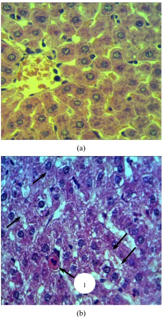
During the study of the liver 3 hours after one-fold in- tramuscular introduction of copper nanoparticles in dosa- ge of 2 mg/kg copper nanoparticles (Figure 1) can be
seen in the vascular part of the periportal hepatocytes and in the Kupffer cells cytoplasm of the experimental ani- mals liver, which disappear within 3 days after the intro- duction. At the same time no structural changes in the organism have been identified.
In the vascular part of the periportal hepatocytes and in the Kupffer cells cytoplasm of the experimental animals liver an amorphous moderately blue staining (indicated with the arrow) of exogenous copper can be seen using the modified benzidine staining on histological liver sec-tions.
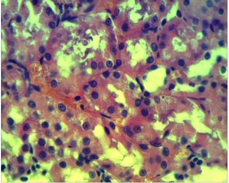
However, in periportal hepatocytes, there are signs of hydropic degeneration, which is not revealed during the study of an organism 7 days after a one-fold introduction of the metal. At a repeated intramuscular introduction of copper one week later nanoparticles are revealed mainly in the vascular part of the periportal hepatocytes. Here-with one day after the second metal introduction signs of hydropic degeneration are revealed in the vascular part of periportal hepatocytes. Apoptotic Kaunsilmen cells also appear among hepatocytes (Figure 2 (a) and (b)).
As compared to the control rats 1) in experimental rats hepatocytes; 2) vacuolation of the cytoplasm (indicated by an arrow) is observed and oxyphilic apoptotic Kaunsi- lmen cells are revealed (1). At the enteral introduction of copper nanoparticles 2 hours after the first watering there structural changes in liver with signs of hydropic degene- ration in the periportal zone hepatocytes could be seen. 3 days after enteral introduction of copper nanoparticles or- gan structure is not changed. There are no nanoparticles in the liver hepatocytes, but they are found in the Kup- ffer’s cells. Studingy liver 2 hours after the third enteral introduction, copper nanoparticles are present in Kupf- fer’s cells and the hepatocytes vascular zone. Herewith there were no signs of degeneration in cells.
Studing spleen structural organization in response to the increased load of copper on the organism we have found that copper nanoparticles could be seen in the red pulp of spleen, mainly in macrophages first day after in- tramuscular metal introduction and remain after subse- quent introductions. One-fold introduction of copper na- noparticles does not affect spleen structural homeostasis, but promotes a moderate increase of functional activity of the white pulp, the increase of its relative mass density, relative mass density of germinal centers, periarterial ly- mphoid clutches and increased rates of the number of ce- lls per unit of area in these zones testify to that fact. Mo- rphometric parameters of the white pulp after 2 and 3 in- jections of nanoparticles also attest to the increase of its functional activity. More significant changes in morpho- metric parameters of the white pulp could be seen 7 days
[image:3.595.341.504.191.329.2]after the repeated introduction of copper nanoparticles. Increase in lymphoid follicles after the second introduce- tion of copper nanoparticles takes place not only due to increase of germinal centers, where there are processes of blast transformation and lymphocytes reproduction, but in the majority of follicles by means of mantle and mar- ginal zones, where processes of differentiation and cell moving into the red pulp take place.
Figure 1. Histological section of the rat liver fragment 3 hours after one-fold intramuscular introduction of copper nano- particles in dosage of 2 mg/kg.
(a)
1
(b)
[image:3.595.340.506.374.698.2]Nanoparticles of copper could be found in cells of pro- ximal kidney tubules during first days after one-fold in- tramuscular metal introduction. Locally there are hya- line-drop and hydropic dystrophy phenomena in indivi- dual tubule cells. However, these phenomena could not be seen within 7 days after the introduction. Kidney glo- merules have not been changed, but macrophages expre- ssing Caspase 3 could be found in the interstitial tissue more frequently than in the control one. After the second and particularly after the third intramuscular injection of copper nanoparticles the number of tubules with signs of protein dystrophy increases. Epithelial cells having ne-crosis appear in the tubules, local damages of the basal - tubules membrane have been also identified (Figure 3).
There are hyaline-drop and hydropic dystrophy phe-nomena (1) in tubule cells and their necrosis in places (indicated with the arrows).
Nanoparticles are found in all proximal tubules after one-fold enteral introduction of nanoparticles as well as after intramuscular introduction. In some of them there are hyaline dropwise and hydropic dystrophy.
After a three-fold enteral introduction, along with pro- tein dystrophy phenomena the necrosis of some epithelial cells in tubules is revealed, but the basal membrane is not damaged that is good for the prediction of the regenera- tion process in these tubules.
Structural changes in the organs of the experimental group in comparison with the control one were not found after one-fold intramuscular introduction of copper nano- particles into the sensomotor zone of cerebral cortex. Be- nzidine staining had no positive results, probably due to the small amount of nanoparticles that entered tissue ele- ments of the nervous system.
These data suggest that copper nanoparticles can easily penetrate into all organs and tissues at both intramuscular and enteral introduction. Specific structural changes are observed depending on the type of tissue, the total dosa- ge of introduced nanoparticles and the response time. In case of copper nanoparticles load increase (total dosage of 4 mg/kg and 6 mg/kg) the phenomena of dystrophy and tissue necrosis are revealed in the studied tissues. Enteral way of nanoparticles introduction is safer than intramuscular one.
3.2. Expression of Apoptosis Marker after the Introduction of Copper Nanoparticles
Changes in the index of cell readiness to apoptosis, pro- grammed necrocytosis, take place together with structu- ral reorganization of tissues under the influence of cop- per nanoparticles. It is known that apoptosis is a form of necrocytosis, during which self-destruction occurrs as a result of DNA damage in a cell nucleus and it could not be resolved by a repair system. A protein encoded by the
[image:4.595.310.537.517.699.2]gene p53 follows this process. If it is impossible to re- solve the DNA damage an apoptosis is activated under the action of protein53 [21]. It is known that trace ele- ments play an important role in apoptosis regulation, which may intensify the effect of deficit and excess of elements, or as a result of vital trace elements imbalance. Thus zinc is a selective inhibitor of apoptosis, manganese, cadmium, lead, gallium, etc. are able to modulate an apo- ptosis, i.e. effect differently depending on the dosage [22]. Such elements as selenium, zinc, iron, copper prevent realization of a gene-damaging action [23,24]. Taking into account these data, we have studied the readiness of va- rious tissue cells to apoptosis, estimated by antigen ex- pression of Caspase 3 in case of copper nanoparticles load increase on the organism. The results are presented in the
Table 1 below.
In the liver of control animals the expression of Cas- pase 3 can be found only among centrolobular hepatocy- tes and it is 0.7‰ ± 0.03‰. Apoptag-positive cells are also seen among centrolobular hepatocytes and they are 0.5‰ ± 0.02‰. After repeated intramuscular introduc-tion of copper nanoparticles the cells expressing Caspase 3 antigen as well as having positive respond on a frag- mented DNA are discovered among the periportal heap- tocytes. The difference of markers expression of cell rea- diness to apoptosis is reliable in comparison to liver of the control group of animals 3 days and 7 days after three-fold intramuscular nanoparticle introduction. Ex-pression indexes increase twice (p < 0.05) 7 days after three-fold intramuscular introduction (Table 1).
During the immunohistochemical study of a prolifera- tive cell activity of lymphoid spleen follicles using anti- bodies Ki-67 it has been revealed that 7 days after the se- cond introduction of nanoparticles the expression Ki-67 has increased two times in comparison with the control
Table 1. The indexes of Caspase 3 antigen expression (*,**) in tissue cells after repeated intramuscular introduction of copper nanoparticles in the dosage of 2 mg/kg (in ‰).
Frequency of introduction of copper nanoparticles
First introduction Second introduction Third introduction
Time after an introduction
Liver Animal
groups
3 hour 1 day 3 day 7 day 3 hour 1 day 3 day 7 day 3 hour 1 day 3 day 7 day
control 0.7 ± 0.02 0.7 ± 0.03 0.7 ± 0.02 0.7 ± 0.02 0.7 ± 0.02 0.7 ± 0.02 0.67 ± 0.02 0.7 ± 0.02 0.7 ± 0.02 0.7 ± 0.02 0.7 ± 0.02 0.7 ± 0.02 experiment 0.7 ± 0.04 0.65 ± 0.04 0.8 ± 0.02 0.75 ± 0.03 0.9 ± 0.05 0.7 ± 0.03 0.6 ± 0.05 0.71 ± 0.05 0.68 ± 0.04 1.0 ± 0.04 1.1 ± 0.03** 1.5 ± 0.06**
Proximal kidney tubules
control 24.1 ± 0.6 24.1 ± 0.6 24.1 ± 0.6 24.1 ± 0.6 24.1 ± 0.6 24.1 ± 0.6 24.1 ± 0.6 24.1 ± 0.6 24.1 ± 0.6 24.1 ± 0.6 24.1 ± 0.6 24.1 ± 0.6
experiment 26.3 ± 0.9 25.8 ± 0.7 27.2 ± 0.5 24.6 ± 0.8 25.5 ± 0.9 27.1 ± 0.2 26.1 ± 0.6 29.1 ± 0.7 32.1 ± 1.8** 36.1 ± 2.6** 44.1 ± 3.8** 60.2 ± 3.4**
Spleen
First introduction Second introduction Twelfth introduction
Time after an introduction
3 hour 1 day 3 day 7 day 3 hour 1 day 3 day 7 day 3 hour 1 day 3 day 7 day control 1.8 ± 0.6 2.0 ± 0.4 1.7 ± 0.5 2.2 ± 0.3 1.9 ± 0.4 2.1 ± 0.2 2.0 ± 0.3 2.3 ± 0.5 2.0 ± 0.1 2.4 ± 0.4 2.3 ± 0.7 2.5 ± 0.7
experiment 2.2 ± 0.4 2.5 ± 0.6 2.0 ± 0.3 3.5 ± 0.5 2.8 ± 0.3 2.6 ± 0.4 3.2 ± 0.5 1.9 ± 0.2 5.1 ± 0.4** 5.7 ± 0.3** 6.9 ± 0.5** 8.3 ± 0.4**
Microgliocytes of sensomotor cortex zone
First introduction
Time after an introduction
1 day 3 day 7 day 14 day
control 1.5 ± 0.4 1.5 ± 0.4 1.5 ± 0.4 1.5 ± 0.4
experiment 1.7 ± 0.2 1.8 ± 0.1 4.6 ± 0.3** 1.4 ± 0.3
*Data presented as: average (X) ± standard error of the average (SE) (n = 1000); **results are statistically significant (p < 0.05).
one and decreased 1.5 times 7 days after the 12th inject- tion of copper nanoparticles in comparison with the con- trol group (p < 0.05). The study of follicular cells readi- ness to apoptosis has revealed a reduction of Caspase 3 antigen expression 7 days after the second injection of nanoparticles. Seven days after the 12th injection the nu- mber of cells of lymphoid spleen follicles expressing Caspase 3 antigen increases, indicating an increased rea- diness of follicular cells to apoptosis (Table 1).
The readiness of kidney tubule epithelial cells to apo- ptosis significantly increases after the second and parti- cularly after the third intramuscular injection of copper nanoparticles. If the antigen expression of Caspase 3 in tubular epithelium of the control animals was 24.1‰ ± 0.6‰, after the third intramuscular injection of copper nanoparticles indexes increase 2.5 times (p < 0.05) in co- mparison with the control (Table 1) ones.
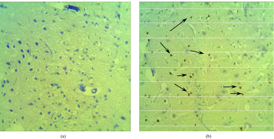
Immunohistochemical detection of cells expressing Сa- spase 3 antigen in the sensomotor cortex zone revealed the readiness of cells to a programmed necrocytosis a- mong microgliocytes (brain macrophages). Nerve cells expressing Caspase 3 antigen at different times after a
one-fold intramuscular introduction of copper nanoparti- cles (1st, 3rd, 7th, 14th day) have not been found. The rate
of Caspase 3 expression of microgliocytes 1st and 3rd day
after the introduction of copper nanoparticles has been unreliably increased in comparison with the control. Ho- wever, 7 days after one-fold intramuscular introduction of copper nanoparticles the expression of Caspase 3 an- tigen has increased three times in comparison with the control group (Figures 4(a) and (b)). The expression of
Caspase 3 in microgliocytes has decreased and corres- ponded to the indexes of the control animals after 14 days (Table 1).
After the enteral introduction of copper nanoparticles in a dosage of 2 mg/kg of an animal body weight the readiness of cells to a liver cells apoptosis after a three- fold enteral introduction (total dosage is 6 mg/kg) unre- liably differs from the rates of apoptosis marker expres- sion in a liver of control group animals (Table 2). At the
[image:5.595.62.539.121.451.2][image:6.595.61.538.83.325.2]
(a) (b)
Figure 4. Histological liver section of the control (a) and the experimental rat (b) 1 day after the repeated intramuscular introduction of copper nanoparticles in the dosage of 2 mg/kg.
Table 2. The indexes of Caspase 3 antigen expression (*) in liver cells after an enteral introduction of copper nanoparticles in the dosage of 2 mg/kg (in ‰).
Frequency of introduction of copper nanoparticles
First introduction Second introduction Third introduction
Time after an introduction Animal
groups
1 hour 2 hour 1 hour 2 hour 1 hour 2 hour
control 0.5 ± 0.04 0.5 ± 0.04 0.5 ± 0.04 0.5 ± 0.04 0.5 ± 0.04 0.5 ± 0.04
experiment 0.6 ± 0.03 0.56 ± 0.02 0.65 ± 0.05 0.58 ± 0.07 0.69 ± 0.02 0.7 ± 0.05
*Data presented as: average (X) ± standard error of the average (SE) (n = 1000).
Microgliocytes expressing Caspase 3 antigen (indicated with arrows) are revealed more in the sensomotor cortex zone of the experimental rat 1) compared with the control; 2) animal.
Therefore, at copper nanoparticles load increase on the organism, besides tissues restructuring, there is a signify- cant increase of Caspase 3 expression in microgliocytes (brain macrophages) of the cortex 7 days after one-fold intramuscular introduction of copper nanoparticles 2 mg/kg), in liver cells-3 days and 7 days after 3-fold intramuscular introduction of copper nanoparticles (total dosage is 6 mg/kg), in proximal kidney tubules it happens 3 hours, 1 days, 3 days, and 7 days after 3-fold intramuscular intro- duction of copper nanoparticles (total dosage is 6 mg/kg), in spleen cells—3 hours, 1, 3, and 7 days after a 12-fold intramuscular introduction of copper nanoparticles (total dosage is 24 mg/kg).
The data indicate a high biological activity of copper
nanoparticles when injected into the organism. Copper nanoparticles in a dosage of 2 mg/kg reveal neurotoxicity. Microgliocytes (brain macrophages) of the sensomotor cortex zone are the most sensitive to copper nanoparti- cles. And though the effect on them at the index of cells readiness to apoptosis was reversible (2 weeks after one- fold introduction of copper nanoparticles in a dosage of 2 mg/kg the level of apoptosis can be compared with that of the control group), its realization in case of further do- sage increase of introduced nanoparticles will lead to the loss of cells that can cause neurodegenerative processes in the cortex.
Hepatotoxicity and nephrotoxicity of copper nanopar- ticles appear in a dosage of 6 mg/kg. Moreover, the rea- diness to apoptosis is observed in peri-portal hepatocytes after 3 days, in kidney tubules epithelial cells 3 hours after 3-fold introduction of copper nanoparticles.
[image:6.595.65.540.394.496.2]copper nanoparticles was: cells of lymphoid follicles in- creased expression of Caspase 3 antigen in response to the introduction of copper nanoparticles in a dosage of 24 mg/kg, i.e. in a dosage close to LD100.
The established quality of copper nanoparticles in non- toxic dosages (2 mg/kg - 6 mg/kg) to increase the cell readiness to apoptosis can be used in the chemotherapy of malignant neoplasms when there is apoptosis inhibit- tion of cells containing mutations. Silver nanoparticles as copper nanoparticles have the same quality to modulate apoptosis, based on that, there are bandages for healing wounds, burns and trophic ulcer. Due to this quality na- nocrystalline silver reduces inflammatory stage of heal- ing and thus eases an initial period of wound healing [1].
In addition, using data received from the conducted re- search, it seems obvious that index of cell readiness to a- poptosis reflects the beginning of irreversible structural changes regardless of tissue peculiarity in response to the introduction of copper nanoparticles. All this allow us to suggest an apoptosis index as a criterion for the assess- ment of safe introduction of copper nanoparticles into organism of living systems. By this index we can judge acceptable dosages of introduced metal, target organs, es- tablish optimal and safe methods of introducing nanopar- ticles into the organism for further use in the composition of drugs and biological preparations.
REFERENCES
[1] J. B. Wright, K. Lam, A. G. Buret, M. E. Olson and R. E. Burrell, “Early Healing Events in a Porcine Model of Contaminated Wounds: Effects of Nanocrystalline Silver on Matrix Metal loproteinases, Cell Apoptosis, and Heal- ing,” Wound Repair and Regeneration, Vol. 10, No. 3, 2002, pp. 141-151.
doi:10.1046/j.1524-475X.2002.10308.x
[2] N. N. Glushchenko, O. A. Bogoslovskaya and I. P. Olkhovs- kaya, “Physical and Chemical Laws of Biological Effect of Superfine Metal Powders,” Chemical Physics, Vol. 21, No. 4, 2002, pp. 79-86.
[3] T. A. Baytukalov, N. N. Glushchenko, O. A. Bogoslovs- kaya, I. P. Olkhovskaya, G. E. Folmanis and I. P. Arsen-tieva, “Wound Healing Composition and Method of Pre- paration,” RF Patent No. 2296571, 2007.
[4] T. A. Baytukalov, N. N. Glushchenko, O. A. Bogoslovs- kaya, I. P. Olkhovskaya, I. O. Leipunsky, A. N. Zhigach and E. A. Shafranovsky, “Drug Accelerating Wound Heal- ing,” RF Patent No.2306141, 2007.
[5] “Publications for 1977-2010 Years on the Web Site of the Laboratory.” http://www. nanobiology.narod.ru
[6] A. A. Shvedova and V. E. Kagan, “The Role of Nanotoxi- cology in Realizing the ‘Helping without Harm’ Para- digm of Nanomedicine: Lessons from Studies of Pulmo- nary Effects of Single-Walled Carbon Nanotubes,” Jour-
nal of Internal Medicine, Vol. 276, No. 1, 2010, pp. 106-
118.
[7] H. S. Sharma, S. Hussain, J. Schlager, S. F. Ali and A. Shar- ma, “Influence of Nanoparticles on Blood-Brain Barrier Permeability and Brain Edema Formation in Rats,” Acta
Neurochirurgica Supplementum, Vol. 106, 2010, pp. 359-
364. doi:10.1007/978-3-211-98811-4_65
[8] Z. Yang, Z. W. Liu, R. P. Allaker, P. Reip, J. Oxford, Z. Ahmad and G. Ren, “A Review of Nanoparticle Func- tionality and Toxicity on the Central Nervous System,”
Journal of the Royal Society Interface, Vol. 7, Suppl. 4,
2010, pp. 411-422.
[9] J. Wang, M. F. Rahman, H. M. Duhart, G. D. Newport, T. A. Patterson, R. C. Murdock, S. M. Hussain, J. J. Schla- ger and S. F. Ali, “Expression Changes of Dopaminergic System-Related Genes in PC12 Cells Induced by Manga- nese, Silver, or Copper Nanoparticles,” Neurotoxicology, Vol. 30, No. 6, 2009, pp. 926-933.
doi:10.1016/j.neuro.2009.09.005
[10] M. Y. Liao, R. H. Lei, C. Q. Wu, B. H. Yang, H. Z. Ma, C. Shi, Q. J. Wang, Q. X. Wang and Y. Yuan, “Integrated Metabolomic Analysis of the Nano-Sized Copper Particle- Induced Hepatotoxicity and Nephrotoxicity in Rats: A Ra-
pid in Vivo Screening Method for Nanotoxicity,” Toxi-
cology and Applied Pharmacology, Vol. 232, No. 2, 2008,
pp. 292-301. doi:10.1016/j.taap.2008.06.026
[11] H. L. Karlsson, P. Cronholm, J. Gustafsson and L. Möller, “Copper Oxide Nanoparticles are Highly Toxic: A Com- parison between Metal Oxide Nanoparticles and Carbon Nanotubes,” Chemical Research in Toxicology, Vol. 21, No. 9, 2008, pp. 1726-1732. doi:10.1021/tx800064j [12] Z. Chen, H. Meng, G. Xing, C. Chen, Y. Zhao, G. Jia, T.
Wang, H. Yuan, C. Ye, F. Zhao, Z. Chai, C. Zhu, X. Fang, B. Ma and L. Wan, “Acute Toxicological Effects of Cop- per Nanoparticles in Vivo,” Toxicology Letters, Vol. 163, No. 2, 2006, pp. 109-120.
doi:10.1016/j.toxlet.2005.10.003
[13] A. R. Murray, E. Kisin, S. S. Leonard, S. H. Young, C. Kom- mineni, V. E. Kagan, V. Castranova and A. A. Shvedova, “Oxidative Stress and Inflammatory Response in Dermal Toxicity of Single-Walled Carbon Nanotubes,” Toxicol- ogy, Vol. 257, No. 3, 2009, pp. 161-171.
doi:10.1016/j.tox.2008.12.023
[14] G. G. Onishchenko, “The Concept of Toxicological Stu- dies, Estimated Risk Methodology, Methods of Identifi- cation and Quantification of Nanomaterials,” Resolution
79, Registered in the Russian Ministry of Justice, Regis- tration No. 10528, 2007.
[15] M. Ya. Gen and A. V. Miller, “Author’s Certificate of the USSR No. 814432,” Bulletin of Discoveries, No. 11, 1981, pp. 25-28.
[16] А. N. Zhigach, I. О. Leipunsky, М. L. Kuskov, N. I. Sto- enko and V. B. Storozhev, “Facility for Producing and Studying Physical and Chemical Properties of Metal Na- noparticles,” Instruments and Experimental Techniques, Vol. 4, No. 6, 2000, pp.122-125.
[17] E. Peirce, “Histochemistry, Theoretical and Applied,” Chur- chill, Moscow, 1962, pp. 962-968.
per Nanoparticles Depending on Their Physical and Che- mical Characteristics,” Russian Nanotechnology, Vol. 5, No. 3-4, 2010, pp. 102-111.
[19] N. N. Glushchenko, I. P. Olkhovskaya, T. V. Pleteneva, L. D. Fatkullina, Yu. A. Ershov and Yu. I. Fedorov, “The Biological Effect of Superfine Metal Powders,” Izvestiya
АN, Vol. 6, No.3, 1989, pp. 415-418.
[20] О. А. Bogoslovskaya, Е. А. Sizova, V. S. Polyakova, S.
А. Miroshnikov, I. О. Leipunsky, I. P. Olkhovskaya and N. N. Glushchenko, “The Study of Safe Introduction of Copper Nanoparticles with Different Physical-Chemical Characteristics into Organisms of Animals,” Bulletin of OSU, Vol. 32, No. 2, 2009, pp. 124-128.
[21] V. А. Galitsky, “The Emergence of Eukaryotic Cells and the Origin of Apoptosis,” Cytology, Vol. 47, No. 2, 2005,
pp. 103-108.
[22] А. V. Kudrin and А. А. Zhavoronkov, “The Role of Minor Constituences and Calcium in the Regulation of Apop- tosis,” Successes of Modern Biology, Vol. 118, 1998, pp. 7-15.
[23] N. I. Kaletina and G. I. Kaletin, “Minor Constituences— Biological Controls,” Academy of Science, No. 1, 2007, pp. 50-60.